Successful Cryoablation of Atrioventricular Nodal
Reentrant Tachycardia and Coexisting Accessory Pathways
without Fluoroscopy
Enes Elvin Gul, MD, Fatma Seyma Ugur, PhD, Celal Akdeniz, MD, and Volkan Tuzcu, MD Department of Pediatric Cardiology/Electrophysiology, Medipol University, Istanbul, Turkey
A B S T R A C T
We report the case of a 14-year-old boy patient admitted to our outpatient clinic with palpitations and documented supraventricular tachycardia. Electrophysiological study and ablation were planned. In the electrophysiological study, two tachycardias with different cycle lengths and morphologies were induced. After elimination of the slow pathway, left posterior accessory pathway was detected and successfully ablated. Another pathway was detected following that ablation. Due to the slow retrograde conduction of this pathway, diltiazem infusion was started to uncover the accessory pathway. The second accessory pathway was at the left posteroseptal region and was successfully ablated. After a 30-minute waiting period, no tachycardia was induced. In addition, no fluoroscopy was used during the procedure.
Key Words. AVNRT; Accessory Pathway; Cryoablation; Fluoroscopy
Introduction
T
he prevalence of coexistent atrioventricular reentrant tachycardia (AVNRT) and AVRT has been rarely reported in the literature. The bypass tract is usually first targeted for ablation because of the concept that dual atrioventricular (AV) nodal physiology is more physiological for the heart than accessory pathway (AP). In this article, we briefly discuss that multiple supraven-tricular arrhythmia substrates can be successfully eliminated without fluoroscopy with the aid of electroanatomical systems.Case Presentation
A 14-year-old boy presented with recurrent palpi-tations, despite the treatment with metoprolol. Twelve-lead resting electrocardiography (Mortara Instrument, Inc., Milwaukee, WI, USA) showed normal sinus rhythm without preexcitation. An event recorder revealed supraventricular tachycar-dia with a rate of 167 bpm. There was no family history of syncope, sudden death, or arrhythmias. Admission physical examination was unremark-able. Echocardiography (Vivid S6, GE Healthcare, Wauwatosa, WI, USA) revealed a structurally normal heart. Electrophysiology study and
abla-tion were recommended due to recurrent tachycar-dia under medical treatment.
An electrophysiological study was performed under general anesthesia without intubation. Quadripolar catheters were positioned at the high atrium, His-bundle area, and right ventricular apex. A steerable decapolar catheter was inserted into the coronary sinus for a positional reference. The EnSite NavX system (St. Jude Medical, St. Paul, MN, USA) was used for three-dimensional mapping and catheter navigation. Right atrial anatomy was reconstructed without fluoroscopy. Baseline electrophysiologic (EP) measurements were obtained. During incremental ventricular pacing, ventriculoatrial conduction was eccentric, and block was at 420 ms. After intravenous orci-prenaline injection, tachycardia with a 375-ms cycle length was induced. During the tachycardia, the earliest ventriculoatrial conduction was from the left posterior wall (Figure 1). Another form of tachycardia with a different cycle length (305 ms) consistent with AVNRT was induced (Figure 2). A 6-mm tip cryoablation catheter (Freezor, Medtronic Inc., MN, USA) was inserted via the right femoral vein. Cryoablation of the slow pathway was initiated, and the tachycardia was ter-minated at 12 seconds of the ablation. Six lesions, © 2012 Wiley Periodicals, Inc. Congenit Heart Dis. 2013;8:E178–E182
240 seconds each, were placed in the slow pathway region. To eliminate the left posterior pathway, a cryocatheter was inserted into the left side via patent foramen ovale. The left concealed acces-sory pathway was eliminated with cryoablation. In addition, Kent spikes were demonstrated during the cryoablation of the left posterior AP. After-ward, during the incremental ventricular pacing, the ventriculoatrial (VA) conduction was still eccentric with indication of a second left-sided pathway. The retrograde conduction was slow, and
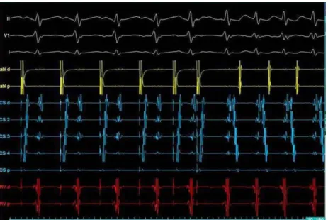
AV nodal conduction was prominent. Therefore, intravenous diltiazem (15 mg) was started to uncover the second AP. After diltiazem, the second AP in the left posteroseptal location started to demonstrate intermittent preexcitation (Figure 3). Successful cryoablation with four lesions, each 240 seconds, were placed for the left posteroseptal AP (Figure 4). After a 30-minute waiting period, there was no inducible tachycardia with or without orciprenaline. In addition, during the AV block obtained after adenosine injection, no preexcita-Figure 1. S1 and S2 stimulations from coronary sinus before ablation. VA conduction after pacing is eccentric: the earliest atrial activation is at distal coronary sinus suggesting left posterior AP. VA, ventriculoatrial; AP, accessory pathway.
tion was manifested. The total procedure time was 330 minutes, and no fluoroscopy was used. No complications occurred.
Discussion
The prevalence of coexistent AVNRT and AVRT has been reported in about 3–4% out of all
patients with supraventricular reentrant tachycar-dias.1,2The bypass tract is usually first targeted for
ablation because of the concept that dual AV nodal physiology is more physiological for the heart than AP. The AV node slow pathway is ablated only when tachycardia is induced during the electro-physiological study. There are some reports that two different tachycardias were treated by the Figure 3. Manifest preexcitation is shown: the earliest atrial activity at CS 4, i.e., at the left posteroseptal region. CS, coronary sinus.
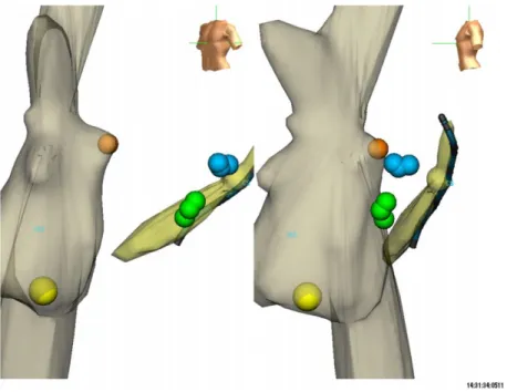
Figure 4. Three-dimensional anatomic mapping showing successful cryoablation sites of AVNRT (yellow lesion) and both APs: left posterior (blue lesions) and left posteroseptal (green lesions). Note that patent foramen ovale (PFO) was crossed. AVNRT, atrioventricular nodal reentrant tachycardia; AP, accessory pathway.
ablation of the slow pathway alone when the APs are in high-risk regions (septal area) for complica-tions.3,4 The success of the abovementioned
reports was due to the fact that the anterograde limb of the AP was the slow AV nodal pathway. However, in our case, we induced supraventricular tachycardia utilizing the left posterior AP follow-ing successful AVNRT ablation. In addition, a second AP with intermittent antegrade conduc-tion was shown in the left posteroseptal area.
To minimize the risk of heart block, cryothermia mapping has been recently used.5Cryoablation is a
safer option for ablation in close proximity to the AV node.6To the best of our knowledge, this is the
first case where successful cryoablation of the AV node slow pathway and coexisting APs (left posterior and left posteroseptal) were performed under the guidance of a nonfluoroscopic, three-dimensional mapping system. Superiorities of cryoablation are as follows: (1) safety, (2) low risk of heart block, (3) better catheter stabilization, and (4) less thrombogenicity. Cryoablation is a newer method of ablation, which has proven to be safe in many studies.7,8An acute success rate of
cryoabla-tion is 95% for AVNRT.9Conventional method for
left-sided AP ablation is radio frequency (RF) abla-tion. However, a recent study demonstrated signifi-cantly improved acute and long-term success rates for the left-sided APs using cryoablation when compared with RF ablation.8
Nonfluoroscopic method is a superiority of this case because radiation exposure during radiofre-quency ablation carries known associated risks.10
Three-dimensional anatomic mapping with the EnSite system mainly eliminates fluoroscopy use in majority of the procedures.11,12 Some recent
studies have demonstrated that children are espe-cially vulnerable for cancer with increasing number of diagnostic and therapeutic radiation exposures and this risk appears to persist through-out life.13Very low doses of radiation may be more
harmful than previously suspected and that the relationship may not, in fact, be linear in the pedi-atric population.14 There are reasonable benefits
to both patients and staff (especially pregnant women) in eliminating radiation exposure. Ortho-pedic complications related to lead aprons are relatively common.15In addition, left brain
malig-nancies were reported among interventional car-diologists.16
In conclusion, this case demonstrates that mul-tiple supraventricular arrhythmia substrates can be successfully eliminated without fluoroscopy with the aid of electroanatomical systems.
Author Contributions
Enes Elvin Gul: manuscript writing and literature search. Fatima S. Ugur: language editing.
Celal Akdeniz: performed ablation.
Volkan Tuzcu: performed ablation, manuscript writing, editing, and supervision.
Corresponding author: Volkan Tuzcu, MD, Depart-ment of Pediatric Cardiology/Electrophysiology, Istan-bul Medipol University, IstanIstan-bul 34093, Turkey. Tel: (+0090) 212-460-7782; Fax: (+90) (212) 460 70 70; E-mail: [email protected]
Conflict of interest and disclosure of funding: The manu-script, as submitted or its essence in another version, is not under consideration for publication elsewhere and will not be published elsewhere while under consider-ation by Congenital Heart Disease journal. The authors have no commercial associations or sources of support that might pose a conflict of interest. All authors have made substantive contributions to the study, and all authors endorse the data and conclusions.
Accepted in final form: November 13, 2012.
References
1 Kuo JY, Tai CT, Chiang CE, et al. Mechanisms of transition between double paroxysmal supraven-tricular tachycardias. J Cardiovasc Electrophysiol. 2001;12:1339–1345.
2 Pritchett EL, Prystowsky EN, Benditt DG, Gal-lagher JJ. “Dual atrioventricular nodal pathways” in patients with Wolff-Parkinson-White syndrome. Br Heart J. 1980;43:7–13.
3 Padanilam BJ, Akula D, Vaughn P, Prystowsky EN. Successful elimination of concealed accessory pathway-mediated tachycardia by ablation of AV nodal slow pathway. J Cardiovasc Electrophysiol. 2006; 17:674–677.
4 Di Biase L, Bai R, Tritto M, Grimaldi M, Biasco MG. Ablation of atrioventricular nodal “slow pathway” for simultaneous treatment of coexisting atrioventricular and nodal reciprocating tachycar-dias. J Interv Card Electrophysiol. 2007;19:143–147. 5 Lanzotti ME, De Ponti R, Tritto M, Spadacini G,
Salerno-Uriarte JA. Successful treatment of anteroseptal accessory pathways by transvenous cryomapping and cryoablation. Ital Heart J. 2002; 3:128–132.
6 Tuzcu V. Cryoablation of accessory pathways in children. Pacing Clin Electrophysiol. 2007;30:1129– 1135.
7 Drago F, De Santis A, Grutter G, Silvetti MS. Trans-venous cryothermal catheter ablation of re-entry
circuit located near the atrioventricular junction in pediatric patients: efficacy, safety, and midterm follow-up. J Am Coll Cardiol. 2005;45:1096–1103. 8 Tuzcu V. Significant reduction of fluoroscopy in
pediatric catheter ablation procedures: long-term experience from a single center. Pacing Clin Electro-physiol. 2012;35:1067–1073.
9 de Sisti A, Tonet J. Cryoablation of atrioventricular nodal reentrant tachycardia: a clinical review. Pacing Clin Electrophysiol. 2012;35:233–240.
10 Lindsay BD, Eichling JO, Ambos HD, Cain ME. Radiation exposure to patients and medical person-nel during radiofrequency catheter ablation for supraventricular tachycardia. Am J Cardiol. 1992; 70:218–223.
11 Smith G, Clark JM. Elimination of fluoroscopy use in a pediatric electrophysiology laboratory utilizing three-dimensional mapping. Pacing Clin Electro-physiol. 2007;30:510–518.
12 Tuzcu V. A nonfluoroscopic approach for electro-physiology and catheter ablation procedures using
a three-dimensional navigation system. Pacing Clin Electrophysiol. 2007;30:519–525.
13 Kleinerman RA. Cancer risks following diagnostic and therapeutic radiation exposure in children. Pediatr Radiol. 2006;36(suppl 2):121–125.
14 Beels L, Bacher K, De Wolf D, Werbrouck J, Thie-rens H. gamma-H2AX foci as a biomarker for patient X-ray exposure in pediatric cardiac catheter-ization: are we underestimating radiation risks? Circulation. 2009;120:1903–1909.
15 Goldstein JA, Balter S, Cowley M, Hodgson J, Klein LW. Occupational hazards of interventional cardiologists: prevalence of orthopedic health prob-lems in contemporary practice. Catheter Cardiovasc Interv. 2004;63:407–411.
16 Roguin A, Goldstein J, Bar O. Brain tumours among interventional cardiologists: a cause for alarm? Report of four new cases from two cities and a review of the literature. EuroIntervention. 2012; 7:1081–1086.