https://doi.org/10.1007/s11696-020-01280-5
ORIGINAL PAPER
An electrochemical label‑free DNA impedimetric sensor
with AuNP‑modified glass fiber/carbonaceous electrode
for the detection of HIV‑1 DNA
Ece Ç. Yeter1 · Samet Şahin2 · M. Oguzhan Caglayan2 · Zafer Üstündağ1
Received: 23 April 2020 / Accepted: 6 July 2020
© Institute of Chemistry, Slovak Academy of Sciences 2020 Abstract
In this study, a highly sensitive, electrochemical, and label-free DNA impedimetric sensor was developed using carbonized glass fiber–coal tar pitch (GF–CTP) electrodes supported with gold nanoparticles (AuNPs) for the detection of HIV-1 gene. Thiol-modified GF–CTP electrodes were prepared using amine crosslinking chemistry and AuNPs were self-assembled obtaining highly conductive nanoelectrodes, GF–CTP–ATP–Au. All steps of electrode modifications were characterized using electrochemical, spectroscopic, and microscopic methods. GF–CTP–ATP–Au electrode was then modified with a capture DNA probe (C-ssDNA) and optimized with a target DNA probe in terms of hybridization time and temperature between 30 and 180 min and 20–50 °C, respectively. Finally, the analytic performance of the developed ssDNA biosensor was evaluated using electrochemical impedance spectroscopy. The calibration of the sensor was obtained between 0.1 pM and 10 nM analyte working range. The limit of detection was calculated using signal to noise ratio of 3 (S/N = 3) as 13 fM. Moreover, interference results for two noncomplementary DNA probes were also tested to demonstrate non-specific ssDNA interactions. An electrochemical label-free DNA impedimetric sensor was successfully developed using a novel GF–CTP– ATP–Au electrode. This study suggests that highly sensitive DNA-based biosensors can be developed using relatively low-cost carbonaceous materials.
Keywords Carbonaceous materials · Impedimetric sensor · DNA sensor · HIV-1 · Coal tar pitch
Introduction
Viruses cause many morbidities and mortalities worldwide as they can spread in many ways. The major viral patho-gens include influenza virus, respiratory syncytial virus, and coronavirus that can cause acute respiratory diseases; on the other hand, some viruses attack the immune system such as human immunodeficiency virus (HIV) (Mahony 2008). World Health Organization estimates that approximately 37.9 million people were living with HIV at the end of
2018 with only 62% receiving lifelong antiretroviral therapy (WHO 2019). HIV infection, therefore, still is a major public health issue in the world; moreover, HIV testing is not acces-sible to everyone. There are two types of HIVs: HIV-1 and HIV-2, but research on HIV mostly focuses on HIV-1 since it is more viral and pandemic (Fatin et al. 2016).
HIV-1 can be detected using several methods. The meth-ods used for HIV-1 detection include enzyme-linked immu-nosorbent assay (Re et al. 2001), western blot (Pagans et al.
2011), immunostaining (Del Valle et al. 2000), fluorescence immunoassay (Yamamoto and Kumar 2000), quartz crys-tal microbalance (Minunni et al. 2004), surface plasmon resonance chip (Tombelli et al. 2005), field-effect transistor (Ruslinda et al. 2013) and biosensors (Laksanasopin et al.
2015). Most of these methods rely on antibody–antigen reac-tion and it provides accurate results; however, it is time-con-suming and costly as well as requires large sample volumes. On the other hand, electrochemical biosensors can be a good alternative due to their simplicity, relatively low cost, high sensitivity, and their potential for point of care use.
* Samet Şahin
[email protected] * Zafer Üstündağ
1 Department of Chemistry, Kütahya Dumlupınar University,
43100 Kütahya, Turkey
2 Department of Bioengineering, Bilecik Şeyh Edebali
Various electrochemical methods have been developed for the detection of the HIV-1 gene recently. Square wave voltammetry (Zhang et al. 2010), differential pulse voltam-metry (Li et al. 2014), amperometry (Gao et al. 2018), elec-trogenerated chemiluminescence (Poorghasem et al. 2016) and electrochemical impedance spectroscopy (EIS) (Gong et al. 2015) techniques were employed to design ultra-sen-sitive HIV-1 DNA biosensors. Most of them resulted in a limit of detection values of below pM concentrations for pM–nM detection ranges. However, some of the mentioned methods require hybridization indicators or redox-labels that bring complexity to sensor design; therefore, a label-free electrochemical biosensor would be desirable. EIS, on the other hand, can produce ultra-sensitive results without the need of a redox or any other labeling for hybridization and electrochemical signal production. Promising results for the detection of HIV-1 gene have been obtained using EIS reaching pM to fM concentrations (Gong et al. 2017). Recently, electrochemical studies based on EIS and differ-ential pulse voltammetry (DPV) have been reported with a limit of detection values of as low as 8.3 fM utilizing carbon nanostructures (Jia et al. 2019; Li et al. 2020). However, the cost and complex synthesis processes of nanostructures such as carbon nanotubes (CNTs) and graphene are still a con-cern. Therefore, more efficient and low-cost ways to produce electrically conductive electrodes are essential for sensor development.
Carbonaceous materials (CMs) such as CNTs and gra-phene have attracted so many researchers for the last 2 dec-ades (Jana et al. 2013). CMs show excellent mechanical properties and electrical conductivity which make them a perfect candidate for industrial applications such as solar cells (Chen et al. 2014), hydrogen storage (Hassan et al.
2007), supercapacitors (Frackowiak et al. 2006) and sen-sors (Wei et al. 2013). However, CNTs and GPH are some of the expensive examples of CMs; on the other hand, low-cost CMs can also be synthesized using low-low-cost alterna-tives such as coal tar pitch (CTP). Carbonized CTP can be a very good alternative with high surface area and excellent electrical conductivity especially for the synthesis of CTP composites (Erkal et al. 2016). GF is a low-cost and com-mercially available material often used in refractor materi-als and furnace insulations due to aluminum silicate in the structure; therefore, its use in bioelectronic applications is limited. However, when carbonized with CTP, its electrical properties could dramatically improve.
DNA immobilization and hybridization efficiencies are very important to construct ultra-sensitive biosensors; therefore, several nanomaterials such as gold nanoparticles (AuNPs) (Cai et al. 2001), ZnO (Yumak et al. 2011), Fe/ Pt/ZSn nanocore–shell (Chang and Wu 2010), a mercap-toaceticacid-modified cadmium sulfide (Sun et al. 2007), and carbon-nanotubes/nanozirconiumdioxide/chitosan
(Yang et al. 2007) have been used to help DNA immo-bilization and hybridization. Among all, AuNPs are the most employed nanomaterial and previous results showed that both immobilization and hybridization qualities are improved using AuNP-supported electrodes compare to their bare form.
In this work, a novel material based on glass fiber (GF) was first developed using CTP, and then a highly sensitive, electrochemical, and label-free DNA impedi-metric sensor was developed using carbonized GF–CTP electrodes supported with AuNPs for the detection of the HIV-1 DNA. Different electrode conformations were pre-pared for electrochemical and surface characterizations. Voltammetric and impedimetric analyses were used for electrochemical characterization; IR spectroscopy, scan-ning electron microscopy–energy-dispersive X-ray spec-troscopy (SEM–EDX), X-ray photoelectron specspec-troscopy (XPS), and transmission electron microscopy (TEM) anal-yses were used for the characterization of the electrodes. The analytic performance of the developed DNA biosensor was evaluated using EIS. This study, to the best of authors’ knowledge, is a pioneer demonstration of the carboniza-tion of GF with CTP for sensor applicacarboniza-tions. Therefore, highly sensitive DNA-based biosensors can be developed using relatively low-cost carbon materials, and more sen-sitive results can be achieved with further modifications.
Experimental
Materials
Tetrachloroauric (III) acid (Sigma-Aldrich, 99%), triso-dium citrate (Merck, > 99%), 1-ethyl-3-(3-dimethylamino-propyl) carbodiimide (EDC, Sigma-Aldrich), N-hydrox-ysuccinimide (NHS, Sigma-Aldrich), 4-aminothiophenol (ATP, Sigma-Aldrich), acetonitrile (Riedel, HPLC grade), ethanol (Merck, absolute), K3Fe(CN)6/K4Fe(CN)6
(Sigma-Aldrich, > 99.9%), DNA probes (Helix Bio), CTP (Orcan Company) and other chemicals of reagent grade were used as received from the supplier. All reagents and standards were prepared in ultrapure water (UPW, Human Power 1 + , Korea). The electrochemical experiments were car-ried out with an Ivium CompactStat (NL). The electro-chemical measurements were performed using a three-electrode system under high-purity argon (99.999%) atmosphere at room temperature (24 ± 1 °C). The reference electrode was an Ag/AgCl (Sat) electrode, the auxiliary electrode a platinum wire. AuNP-modified glass fiber/car-bonaceous disk electrode was used as a working electrode. The electrode surface area was adjusted as 0.071 cm2 with
Preparation of thiol‑modified carbon disk electrodes
The preparation of carbonized sensor materials and its sen-sor application was performed accordingly to the procedure described in E. C. Yeter’s M.Sc. thesis (Yeter 2018). Thiol (SH-)-terminated CTP-coated glass fiber (GF) electrodes were prepared according to the procedure described else-where (Güzel et al. 2010) using amine cross-linking chem-istry. Briefly, GF was washed with UPW, ethanol, and ace-tonitrile, respectively, for 10 min in an ultrasonic bath. 10 g of dried GF was then mixed and homogenized with 10 g of CTP in acetone. Carbonization of the GF–CTP mixture was carried out in a tube furnace (Protherm, USA) at 1000 °C with a heating rate of 10 °C/min at under nitrogen atmos-phere. Carbonized GF–CTP was ground into fine powder in a planetary ball mill (Retcsch/PM100, Germany) for 1 h under 500 rpm. Disk electrodes were prepared under pres-sure using a ‘kamalak’ (Cedrus libani A. Rich.) resin in ethanol as a binder with GF–CTP and CTP (Üstündağ and Erkal 2017).
GF–CTP electrodes were then prepared for AuNP modification.
The schematic representation of the modification process for AuNP terminated carbonaceous material is shown in Fig. 1. First, GF–CTP was treated with 4 M HNO3 for 12 h
to obtain carboxylic acid groups on the surface (denoted as
GF–CTP–COOH). GF–CTP–COOH was then washed with UPW and COOH groups were activated with 0.01 M EDC and NHS mixture in 0.1 M potassium phosphate buffered solution (PBS) at pH 7 for 12 h. Activated COOH groups were modified with 1 mM ATP, 0.1 M PBS, pH 7, for 8 h and denoted as GF–CTP–ATP.
Preparation of gold nanoparticles (AuNPs) and AuNP‑modified carbonaceous disk electrodes
250 µL of tetrachloroauric (III) acid (25 mM, HAuCl4.3H2O)
solution was first diluted to 50 mL, and 5 mL of 1.5% triso-dium citrate solution was slowly added under 500 rpm vig-orous stirring at 98 °C. Upon color change to wine red, the colloidal solution was cooled down to 4 °C and centrifuged (Hermle, Z36HK, Germany) at 20,000 rpm. The obtained AuNPs were then washed twice with UPW and absolute ethanol, respectively, and kept at 4 °C in ethanol when not in use. GF–CTP–ATP electrodes were then immersed in an ethanoic AuNP solution for 8 h to obtain self-assembled monolayers (SAMs) on the electrode surface. Final electrode conformation was denoted as GF–CTP–ATP––ATP–Au.
Characterization of the carbonaceous electrodes
GF–CTP–ATP––ATP–Au, GF–CTP–ATP, GF–CTP, CTP, and glassy carbon (GC) electrodes were first characterized using linear sweep voltammetry (LSV) and electrochemical impedance spectroscopy (EIS). All tested electrodes have a Fig. 1 Schematic diagram of the
AuNP modification on GF–CTP electrode
surface area of 0.071 cm2 and all electrochemical tests were
performed using 2 mM K3Fe(CN)6/K4Fe(CN)6 redox probe
in 0.1 M KCl. IR spectroscopy (Bruker, Tensor-27, Ger-many), SEM–EDX (Nova, NanoSEM-650, Belgium), XPS (PHI 5000-Versa Probe, Φ ULVAC-PHI.Inc., Japan/USA) and TEM (JEOL Ltd., Tokyo, Japan) analyses were used for the characterization of the prepared electrodes and AuNPs.
ssDNA‑based impedimetric sensor characterization
Table 1 shows a summary of the single-strand DNA (ssDNA) oligonucleotides used in this study. All ssDNA oligonucleotide solutions were prepared using 20 mM NaCl at pH 7.4 and kept at − 20 °C when not in use.
GF–CTP–ATP–Au electrode surface was first modified with 1 µM of C-ssDNA (Ella Biotech GmbH, Germany) for 1 h following by a washing step with UPW to remove unbound C-ssDNA molecules. This conformation was denoted as GF–CTP–ATP–Au/C-ssDNA. Optimization of the ss-DNA-based impedimetric sensor was performed using EIS in 2 mM K3Fe(CN)6/K4Fe(CN)6 redox couple (0.1 M KCl) for hybridization time and temperature parameters. Hybridization time is defined as the interaction time between C-ssDNA and T-ssDNA. Hybridization time and tempera-ture ranges were chosen as 30–180 min and 20–50 °C, respectively. The sensor calibration range was chosen as 0.1 pM – 10 nM. The schematic representation of the detect-ing principle for T-ssDNA is given in Fig. 2. The stability of the sensor was calculated using 1 nM T-ssDNA and accu-racy tests were conducted using 100 pM and 1 nM T-ssDNA for 7 days. Finally, selectivity was determined using two different ss-DNA strands for 10 nM T-ssDNA.
Results and discussion
To evaluate the effect of the modifications performed on disk electrode surfaces, LSV and EIS tests were first conducted. Figure 3a shows that the GF–CTP–ATP–Au electrode has the highest oxidation peak potential reaching around ca. 30 µA current response indicating enhanced signal response compared to CTP electrode and commercial GC electrode. This behavior was further confirmed using EIS technique
(frequency range from 100 to 0.1 kHz with 10 mV wave amplitude at a DC potential of 0.115 V in 2 mM K3Fe(CN)6/
K4Fe(CN)6 redox probe in 0.1 M KCl) and the Nyquist plots
of the redox couple for each electrode are shown in Fig. 3b. Charge transfer resistance (Rct) values were calculated for each electrode by fitting the EIS data using a War-burg-effected diffusion-controlled type equivalent circuit (Randviir and Banks 2013). A sample equivalent circuit and fitted experimental data can be seen from Fig. 3c. The Rct values for GC, CTP, GF–CTP, GF–CTP–ATP,
and GF–CTP–ATP–Au were calculated as 9.41 ± 0.67, 8.83 ± 0.74, 6.44 ± 0.47, 5.57 ± 0.41, and 1.83 ± 0.17 kΩ, respectively. Impedance analysis showed that GF–CTP–ATP–Au electrode has the lowest charge trans-fer resistance indicating fast electron transtrans-fer type kinetics for the redox probes compared to other tested electrodes. Voltammetry and impedance tests, therefore, confirmed that AuNPs enhanced the electron transfer rate of redox probes providing minimum charge transfer resistance among pre-pared electrodes.
Figure 3d shows the stability of the GF–CTP–ATP–Au electrode when kept in open-air conditions at room tem-perature for 10 days. The oxidation peak current for 2 mM K3Fe(CN)6/K4Fe(CN)6 redox probe was changed from 30.33 ± 0.35 µA to 30.27 ± 0.41 µA showing that the GF–CTP–ATP–Au electrode has very good stability. This superior electrochemical properties and good stability of GF–CTP–ATP–Au could be due to the tunneling effect of the nanoparticle-supported carbonaceous structure (Sayed et al. 2012).
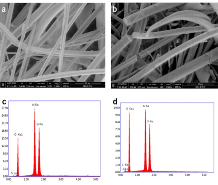
Figure 4 also shows SEM and EDX analyses of the GF electrode before and after carbonization. Figure 4a, b does not show distinctive differences between GF and GF–CTP; however, the EDX spectrum (Fig. 4c) confirms that GF is coated with CTP confirmed with the appearance of C (Kα1). Furthermore, EDX results revealed that no C was found in GF; on the other hand, GF–CTP electrode has 2% C by weight.
Figure 5a shows the TEM image of synthesized AuNP confirming 10–60 nm size spherical-like AuNPs was suc-cessfully synthesized. IR analysis of GF–CTP–ATP (Fig. 5b) shows that GF has its characteristic peaks at 950–1500 cm−1
and 550–800 cm−1 for Al–O–Si deformation and Si–O
Table 1 Summary of the DNA oligonucleotides used in this study
ssDNA ssDNA Sequence
Capture ssDNA (C-ssDNA) 5′-HS-(CH2)4-GAT CGC CCT TTT CCA TTT CCA TAC
ATA TTT CTG TTA -3’
Target ssDNA (T-ssDNA) 3′-GGA AAA GGT AAA GGT ATG TAT AAA GAC AAT -5’
Non-specific ssDNA-1
(NC1-ssDNA) 3′-ATA ATC CAC CTA TCC CAG TAG GAG AAAT-5’
Non-specific DNA-2
quartz, respectively. Moreover, Si–O–Si and H–O–H con-formations can also be seen at 475 cm−1 and 1650 cm−1,
respectively (Rondón et al. 2013; Vaculikova et al. 2011). After the carbonization process (GF–CTP), we can observe aliphatic and aromatic C–H interactions at 2700–3000 cm−1
and 3000–3100 cm−1, respectively. O–H stretching was
seen at 3000–3500 cm−1 after the carbonization process.
Besides, aromatic C–H bending can be seen between 650 and 1000 cm−1. Aromatic ring mode can also be seen around
1600 cm−1. Finally, upon amine functionalization, -C = O
(carbonyl) peaks appeared at 1720 cm−1 and N–H groups
can be seen at 3400–3700 cm−1 (Cheng et al. 2014; Çelik
et al. 2016; Lin et al. 2005).
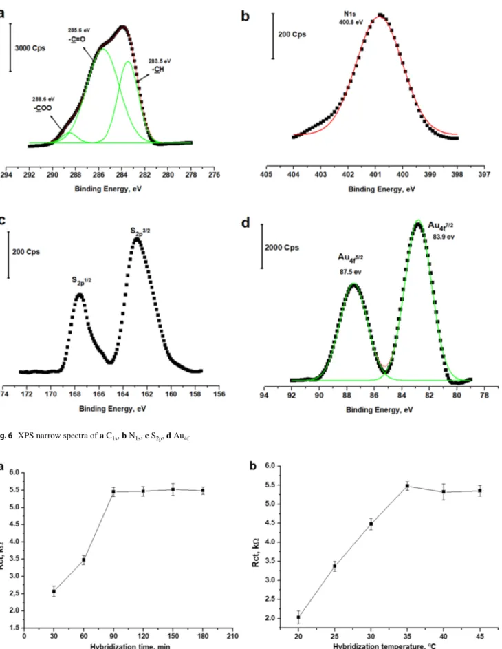
H i g h - r e s o l u t i o n n a r r o w s p e c t r a o f GF–CTP–ATP–Au electrode for C1s, N1s, S2p, and Au4f are
given in Fig. 6. High-resolution C1s narrow spectrum of the GF–CTP–ATP–Au electrode is given in Fig. 6a. When the C1s region is fitted, three main peaks regarding -COO,
-C = O and -CH were obtained at 288.6 eV, 285.6 eV, and 283.5 eV, respectively. Similarly, N1s binding energy was
obtained at 400.8 eV. Furthermore, S2p1/2 and S
2p3/2 binding
energies were obtained at 167.8 eV and 162.7 eV respec-tively. Finally, Au4f5/2 and Au4f7/2 binding energies were
obtained at 87.5 eV and 83.9 eV, respectively. As a result, XPS analysis confirmed the formation of a carbonaceous electrode with ATP amidization and the functionalization of AuNPs on -SH groups.
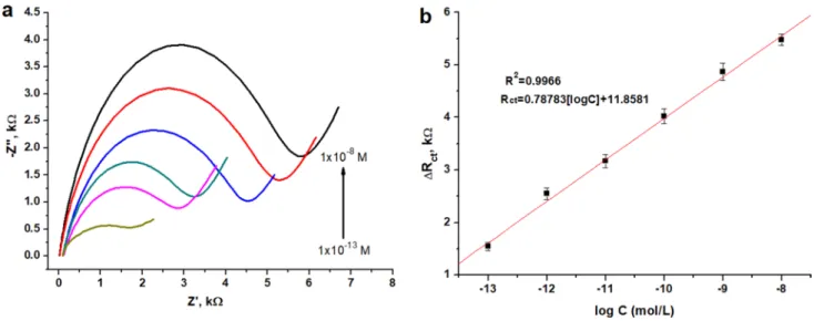
Optimization studies were carried out to confirm the hybridization time and temperature for GF–CTP–ATP–Au. First, the electrode was modified with C-ssDNA for 1 h Fig. 2 Schematic diagram of the
and washed excessively with UPW to remove unbounded ssDNA. GF–CTP–ATP–Au/C-ssDNA was then exposed to 10 nM T-ssDNA for 30 to 180 min from 20 to 50 ºC. Fig-ure 7 shows the optimization results for (1) hybridization time and (2) temperature using Rct data obtained from EIS
analysis. Figure 7 shows that hybridization time and the tem-perature reached a plateau at 90 min and 35 ºC, respectively. Therefore, these parameters were used to obtain calibration curves for the ssDNA sensor.
The calibration of the ssDNA sensor was obtained using the EIS technique between 0.1 pM and 10 nM analyte work-ing range. Figure 8 shows the Nyquist data and calibration curve for the developed ssDNA sensor. It can be seen that the Rct values increased upon the increasing concentration
of T-ssDNA indicating that the electron transfer rate of the redox couple becomes dramatically limited. This type of behavior from DNA hybridization in impedance analysis can
also be seen from similar studies in the literature (Bonanni and Pumera 2011; Riedel et al. 2014). The limit of detec-tion (LOD) was calculated using a signal to noise ratio of 3 (S/N = 3) as 13 fM. This is one of the highest LOD values with a wide range of detection range among various DNA-based sensors (Gupta et al. 2013; Liu et al. 2010; Yang et al.
2012; Zhang et al. 2011; Zhu et al. 2005).
The stability of the prepared C-ssDNA sensor for T-ssDNA hybridization is presented in Table 2 for 7-day tests. The prepared sensor showed very good stability over 7 days changing between 96.7% and 101.4% for minimum and maximum obtained recoveries, respectively. When considered for 95% confidence level, the obtained stability results do not show a significant difference during 7 days.
Precision (RSD %) and accuracy measurements were conducted for two different concentrations of T-ssDNA (100 pM and 1 nM). Experiments were performed in two Fig. 3 a LSV scans and b Nyquist plots of different electrode
confor-mations in 2 mM K3Fe(CN)6/K4Fe(CN)6 redox probe (in 0.1 M KCl). c Equivalent circuit and fitted experimental data for GF–CTP–ATP–
Au electrode. d Stability of the GF–CTP–ATP–Au electrode when kept in open-air conditions at room temperature for 10 days, tested in 2 mM K3Fe(CN)6/K4Fe(CN)6 redox probe (in 0.1 M KCl)
Fig. 4 SEM imaging of a GF and b GF–CTP and EDX spectra of c GF and d GF–CTP
Fig. 6 XPS narrow spectra of a C1s, b N1s, c S2p, d Au4f
Fig. 7 Optimization of a hybridization time and b temperature for GF–CTP–ATP–Au/C-ssDNA after exposed to 10 nM T-ssDNA, tested in 2 mM K3Fe(CN)6/K4Fe(CN)6 redox probe (in 0.1 M KCl).
Hybridization time tests were conducted at 35 ºC. Error bars are sam-ple standard deviations (N = 5 samsam-ples)
different modes: intra-day and inter-day precision for five independent samples. Moreover, inter-day precision meas-urements were performed for seven consecutive days. The obtained results are given in Table 3. RSD % and accu-racy results show that the proposed method provides RSD % and accuracy % values not more than around ≤ 2.2% and ≥ − 2%, respectively, for both intra-day and inter-day measurements. This indicates very good precision and accuracy for the developed ssDNA sensor.
Table 4 shows the interference results for two possible interferences, NC1-ssDNA and NC2-ssDNA to test
non-specific ssDNA interactions. It can be seen from Table 4
that the change in the value of Rct is negligible compared
to the values obtained for the T-ssDNA for the interfer-ing ssDNAs. Therefore, no specific interaction from the selected ssDNAs was observed.
The proposed ssDNA sensor shows a lower LOD with a broad detection range for HIV-1 DNA detection than recently published studies using EIS (Table 5). This is mainly because of the highly conductive carbon fiber structures obtained after the carbonization process and the contribution of Au on the electrode surface. Moreover, the carbon fiber structure also provides a very high active sur-face area. DPV and amperometry are other electrochemical methods that provide a range of limits of detection val-ues among which a recent study based on DPV performs similar to this study. The main advantages of this work are that (1) a low-cost raw material (GF) was converted into valuable conducting electrode material (GF–CTP) and (2) its performance was further improved using a simple approach. As a result, a highly sensitive impedimetric ss-DNA sensor for HIV-1 ss-DNA was successfully developed. Fig. 8 The Nyquist data and b calibration curve for the developed ssDNA sensor. Error bars are sample standard deviations (N = 5 samples)
Table 2 Stability of the ssDNA sensor for T-ssDNA hybridization. Error bars are sample standard deviations (N = 5 samples) Day Rct, kΩ RSD % 1 4.87 ± 0.6 – 2 4.79 ± 0.11 98.4 3 4.85 ± 0.15 99.6 4 4.71 ± 0.15 96.7 5 4.94 ± 0.11 101.4 6 4.83 ± 0.17 99.2 7 4.80 ± 0.16 98.6
Table 3 Precision and accuracy of the ssDNA sensor
Error bars are sample standard deviations (N = 5 samples)
Found value Added T-DNA
100.0 pM 1.00 nM
Intra-day Found value 98.6 ± 2.18 1.02 ± 0.01
RSD % 2.211 0.980
Accuracy % − 1.4 2.0
Inter-day Found value 99.16 ± 1.74 0.98 ± 0.01
RSD % 1.755 1.020
Accuracy % − 0.84 − 2.0
Table 4 The effect of possible interfering ssDNAs on the sensor Interfering ssDNA Concentration, (μM) Rct change %
NC1-DNA 1.0 − 1.24
Conclusions
A novel and sensitive carbonaceous electrode was designed for HIV-1 detection using a label-free DNA probe. A low-cost and commercially available GF is utilized to obtain highly conductive and high-surface area electrode material due to its fiber structure and carbonization process. Moreo-ver, this structure was further improved using self-ordered AuNPs attached to the thiol-modified GF–CTP electrode. Electrochemical investigations showed successful attach-ment of each modification layer on electrodes. XPS and IR results also confirmed the structure of the Au-attached carbonized electrode. Optimization studies revealed that hybridization time and temperature of C-ssDNA and T-ssDNA were stable after 90 min and 35 ºC, respectively. The calibration of the sensor was obtained using EIS between 0.1 pM and 10 nM (1 × 10⁻¹³ and 1 × 10⁻⁸ M) with
13 fM (1.3 × 10⁻¹⁴M) detection limit. No significant
non-specific ssDNA interactions were observed for two different noncomplementary DNA probes chosen. Furthermore, very good precision and accuracy for the ssDNA sensor were obtained providing RSD % and accuracy % values between 2.2% and − 2%, respectively, for both intra-day and inter-day measurements. This study shows one of the best analytic performance parameters compared to similar ssDNA-based sensors in the literature providing a low-cost and easy elec-trode preparation methodology that could also be used in different analyte-sensing strategies.
Compliance with ethical standards
Conflict of interest On behalf of all authors, the corresponding authors state that there is no conflictof interest.
References
Bonanni A, Pumera M (2011) Graphene platform for hairpin-DNA-based impedimetric genosensing. ACS Nano 5:2356–2361. https ://doi.org/10.1021/nn200 091p
Cai H, Xu C, He P, Fang Y (2001) Colloid Au-enhanced DNA immo-bilization for the electrochemical detection of sequence-specific DNA. J Electroanal Chem 510(78):85
Chang H, Wu S-h (2010) Hybridization of FePt/ZnS nanocore-shell structure with DNAs of different sequences. Mater Trans 51:2094–2098
Chen J-G, Vittal R, Yeh M-H, Chen C-Y, Wu C-G, Ho K-C (2014) Carbonaceous allotropes modified ionic liquid electrolytes for efficient quasi-solid-state dye-sensitized solar cells. Electrochim Acta 130:587–593
Cheng Y, Fang C, Su J, Yu R, Li T (2014) Carbonization behavior and mesophase conversion kinetics of coal tar pitch using a low temperature molten salt method. J Anal Appl Pyrol 109:90–97 Çelik GK, Üzdürmez AF, Erkal A, Kılıç E, Solak AO, Üstündağ Z
(2016) 3, 8-Diaminobenzo [c] cinnoline derivatived graphene oxide modified graphene oxide sensor for the voltammetric deter-mination of Cd 2+ and Pb 2+. Electrocatalysis 7:207–214 Del Valle L, Croul S, Morgello S, Amini S, Rappaport J, Khalili K
(2000) Detection of HIV-1 Tat and JCV capsid protein, VP1, in AIDS brain with progressive multifocal leukoencephalopathy. J Neurovirol 6:221–228
Erkal A, Aşık İ, Yavuz S, Kariper A, Üstündağ Z (2016) Biosensor application of carbonaceous nanocoil material: preparation, char-acterization, and determination of dopamine and uric acid in the presence of ascorbic acid. J Electrochem Soc 163:H269–H277 Fatin M et al (2016) HIV-1 Tat biosensor: current development and
trends for early detection strategies. Biosens Bioelect 78:358–366 Frackowiak E, Machnikowski J, Béguin F (2006) Novel carbonaceous materials for application in the electrochemical supercapacitors. In: new carbon based materials for electrochemical energy stor-age systems: Batteries, Supercapacitors and Fuel Cells. Springer, pp 5–20
Gao X, Wang X, Li Y, He J, Yu H-Z (2018) Exonuclease I-hydrolysis assisted electrochemical quantitation of surface-immobilized DNA hairpins and improved HIV-1 gene detection. Anal Chem 90:8147–8153
Gong Q, Wang Y, Yang H (2017) A sensitive impedimetric DNA bio-sensor for the determination of the HIV gene based on graphene-Nafion composite film. Biosens Bioelectron 89:565–569 Gong Q, Yang H, Dong Y, Zhang W (2015) A sensitive
impedimet-ric DNA biosensor for the determination of the HIV gene based on electrochemically reduced graphene oxide. Anal Methods 7:2554–2562
Gupta VK, Yola ML, Qureshi MS, Solak AO, Atar N, Üstündağ Z (2013) A novel impedimetric biosensor based on graphene oxide/ gold nanoplatform for detection of DNA arrays. Sens Actuators B Chem 188:1201–1211
Güzel R et al (2010) Effect of Au and Au@ Ag core–shell nanoparti-cles on the SERS of bridging organic molecules. J Coll Inter Sci 351:35–42
Hassan NHA, Mohamed AR, Zein SHS (2007) Study of hydrogen stor-age by carbonaceous material at room temperature. Diam Relat Mat 16:1517–1523
Jana P, Fierro V, Celzard A (2013) Ultralow cost reticulated carbon foams from household cleaning pad wastes. Carbon 62:517–520 Jia Z et al (2019) NiCo2O4 spinel embedded with carbon nanotubes
derived from bimetallic NiCo metal-organic framework for the Table 5 Analytical performance
of some of the recent studies in the literature based on electrochemical HIV DNA detection
Electrode Technique Linear range LOD References
hpDNA/Au Amperometry 10 pM–50 nM 150 fM (Gao et al. 2018)
NH2-rGO/beta-CD/GCE DPV 0.05 pM–1 pM 8.7 fM (Li et al. 2020)
NiCo2O4/CoO@CNTs EIS 0.1 pM–20 nM 16.7 fM (Jia et al. 2019)
GN/Nafion/GCE EIS 0.1 pM –100 nM 23 fM (Gong et al. 2017)
ultrasensitive detection of human immune deficiency virus-1 gene. Biosens Bioelect 133:55–63
Laksanasopin T et al (2015) A smartphone dongle for diagno-sis of infectious diseases at the point of care. Sci Trans Med 7:273–re271
Li B et al (2014) Sensitive HIV-1 detection in a homogeneous solution based on an electrochemical molecular beacon coupled with a nafion–graphene composite film modified screen-printed carbon electrode. Biosens Bioelectron 52:330–336
Li J, Jin X, Feng M, Huang S, Feng J (2020) Ultrasensitive and highly selective electrochemical biosensor for hiv gene detection based on amino-reduced graphene oxide and β-cyclodextrin modified glassy carbon electrode. Int J Electrochem Sci 15:2727–2738 Lin Q, Li T, Ji Y, Wang W, Wang X (2005) Study of the modification
of coal-tar pitch with p-methyl benzaldehyde. Fuel 84:177–182 Liu S, Liu J, Wang L, Zhao F (2010) Development of electrochemical
DNA biosensor based on gold nanoparticle modified electrode by electroless deposition. Bioelectrochemistry 79:37–42
Mahony JB (2008) Detection of respiratory viruses by molecular meth-ods. Clin Microbiol Rev 21:716–747
Minunni M, Tombelli S, Gullotto A, Luzi E, Mascini M (2004) Devel-opment of biosensors with aptamers as bio-recognition element: the case of HIV-1 Tat protein. Biosens Bioelect 20:1149–1156 Pagans S, Sakane N, Schnölzer M, Ott M (2011) Characterization of
HIV Tat modifications using novel methyl-lysine-specific antibod-ies. Methods 53:91–96
Poorghasem R, Saberi RS, Shayan M, Mehrgardi MA, Kiani A (2016) Closed bipolar electrochemistry for the detection of human immunodeficiency virus short oligonucleotide. Electrochim Acta 222:1483–1490
Randviir EP, Banks CE (2013) Electrochemical impedance spectros-copy: an overview of bioanalytical applications. Anal Methods 5:1098–1115
Re M, Vignoli M, Furlini G, Gibellini D, Colangeli V, Vitone F, La Placa M (2001) Antibodies against full-length Tat protein and some low-molecular-weight Tat-peptides correlate with low or undetectable viral load in HIV-1 seropositive patients. J Clin Virol 21:81–89
Riedel M, Kartchemnik J, Schöning MJ, Lisdat F (2014) Impedimetric DNA detection-steps forward to sensorial application Anal Chem 86:7867–7874
Rondón W, Freire D, Benzo Z, Sifontes A, González Y, Valero M, Brito J (2013) Application of 3A zeolite prepared from venezuelan kaolin for removal of Pb (II) from wastewater and its determina-tion by flame atomic absorpdetermina-tion spectrometry. Am J Anal Chem 4(10):584-593. https ://doi.org/10.4236/ajac.2013.41006 9 Ruslinda AR, Tanabe K, Ibori S, Wang X, Kawarada H (2013) Effects
of diamond-FET-based RNA aptamer sensing for detection of real sample of HIV-1 Tat protein. Biosens Bioelect 40:277–282 Sayed SY, Fereiro JA, Yan H, McCreery RL, Bergren AJ (2012) Charge
transport in molecular electronic junctions: Compression of the molecular tunnel barrier in the strong coupling regime. Proc Natl Acad Sci 109:11498–11503
Sun W, Zhong J, Zhang B, Jiao K (2007) Application of cadmium sulfide nanoparticles as oligonucleotide labels for the electro-chemical detection of NOS terminator gene sequences. Anal Bioanal Chem 389:2179–2184
Tombelli S, Minunni M, Luzi E, Mascini M (2005) Aptamer-based biosensors for the detection of HIV-1 Tat protein. Bioelectrochem-istry 67:135–141
Üstündağ İ, Erkal A (2017) Determination of dopamine in the presence of ascorbic acid on digitonin-doped coal tar pitch carbonaceous electrode. Sens Mat 29:85–94
Vaculíková L, Plevová E, Vallová S, Koutnik I (2011) Characteriza-tion and differentiaCharacteriza-tion of kaolinites from selected Czech deposits using infrared spectroscopy and differential thermal analysis. Acta Geodynamica et Geomaterialia 8(1):59–67 [cit. 2020-07-07] Wei Y, Yang R, Liu J-H, Huang X-J (2013) Selective detection toward
Hg (II) and Pb (II) using polypyrrole/carbonaceous nanospheres modified screen-printed electrode. Electrochim Acta 105:218–223 WHO, HIV/AIDS. https ://www.who.int/news-room/fact-sheet s/detai l/
hiv-aids. Accessed 19 Nov 2019
Yamamoto R, Kumar PK (2000) Molecular beacon aptamer fluoresces in the presence of Tat protein of HIV-1. Gene Cells 5:389–396 Yang J, Wang X, Shi H (2012) An electrochemical DNA biosensor for
highly sensitive detection of phosphinothricin acetyltransferase gene sequence based on polyaniline-(mesoporous nanozirconia)/ poly-tyrosine film. Sens Acta B Chem 162:178–183
Yang Y, Wang Z, Yang M, Li J, Zheng F, Shen G, Yu R (2007) Elec-trical detection of deoxyribonucleic acid hybridization based on carbon-nanotubes/nano zirconium dioxide/chitosan-modified elec-trodes. Anal Chim Acta 584:268–274
Yeter EÇ (2018) Preparation of Glass Fiber Electrode for Impedimetric DNA Analysis and its Application. Kütahya Dumlupınar Univer-sity, M.Sc.
Yumak T, Kuralay F, Muti M, Sinag A, Erdem A, Abaci S (2011) Preparation and characterization of zinc oxide nanoparticles and their sensor applications for electrochemical monitoring of nucleic acid hybridization. Colloids Surf B 86:397–403
Zhang D, Peng Y, Qi H, Gao Q, Zhang C (2010) Label-free electro-chemical DNA biosensor array for simultaneous detection of the HIV-1 and HIV-2 oligonucleotides incorporating different hairpin-DNA probes and redox indicator. Biosens Bioelect 25:1088–1094 Zhang Y et al (2011) An electrochemical DNA sensor based on a lay-ers–film construction modified electrode. Analyst 136:4204–4210 Zhu N, Chang Z, He P, Fang Y (2005) Electrochemical DNA biosen-sors based on platinum nanoparticles combined carbon nanotubes. Anal Chim Acta 545:21–26
Publisher’s Note Springer Nature remains neutral with regard to jurisdictional claims in published maps and institutional affiliations.