* Corresponding Author DOI: 10.37094/adyujsci.729426
Histopathological and Biochemical Effects of Eugenol on Alcohol-Treated Rat
Liver
Hasan YILDIZ1,*, Eaylettin ÖZTÜRK2
1Hatay Mustafa Kemal University, Arts and Sciences Faculty, Department of Biology, Hatay, Turkey [email protected], ORCID: 0000-0001-5486-4005
2Hatay Mustafa Kemal University, Graduate School of Natural and Applied Sciences, Department of Biology, Hatay, Turkey
[email protected], ORCID: 0000-0002-4412-8087
Received: 29.04.2020 Accepted: 18.05.2020 Published: 25.06.2020
Abstract
The effects of eugenol, which has a high antioxidant capacity, alone and together with ethyl alcohol, an oxidative stress factor, were evaluated histopathologically and biochemically. In this study, 40 Wistar albino female rats weighing 300-390 g were used.Rats were randomly divided into 4 groups of 10 and fed with standard pellet type feed as ad libitum.Group 1 (Control group) with 3 ml Serum physiological (Sf)/day via gavage to create the same stress as other groups, Group 2 (Ethyl alcohol group), 40% Ethyl alcohol with 3 ml/day via gavage,Group 3 (Eugenol Group) with 50 mg/kg/day via gavage, Group 4 (Ethical alcohol + Eugenol group) was given for 30 days as 40% Ethyl alcohol 3 ml/day via gavage + 50 mg/kg/day via gavage eugenol 3 ml/day via gavage. At the end of the study, biochemical analyzes and histological preparations were made in blood and liver tissue from rats. When eugenol is consumed together with ethyl alcohol, it was found that ethanol reduces hepatotoxicity on the liver. A statistically significant difference was found in AST, ALT, ALP, LDH and TRIG levels in the eugenol rats compared to the alcohol group rats (p < 0.05). However, no significant difference was found between the groups at BILD, BILT, CHOL, AFP and CEA125 levels (p < 0.05). The use of eugenol alone increased the value of TAS. It was determined that the use of eugenol alone decreased the TOS values while
increasing the TAS value. According to the biochemical data we obtained, it is possible to say that eugenol reduces the hepatotoxicity formed as a result of ethanol application by changing the oxidant-antioxidant balance in favor of oxidants. The results obtained from histopathological examination of the liver support biochemical data and eugenol have been shown to reduce hepatotoxic effects on the liver when consumed with ethanol.
Keywords: Liver; Eugenol; Ethanol; Hepatotoxicity; Antioxidant; Oxidative stress. Öjenolün Alkol ile Muamele Edilen Sıçan Karaciğeri Üzerindeki
Histopatolojik ve Biyokimyasal Etkileri Öz
Yüksek antioksidan kapasiteye sahip olan Öjenol maddesinin tek başına ve bir oksidatif stres etkeni olan etil alkol ile beraber tüketilmesi sonucu karaciğer hasarlanması ve oksidatif stres düzeyindeki etkileri histopatolojik ve biyokimyasal olarak değerlendirildi. Bu çalışmada, 300-390 g ağırlığında 40 adet Wistar albino dişi sıçan kullanıldı. Sıçanlar rastgele 10’arlı 4 gruba ayrıldı ve standart pellet tipi yem ile ad libitum olarak beslendi. 1. Grup (Kontrol grubu), diğer gruplar ile aynı stresi oluşturmak amacı ile 3 ml Serum fizyolojik (Sf)/gün gavaj ile 2. Grup (Etil alkol grubu), %40’lık Etil alkol 3 ml/gün gavaj ile 3. Grup (Öjenol Grubu), 50 mg/kg/gün gavaj ile, 4. Grup (Etik alkol + Öjenol grubu), %40’lık Etil alkol 3 ml/gün gavaj + 50 mg/kg/gün gavaj ile öjenol 3 ml/gün gavaj yoluyla olacak şekilde 30 gün boyunca verildi. Çalışma sonunda sıçanlardan alınan kan ve karaciğer dokusunda biyokimyasal analizler ve histolojik preparatlar hazırlandı. Öjenol, etil alkol ile beraber tüketildiğinde karaciğer üzerinde etanolün oluşturduğu hepatotoksisiteyi azalttığı tespit edildi. Öjenol grubu sıçanlarda alkol grubu sıçanlara kıyasla AST, ALT, ALP, LDH ve TRİG seviyelerinde istatistiksel olarak anlamlı bir fark bulundu (p < 0.05). Ancak BİLD, BİLT, CHOL, AFP ve CEA125 seviyelerinde gruplar arasında anlamlı bir fark bulunamadı (p < 0.05). Tek başına Öjenol kullanımı TAS değerini arttırırken TOS değerlerini azalttığı belirlendi. Elde ettiğimiz biyokimyasal verilere göre, öjenolün, oksidan-antioksidan dengesini oksidanlar lehine değiştirerek etanol uygulaması sonucunda oluşan hepatotoksisiteyi azalttığını söylemek mümkündür. Karaciğerin histopatolojik incelemesinden elde edilen sonuçlar, biyokimyasal verileri destekler nitelikte olup Öjenolün, etanol ile beraber tüketildiğinde karaciğer üzerinde hepatotoksik etkileri azalttığı görüldü.
Anahtar Kelimeler: Karaciğer; Öjenol; Etanol; Hepatotoksisite; Antioksidan; Oksidatif
1. Introduction
Since ancient times, the relaxing effect of alcohol has been discovered by humans and consumed through the ages. Use of alcohol causes significant, structural, and functional changes especially in the liver depending on the dose taken, duration, individual endurance, diet, and other factors. Oral absorption of alcohol is made through the stomach and small intestine and easily passes into all fluids of the body. Alcohol-induced oxidative stress is much more effective in the liver because alcohol metabolism is primarily carried on in the liver. Alcohol shows its effect depending on destruction product acetaldehyde by various mechanisms and most importantly by increasing lipid peroxidation and free radical levels [1]. Excessive use of alcohol causes 3 different diseases pathologically in the liver: fatty liver, alcoholic hepatitis and cirrhosis. Some enzymes normally found in hepatocytes are mixed into the blood in case of damage to the liver. The levels of enzymes such as aspartate aminotransferase (AST), alanine aminotransferase (ALT) and alkaline phosphatase (ALP) in the blood are important in the context of the indicator of liver damage [2, 3]. The most commonly used liver function tests are serum total bilirubin (BILT), direct bilirubin (BILD), AST, ALT and albumin (ALB) measurements and prothrombin time [4]. Triglyceride (TRIG), cholesterol (CHOL), lactate dehydrogenase (LDH) enzymes are also important parameters that change in ethyl alcohol stress. Synthetic drugs used in the treatment of liver diseases such as corticosteroids, antiviral and immunosuppressive agents can cause negative effects up to serious hepatic damage [5]. Therefore, it is imperative to find more effective and safe agents for the treatment of liver disease. In addition to some natural products (garlic, green tea, grapes, walnuts, etc.), numerous research have been done for the roles of compounds such as L-theanine, vitamin E, N-acetyl cysteine, raksofelast, and betaine in eliminating oxidative stress [6]. The name Eugenol comes from the clove plant (Eugenia caryophyllata, Synonym: Syzygium
aromaticum, Turkish: Karanfil). Clove is one of the most popular and used spices all over the
world. It has been a type of spice used in culinary and medical fields for centuries because of its unique smell, taste and aroma as well as containing vitamins, minerals and other nutrients necessary for the body. Eugenol is one of the antioxidative elements that make up a large part of clove extract. It has a colorless, pale yellow, oily appearance and gives the clove its characteristic smell. Eugenol is a member of the allylbenzene class and its molecular weight is 164.20 g/mold. Its closed formula is in the form of C10H12O2 and its open formula is shown in Fig. 1. Eugenol is
easily soluble in alcohol or oil but partially dissolved in water [7]. Eugenol is called with more than 44 names and some synonyms; “4-allyl-methoxyphenol, 4-allyl-methoxy-phenol, 2-methoxy-4- (2-propenyl) -phenol, 2-2-methoxy-4- (prop-2-en-1-yl) -phenol and 1-Hydroxy-2-Methoxy-4-β-propenylbenzol” [8].
Figure 1: The formula of eugenol [9]
Eugenol can be isolated from the buds, roots and leaves of cloves. It is found mostly in clove oil (80-90%) in nature [10]. In addition, its presence has been shown in studies that have been done using various plants such as Laurel (Pimenta racemosa), Tarragon (Artemisia
dracunculus L.), Cinnamon (Cinnamomum tamala), Coconut (Myristica fragrans), Basil
(Ocimum basilicum L. and Ocimum gratissimum), Japanese star anise (Illicium anisatum) [11-17].
Scientific studies on eugenol and its derivatives have shown that they have anesthetic, analgesic, antimicrobial, antioxidant, antifungal, anti-inflammatory and anticonvulsant, anticancergenic, antimutagenic, and antifumigant properties [18-21]. Because of these properties, it is widely used in medicine, pharmacology, dental care industry, cosmetics, food, agriculture and active packaging materials [22]. It has been reported that the Environment and Working Group (EWG) contains 379 products in different concentrations of eugenol in the cosmetic and personal care database list [23].
In this study, it is aimed to investigate liver damage and changes in oxidative stress level resulting from the consumption of eugenol substance with high antioxidant capacity alone and together with oxidative stress factor (ethanol).
2. Materials and Methods 2.1. Animal material
The rats used in the study obtained from Hatay Mustafa Kemal University Research Experimental Research and Application Center (HMKÜ-DAM). Additionally, the study was approved by the same institution’s ethics committee. Date and number of the ethics committee approval is following: 28 December 2017 and 2017/12-2, respectively. In the study, 40 Wistar albino female rats weighing approximately 300-390 g were used. The rats were randomly divided into 4 groups of 10 and fed with standard pellet type feed as ad libitum.
2.2. Eugenol
Eugenol with 99% purity was obtained from commercial companies (Sigma-Aldrich Co., USA). The required mixture during the working period was prepared freshly everytime before
use. The eugenol amount calculated for the gavage was mixed homogeneously with the help of the vortex device in sterile water and followed by its application to the animals. This process was repeated before each gavage and the homogenous distribution of eugenol was tried to be provided.
2.3. Ethanol
Pure ethanol used to generate oxidative stress in rats was obtained from commercial companies (Sigma-Aldrich Co., USA) and daily mixed with distilled water (20 ml pure ethanol + 30 ml distilled water) in order to prepare fresh 40% ethanol.
2.4. Experimental application
Rats were randomly divided into 4 groups of 10, and were kept at 25 ± 1 oC room
temperature for 12 hours light/12 hours dark light period. Rats were adapted to the environment for 4 days and ad libitum was fed during the study.Control group (Cont): In order to create the same stress with other groups, 3 ml of saline was administered by gavage. Ethyl alcohol group (Eth): Ethyl alcohol (40%, 3 ml/day) was given by gavage [24]. Eugenol group (Eu): Eugenol was given by gavage at 50 mg/kg bw [25]. Ethyl alcohol and Eugenol group (EthEu): Ethyl alcohol + Eugenol (40% 3ml Ethyl alcohol and 50 mg/kg bw eugenol) were given daily by gavage. Eugenol, prepared freshly every day, was given to the specified study group in warm water and mixed well on the shaker. After physical examination and weighing at the end of the thirtieth day, blood and liver tissues were collected from rats under ketamine/xylazine anesthesia. Vacuum tubes with EDTA were used for complete blood count and analyzes were performed immediately. Vacuum plastic gel tubes were used for biochemical analysis. After centrifugation at 4000 rpm, 15 minutes, + 4ºC after coagulation, the serum was separated and kept at -80ºC until the day of analysis. Liver tissues were placed in 10% formaldehyde for histopathological examination and were subjected to fixation.
2.5. Biochemical analyzes
Determination of AST, ALT, ALP, BILD, BILT, LDH, Gamma glutamyl transferase (GGT), TRIG, Total Chol, Alpha-fetoprotein (AFP) and cancer antigen 125 (CA125) from commercial blood using commercial kits (commercially available) (Advia-1800 and Centaur XP).
2.6. Total antioxidant status (TAS) and total oxidant status (TOS) measurements Total antioxidant capacity assay developed by Erel Rel brand kit (Rel Assay Kit Diagnostics, Turkey) were measured using. Trolox, a water-soluble analog of vitamin E, was used as a calibrator. The results were expressed as mmol Torolox equiv./lt [26]. Total oxidant capacity
developed by the brand Erel Rel Assay kit (Rel Assay Kit Diagnostics, Turkey) was measured using. Hydrogen peroxide was used as a calibrator. The results were expressed as µmol H2O2
equiv./lt [27].
When calculating the Oxydative Stress Index (OSI), which is expressed as the percentage of the ratio of total oxidant capacity to total antioxidant capacity, the mmol value in the TAS result display was converted to µmol in the TOS result display. Results are expressed as “arbitary unit” (AU) and calculated using the formula below [27].
𝑂𝑆𝐼 = !"#, &'() *! +! ,-./0./)3
!4#, ''() !5()(6 ,-./0./)3 × 8+
2.7. Histopathological examinations
Samples from experimental animals were fixed in 10% formaldehyde solutions. After routine follow-up, 5 μm thick tissue sections were taken from paraffin blocks and stained with Hemotoxylin-Eosin (H&E) and examined under Olympus BX-51 light microscope.
2.8. Statistical analysis
For statistical calculations, ANOVA multiple comparison and Student Newman-Keuls (SNK) tests were used to compare the changes in experimental groups against the Cont groups. Results were determined as mean ± standard deviation (X ± SD) and showed statistical difference of p < 0.05. All calculations were made using the Prostate Version 5.04 package program.
3. Results
Greater biological sensitivity to alcohol triggered the use of a female rat in our study. Studies have shown that rats and mice fed a long-term alcoholic diet have more weight loss compared to the Cont group [28, 29
].
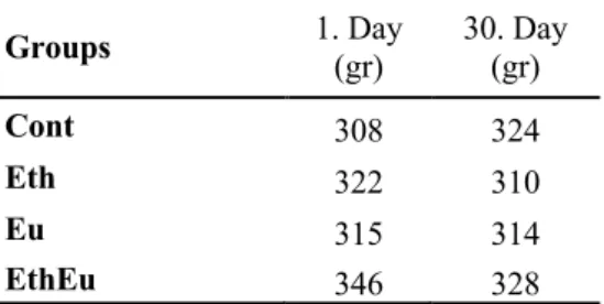
In our study, weight gain was observed in the Cont group at the end of the 30th day, while weight loss was observed in other groups (Table 1). In the alcoholconsuming group, weight loss is thought to be due to possible decreased appetite and decreased food intake as a result of subsequent liver damage.
Table 1: Weights of experimental groups
Groups 1. Day (gr) 30. Day (gr)
Cont 308 324
Eth 322 310
Eu 315 314
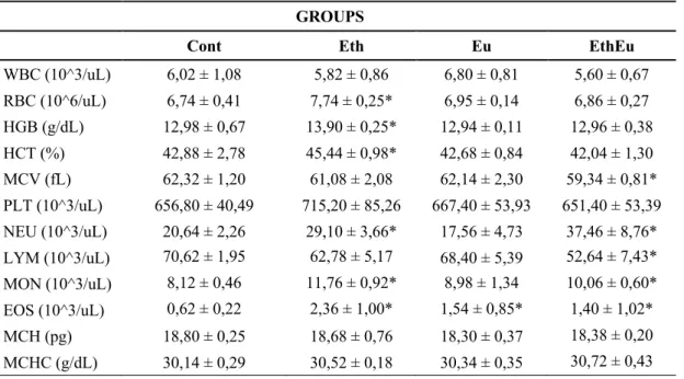
Alcohol consumption causes different negative effects on blood cells. Individuals consuming alcohol may suffer from moderate anemia characterized by structurally abnormal RBCs, slightly decreased WBCs, especially neutrophils and moderately decreased platelet count [30]. As a result of the complete blood count, there was a significant difference between the groups in terms of RBC, HGB, HCT MCV, NEU, LYM, MON, EOS parameters, but no significant difference in terms of WBC, PLT, BAS, MCH and MCHC parameters. As shown in Table 2, while RBC, HGB, HCT and PLT values increased in Eth group compared to the Cont group, there was a decrease in WBC, LYM and MCV values. Blood values of the Eug group and the Cont group were close to each other. Our NEU, LYM and MON data are in line with other studies [31, 32].
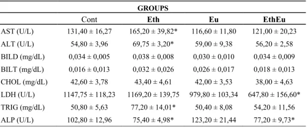
Biochemical analysis results are presented in Table 3. While there was a statistically significant difference in serum AST, ALT, ALP, LDH and TRIG levels between the Cont group and Eth, Eu and EthEu groups, no significant difference was found in the levels of BILD, BILT, CHOL, AFP and CEA125 (p < 0.05). The mean and standard deviation (mean and ± SD) values of AST values in the Cont group were found to be 131.40 ± 16.27. Eth group has the highest AST value with 165.20 ± 39.82, while Eu group has the lowest AST values with 116.60 ± 11.80.
Table 2: Complete blood count parameters of Control, Ethanol, Eugenol, Ethanol and Eugenol groups
(*) Statistically importance compared with the control group, p < 0.05. GROUPS
Cont Eth Eu EthEu
WBC (10^3/uL) 6,02 ± 1,08 5,82 ± 0,86 6,80 ± 0,81 5,60 ± 0,67 RBC (10^6/uL) 6,74 ± 0,41 7,74 ± 0,25* 6,95 ± 0,14 6,86 ± 0,27 HGB (g/dL) 12,98 ± 0,67 13,90 ± 0,25* 12,94 ± 0,11 12,96 ± 0,38 HCT (%) 42,88 ± 2,78 45,44 ± 0,98* 42,68 ± 0,84 42,04 ± 1,30 MCV (fL) 62,32 ± 1,20 61,08 ± 2,08 62,14 ± 2,30 59,34 ± 0,81* PLT (10^3/uL) 656,80 ± 40,49 715,20 ± 85,26 667,40 ± 53,93 651,40 ± 53,39 NEU (10^3/uL) 20,64 ± 2,26 29,10 ± 3,66* 17,56 ± 4,73 37,46 ± 8,76* LYM (10^3/uL) 70,62 ± 1,95 62,78 ± 5,17 68,40 ± 5,39 52,64 ± 7,43* MON (10^3/uL) 8,12 ± 0,46 11,76 ± 0,92* 8,98 ± 1,34 10,06 ± 0,60* EOS (10^3/uL) 0,62 ± 0,22 2,36 ± 1,00* 1,54 ± 0,85* 1,40 ± 1,02* MCH (pg) 18,80 ± 0,25 18,68 ± 0,76 18,30 ± 0,37 18,38 ± 0,20 MCHC (g/dL) 30,14 ± 0,29 30,52 ± 0,18 30,34 ± 0,35 30,72 ± 0,43
Table 3: Some biochemical parameters ın the blood serum of the Control, Ethanol, Eugenol, Ethanol and
Eugenol groups
(*) Statistically importance compared with the control group, p < 0.05.
There was a significant increase in ALT in Eth, Eug and EthEug groups compared to the Cont group (p < 0.05). While the highest ALT level belonged to the Eth group to be 69.75 ± 3.20, the lowest ALT level was found in the Cont group to be 54.80 ± 3.96. It was determined that the ALT level decreased in the Eug group compared to the Eth group and increased in comparison with the Cont group. The change between the groups was found to be significant for ALT. Compared to the Cont group, while the BILD level decreased in the Eug group, it did not increase in the Eth group and did not change much in the EthEug group. BILT level increased in all groups except the Cont group. The highest BILT value was found in Eth group with 0.032 ± 0.026. CHOL level did not change significantly between groups however, Eth was the highest group with a value of 43.40 ± 4.61.
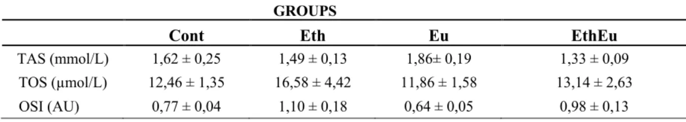
There was a significant difference between the groups at the LDH level and Eth group was the highest group with the value of 1169.20 ± 139.75. There was also a significant change in TRIG levels in all groups. When TRIG level was compared, it was the highest Eth group and the lowest group was Eug group. The ALP level was found in the highest Eug group and the lowest in the Eth group. There was a statistically significant difference between the groups for TAS level. Eth group's TAS level was lower than the Cont group. The eugenol group has the highest value with 1.86 ± 0.19, while the EthEug group has the lowest value. Although no significant difference was found between the groups for the TOS level, the TOS level was found to be the highest in the ethanol-treated group with a value of 16.58 ± 4.42 (Table 4).
GROUPS
Cont Eth Eu EthEu
AST (U/L) 131,40 ± 16,27 165,20 ± 39,82* 116,60 ± 11,80 121,00 ± 20,23 ALT (U/L) 54,80 ± 3,96 69,75 ± 3,20* 59,00 ± 9,38 56,20 ± 2,58 BILD (mg/dL) 0,034 ± 0,005 0,038 ± 0,008 0,030 ± 0,010 0,034 ± 0,009 BILT (mg/dL) 0,016 ± 0,013 0,032 ± 0,026 0,026 ± 0,017 0,018 ± 0,013 CHOL (mg/dL) 42,60 ± 3,78 43,40 ± 4,61 42,00 ± 3,53 38,00 ± 4,63 LDH (U/L) 1147,75 ± 118,23 1169,20 ± 139,75 979,80 ± 103,34 647,80 ± 156,60* TRIG (mg/dL) 50,80 ± 5,63 77,20 ± 14,01* 50,40 ± 8,08 54,20 ± 11,56 ALP (U/L) 102,80 ± 12,96 75,40 ± 4,98* 123,20 ± 21,44 77,20 ± 9,73*
Figure 2: Liver histopathology of Control (A), Eugenol (B), Ethanol (C), Ethanol and Eugenol (D)groups
(H&E, X200)
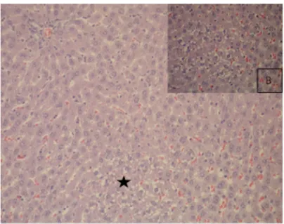
Figure 3: Microvesicular fatty change observed in the liver of Ethanol group rats (asterisk), (H&E, X200),
Changes in liver histology due to alcohol use, macrovesicular and microvesicular fat accumulation, hepatocyte ballooning, cell infiltration, Mallory-Equivalent body, neutrophilic inflammation, pericular fibrosis, hyaline sclerosis, fibrosis and cholestasis are observed [33]. When the groups are compared histopathologically, there is a significant difference between the Cont group and other groups in terms of degeneration levels (Fig. 2). In the Cont group rats, the liver was observed to have a normal histological structure. Severe diffuse degeneration and microvesicular fatty changes were observed in liver hepatocytes of Eth group rats (Fig. 3). Compared with the Cont group, in the Eth group, intracellular macro and microvesicular fatty accumulation due to alcohol, dense mononuclear cell infiltration, mostly lymphocytes, hydropic degeneration, dual-core structures in some hepatocyte cells, and the presence of erythrocytes in the synophoidal spaces were observed. It was found that degenerations decreased in the group with eugenol added.
Table 4: TAS and TOS parameters in the blood serum of Control, Ethanol, Eugenol, Ethanol and Eugenol
groups
GROUPS
Cont Eth Eu EthEu
TAS (mmol/L) 1,62 ± 0,25 1,49 ± 0,13 1,86± 0,19 1,33 ± 0,09 TOS (µmol/L) 12,46 ± 1,35 16,58 ± 4,42 11,86 ± 1,58 13,14 ± 2,63
OSI (AU) 0,77 ± 0,04 1,10 ± 0,18 0,64 ± 0,05 0,98 ± 0,13
(*) Statistically importance compared with the control group, p < 0.05.
4. Discussion
The extrahepatic metabolism of alcohol is very low. Since 90-95% of alcohol is metabolized in the liver, the organ that is most exposed to harmful effects is the liver. These harmful effects of alcohol are mostly caused by disruption of oxidant and antioxidant balance. Alcohol is metabolized to acetaldehyde by alcoholdehydrogenase in cytosol, cytochrome P450 in microsomes and catalase in peroxisomes [34] . When alcohol is advanced oxidized, it increases the free radical production and disrupts the oxidant-antioxidant system's balance. In order to eliminate or minimize the harmful effects of alcohol, some research have been done on many substances that are synthesized or taken daily and have antioxidant properties. According to literature data, although medicines that help advance modern medicine and renew hepatic cells and protect the liver, many herbal extracts are used to prevent liver diseases [35].
Because of antioxidant feature of eugenol, it is used as a preservative and aroma additive in the food industry and cosmetics industry [36]. In our study, we started from the fact that the
eugenol substance has a high antioxidant activity like olive oil. We aimed to determine what effects it has on the liver when applied directly, without dissolving it in some other solvents. Guenette et al. reported that in rats, the half-life (t1/2) of eugenol in plasma was approximately 14 hours and 18 hours in blood [37].
Depending on alcohol use, moderate anemia characterized by structurally abnormal RBCs, mild deficiency of WBCs, especially neutrophils, and moderately decreased platelet counts have been reported [38, 39].
Yalçın and Yağcı reported that high levels of ethanol intake (15% v/v) in rats decreased the white blood cell and red blood cell counts, hemoglobin concentrations, erythrocyte diameters, erythrocyte sodium and potassium levels compared to the Cont group [40]. Similarly, in our study, WBC, MCV and LYM values in Eth group decreased compared to the Cont group, while RBC, HGB, HCT, PLT, MCH, NEU, MON and other parameters increased.
Gülçin et al. examined the antioxidant activity of clove oil in comparison with synthetic antioxidants. They determined the antioxidant activity of clove oil, BHA, BHT, a-tocopherol and trolox by ferric thiocyanate method in linoleic acid system. As a result, clove oil found 99.7%, under the same conditions, standard antioxidant compounds such as butylated hydroxyanisole (BHA), butylated hydroxytoluene (BHT), a-tocopherol and trolox showed 95.5%, 99.7%, 84.6%, and 95.6% inhibition respectively [41].
Anbu et al examined the effect of ethanol and eugenol administration on lipid levels, lipid peroxidation, enzymatic and non-enzymatic antioxidants. They showed that the levels of SOD, CAT, GST and GPx enzymes increased more than the other groups in the eugenol applied group compared to the other groups. They stated that ethanol intake significantly increased some liver enzymes (AST, ALT, GGT and ALP). In this study, olive oil was used as a solvent agent for eugenol and it was given to other groups in equal amounts except the ethanol group [42]. In another study, extracts of about 30 plants were investigated for their antioxidant properties using DPPH and ABTS radical scavenging capacity test, oxygen radical absorbance capacity (ORAC) test, SOD analysis and ferric reductive antioxidant potential (FRAP) analysis. As a result of the study, researchers showed that clove has strong antioxidant properties and high phenolic content (200 mg GAE/g) [43].
Yogalakshmi et al. pre-treatment with eugenol (10.7 mg/kg bw per day) in rats for 15 days, lipid peroxidation indices, protein oxidation and inflammatory markers and glutathione peroxidase (GPx), superoxide dismutase (SOD), catalase (CAT) and glutathione -S-transferase (GST) has been shown to improve antioxidant status by protecting antioxidants [44]. Binu and his team found that when eugenol (5 mg/kg bw) and arsenic trioxide (As2O3) were treated
together, AST and ALT levels were normalized compared to the Cont group and showed hepatoprotective effects with decreased arsenic accumulation [45]. Thuwaini et al., examined the effects of clove extract on liver damage caused by paracetamol and stated that there was a decrease in the amount of albumin in the blood serum and an increase in bilirubin, ALT, AST and ALP levels compared to the Cont group [46]. Muller et al. investigated the liver protective activity of Pecan coconut extract against liver damage caused by ethanol and found increased levels of plasmatic transaminases (ALT, AST) at the end of the study in chronically treated rats with ethanol. As a result, it has been stated that it can be an economical agent to treat liver diseases related to consumption of ethanol from coconut shells [47]. The findings in these studies support our study and similarly, we found a significant increase in AST and ALT levels in Eth group rats (p<0.05). ALT distribution among other groups is shown in Fig. 2. AST/ALT ratio decreases in non-alcoholic liver diseases, while AST/ALT ratio increases in alcoholic liver injury [48]. In our study, while AST/ALT ratio in Eth group increased compared to Eug and EthEug group, no increase was found compared to the Cont group.
LDH is an enzyme that is found in many tissues including liver tissue and goes into serum if these tissues are damaged. Abd El Motteleb et al., have shown in their studies that low eugenol dose (10 mg eugenol/kg per day) can protect the liver from ischemia/reperfusion (I/R) damage by reducing lipid peroxidation levels. They found that AST, ALT and LDH levels decreased significantly compared to the I/R group in rats treated with 10mg eugenol [49]. In our study, there was a significant decrease in the LDH level in the Eug and EthEug groups compared to the Eth group. It is possible to state that with this decrease in the LDH level belonging to the Eug and EthEug groups, the tissue damage caused by alcohol decreased.
The fact that the Eth group was found lower than TAS level compared to the Cont group shows the toxic effect of ethanol on antioxidants [50]. The highest TAS level was found in the Eug group. When the TOS level taken into consideration, the Eth group was found to be the highest group, while in the EthEug group was close to the Cont group. This increase in TOS value is due to the strong antioxidant properties of eugenol.
5. Conclusions
In histopathological examination, intracellular macro and microvesicular fat accumulation due to alcohol in the Eth group was observed. Moreover, intense mononuclear cell infiltration, mostly lymphocytes, hydropic degeneration and the presence of erythrocytes in places in the synosoid spaces were also observed. It was found that degenerations decreased in the Eu group.
Our biochemical results, which express the presence of the antioxidant effect of eugenol on liver tissue, supported with histopathologic results.
As a result, an increase in antioxidant parameters was observed while reducing the oxidative stress effects on biochemical parameters and histopathological findings when using only eugenol as an antioxidant agent on the oxidative stress caused by ethanol in the liver of rats. Biochemical and histopathological findings can be stated that eugenol significantly prevents liver damage. Based on the information obtained from our study, we suggest that Eugenol can be used as a drug additive or nutritional supplement to protect health (particularly the liver) against alcohol damage.
Acknowledgement
This work was supported by Hatay Mustafa Kemal University, Scientific Research Projects coordinator (HMKU-BAP) within the scope of project number 18.D.008.
References
[1] Pandanaboina, S.C., Kondeti, S.R., Rajbanshi, S.L., Kunala, P.N., Pandanaboina, S., Pandanaboina, M.M., Wudayagiri, R., Alterations in antioxidant enzyme activities and oxidative
damage in alcoholic rat tissues: protective role of Thespesia populnea, Food Chem, 132(1),
150-159, 2012.
[2] Gencer, M., Ceylan, E., Aksoy, N., Uzun, K., Oksidatif stres benign ve malign akciğer
hastalıklarının ayırıcı tanısında belirteç olabilir mi?, Turkiye Klinikleri Archives of Lung, 6(3),
89-92, 2005.
[3] Guyton, A.C., Hall, J.E., Tıbbi Fizyoloji, Nobel Tıp Kitapevi, 12. Baskı, İstanbul. 937-942, 2013.
[4] Ghany M, H.J., Approach to the patient with liver disease, Harrison's Principles of Internal Medicine, Fauci AS (ed). 17th Edition, 1918-1923, 2008.
[5] El-Newary, S.A., Shaffie, N.M., ve Omer, E. A., The protection of Thymus vulgaris
leaves alcoholic extract against hepatotoxicity of alcohol in rats, Asian Pacific Journal of
Tropical Medicine, 10(4), 361-371, 2017.
[6] Li, S., Tan, H.-Y., Wang, N., Zhang, Z.-J., Lao, L., Wong, C.-W., Feng, Y., The role of
oxidative stress and antioxidants in liver diseases, Int J Mol Sci, 16(11), 26087-26124, 2015.
[7] NCBI., https://pubchem.ncbi.nlm.nih.gov/compound/3314, Date of access: Mar. 29, 2018.
[8] TOXNET, T.D.N., Eugenol [USP], Date of access: 15.03.2018, from National Library of Medicine, 2018.
[9] Kamatou, G.P., Vermaak, I., Viljoen, A.M., Eugenol—from the remote maluku islands
to the international market place: A review of a remarkable and versatile molecule, Molecules,
17(6), 6953, 2012.
[10] Tripathi, A.K., Mishra, S., Plant Monoterpenoids (Prospective Pesticides) Ecofriendly Pest Management for Food Security, Chapter 16, 507-524, San Diego: Academic Press, 2016.
[11] Bennett, A., Stamford, I.F., Tavares, I.A., Jacobs, S., Capasso, F., Mascolo, N., Carlo, G. D., The biological activity of eugenol, a major constituent of nutmeg (Myristica fragrans):
Studies on prostaglandins, the intestine and other tissues, Phytotherapy Research, 2(3),
124-130,1988.
[12] Johnson, C.B., Kirby, J., Naxakis, G., Pearson, S., Substantial UV-B-mediated
induction of essential oils in sweet basil (Ocimum basilicum L.), Phytochemistry, 51(4), 507-510,
1999.
[13] Dighe, V., Gursale, A., Sane, R., Menon, S., Raje, S., Quantification of eugenol in
Cinnamomum tamala nees and eberm. Leaf powder by high-performance thin-layer chromatography, JPC - Journal of Planar Chromatography - Modern TLC 18(104), 305-307,
2005.
[14] Ueda-Nakamura, T., Mendonça-Filho, R.R., Morgado-Díaz, J.A., Korehisa Maza, P., Prado Dias Filho, B., Aparício Garcia Cortez, D., Nakamura, C. V., Antileishmanial activity of Eugenol-rich essential oil from Ocimum gratissimum, Parasitology International, 55(2), 99-105, 2006.
[15] Lopes-Lutz, D., Alviano, D.S., Alviano, C.S., Kolodziejczyk, P.P., Screening of chemical composition, antimicrobial and antioxidant activities of Artemisia essential oils, Phytochemistry, 69(8), 1732-1738, 2008.
[16] Howes, M.J., Kite, G.C., Simmonds, M.S., Distinguishing chinese star anise from
Japanese star anise using thermal desorption-gas chromatography-mass spectrometry, J Agric
Food Chem, 57(13), 5783-5789, 2009.
[17] Pragadheesh, V., Yadav, A., Singh, S., Gupta, N., Chanotiya, C., Leaf Essential Oil of Cultivated Pimenta Racemosa (Mill.) J.W. Moore from North India: Distribution of Phenylpropanoids and Chiral Terpenoids, Medicinal & Aromatic Plants, 2(1), 2-4, 2013.
[18] Jaganathan, S.K., Supriyanto, E., Antiproliferative and molecular mechanism of
eugenol-induced apoptosis in cancer cells, Molecules, 17(6), 6290-6304, 2012.
[19] Guimarães, A.C., Meireles, L.M., Lemos, M.F., Guimarães, M.C.C., Endringer, D.C., Fronza, M., Scherer, R., Antibacterial activity of terpenes and terpenoids present in essential oils, Molecules, 24(13), 2471, 2019.
[20] Li, Z., Veeraraghavan, V.P., Mohan, S.K., Bolla, S.R., Lakshmanan, H., Kumaran, S., Chinnathambi, A., Apoptotic induction and anti-metastatic activity of eugenol encapsulated
chitosan nanopolymer on rat glioma C6 cells via alleviating the MMP signaling pathway, J
[21] Qian, W., Sun, Z., Wang, T., Yang, M., Liu, M., Zhang, J., Li, Y., Antimicrobial
activity of eugenol against carbapenem-resistant Klebsiella pneumoniae and its effect on biofilms,
Microbial Pathogenesis, 139, 103924, 2020.
[22] Matan, N., Rimkeeree, H., Mawson, A.J., Chompreeda, P., Haruthaithanasan, V., Parker, M., Antimicrobial activity of cinnamon and clove oils under modified atmosphere
conditions, International Journal of Food Microbiology, 107(2), 180-185, 2006.
[23] EWG, http://www.ewg.org/skindeep/search.php?query=eugenol&h=Search#.Wr3 6zeZrOIV, Date of Access: 10.02.2018, from Environmet Working Group.
[24] Yan, S.L., Wang, Z.H., Yen, H.F., Lee, Y.J., Yin, M.C., Reversal of ethanol-induced
hepatotoxicity by cinnamic and syringic acids in mice, Food and Chemical Toxicology, 98(B),
119-126, 2016.
[25] Mnafgui, K., Hajji R., Derbali, F., Gammoudi, A., Khabbabi, G., Ellefi, H., Allouche, N., Kadri, A., Gharsallah, N., Anti-inflammatory, antithrombotic and cardiac remodeling
preventive effects of eugenol in isoproterenol-induced myocardial infarction in wistar rat,
Cardiovascular Toxicology, 16(4), 336–344, 2016.
[26] Erel, O., A novel automated direct measurement method for total antioxidant capacity
using a new generation, more stable ABTS radical cation, Clinical Biochemistry, 37(4), 277-285,
2004.
[27] Erel, O., A new automated colorimetric method for measuring total oxidant status, Clin Biochem, 38(12), 1103-1111, 2005.
[28] Larue-Achagiotis, C., Poussard, A.M., Louis-Sylvestre, J., Alcohol drinking, food and
fluid intakes and body weight gain in rats, Physiol Behav, 47(3), 545-548, 1990.
[29] Bertola, A., Mathews, S., Ki, S.H., Wang, H., Gao, B., Mouse model of chronic and
binge ethanol feeding (the NIAAA model), Nature protocols, 8(3), 627-637, 2013.
[30] Elanchezhian, T., Y., Mayilsamy, S., Radhakrishnan, S., Comparison of
haematological parameters between alcoholics and non-alcoholics, Int J Res Med Sci 5(11),
5041-5407, 2017.
[31] Kawashima, Y., Someya, Y., Shirato, K., Sato, S., Ideno, H., Kobayashi, K., Imaizumi, K., Single administration effects of ethanol on the distribution of white blood cells in rats, J Toxicol Sci, 36(3), 347-355, 2011.
[32] Sonila, B., Klodeta, M., Andrin, T., Irida, P., Changes of some blood count variables
in correlation with the time of alcohol abuse, Journal of Addiction Research & Therapy, 6(2),
221, 2015.
[33] Zhang, M.Q., Ren, X., Zhao, Q., Yue, S.-J., Fu, X.-M., Li, X., Wang, C.-Y.,
Hepatoprotective effects of total phenylethanoid glycosides from Acanthus ilicifolius L. against carbon tetrachloride-induced hepatotoxicity, Journal of Ethnopharmacology, (256), 112795,
[34] Gao, B., Bataller, R., Alcoholic liver disease: pathogenesis and new therapeutic
targets, Gastroenterology, 141(5), 1572-1585, 2011.
[35] Chattopadhyay, R.R., Possible mechanism of hepatoprotective activity of Azadirachta
indica leaf extract: part II, J Ethnopharmacol, 89(2-3), 217-219, 2003.
[36] Barboza, J.N., da Silva Maia Bezerra Filho, C., Silva, R.O., Medeiros, J.V.R., de Sousa, D.P., An overview on the anti-inflammatory potential and antioxidant profile of eugenol, Oxidative medicine and cellular longevity, 3957262, 2018.
[37] Guenette, S.A., Ross, A., Marier, J.F., Beaudry, F., Vachon, P., Pharmacokinetics of
eugenol and its effects on thermal hypersensitivity in rats, Eur J Pharmacol, 562(1-2), 60-67,
2007.
[38] Ballard, H.S., The hematological complications of alcoholism, Alcohol Health Res World, 21(1), 42-52, 1997.
[39] Xiao, W., Study on the joint effects of acrylonitrile and alcohol in rats, Wei Sheng Yan Jiu, 27(5), 295-296 1998.
[40] Yalçın, M., Yağcı, A., Alterations in some blood parameters after high level ethanol
intake, Uludag Univ. J. Fac. Vet. Med., 24( 1-2-3-4), 47-52, 2005.
[41] Gülçin, İ., Elmastaş, M., Aboul-Enein, H.Y., Antioxidant activity of clove oil – A
powerful antioxidant source, Arabian Journal of Chemistry, 5(4), 489-499, 2012.
[42] Anbu, S., Anuradha, C.V., Protective effect of eugenol against alcohol-induced
biochemical changes in rats, International Journal of Research in Biotechnology and
Biochemistry, 2(2), 13-18, 2012.
[43] Dudonne, S., Vitrac, X., Coutiere, P., Woillez, M., Merillon, J.M., Comparative study
of antioxidant properties and total phenolic content of 30 plant extracts of industrial interest using DPPH, ABTS, FRAP, SOD, and ORAC assays, J Agric Food Chem, 57(5), 1768-1774, 2009.
[44] Yogalakshmi, B., Viswanathan, P., Anuradha, C.V., Investigation of antioxidant,
anti-inflammatory and DNA-protective properties of eugenol in thioacetamide-induced liver injury in rats, Toxicology, 268(3), 204-212, 2010.
[45] Binu, P.P., Priya, N.M., Abhilash, S.P., Vineetha, R.C.P., Nair, H.P., Protective effects
of eugenol against hepatotoxicity induced by arsenic trioxide: An antileukemic drug, Iranian
journal of medical sciences, 43(3), 305-312, 2018.
[46] Thuwaini, M., Abdul Mounther, M., Kadhem, H., Hepatoprotective effects of the
aqueous extract of clove (Syzygium aromaticum) against paracetamol-induced hepatotoxicity and oxidative stress in rats, EJPMR, 3(8), 36-42, 2016.
[47] Muller, L.G., Pase, C.S., Reckziegel, P., Barcelos, R.C., Boufleur, N., Prado, A.C., Burger, M.E., Hepatoprotective effects of pecan nut shells on ethanol-induced liver damage,
[48] Nyblom, H., Berggren, U., Balldin, J., Olsson, R., High AST/ALT ratio may indicate
advanced alcoholic liver disease rather than heavy drinking, Alcohol, 39(4), 336-339, 2004.
[49] Abd El Motteleb, D.M., Selim, S.A., Mohamed, A.M., Differential effects of eugenol
against hepatic inflammation and overall damage induced by ischemia/re-perfusion injury, J
Immunotoxicol, 11(3), 238-245, 2014.
[50] Basarslan, S.K., Osun, A., Senol, S., Korkmaz, M., Ozkan, U., Kaplan, I., Protective
effects of intralipid and caffeic acid phenyl esther (CAPE) on neurotoxicity induced by ethanol in rats, Turk Neurosurg, 27(1), 66-73, 2017.