ISSN 0015–5659 journals.viamedica.pl
Address for correspondence: Associate Professor M.O. Dayan, Department of Anatomy, Faculty of Veterinary, Selçuk University, Konya, Turkey e-mail: [email protected]
Three-dimensional modelling of the femur and
humerus in adult male guinea pigs (guinea pig)
with computed tomography and some biometric
measurement values
M.O. Dayan, K. Beşoluk, E. Eken, S. Aydoğdu, N. Turgut
Department of Anatomy, Faculty of Veterinary, Selçuk University, Konya, Turkey [Received: 16 October 2018; Accepted: 5 December 2018]Background: Guinea pig is a species belonging to the Caviidae family of the Ro-dentia order and is frequently used in experimental studies. Biomedical imaging methods are used in the diagnosis and treatment of many diseases in medicine. Among these methods, computed tomography (CT) is one of the most important imaging methods. In this study, it was aimed to perform the three-dimensional (3D) modelling of the CT images, obtained from the humerus and femur in the guinea pigs, via the MIMICS programme, and to make some biometric measure-ments regarding the bones over these models.
Materials and methods: In the present study, 12 male adult guinea pigs were used. The soft tissue on the humerus and femur bones of the guinea pigs was removed. After this procedure, CT images at a 0.5 mm-thickness were obtained from the animals. The images were recorded in DICOM format. Then, the recon-struction process was performed from the images by using the 3D modelling programme MIMICS® 13.1. On the 3D model of the humerus and femur (right-left), volumes, surface areas and lengths as well as other biometric parameters were measured separately, and the values were recorded. In addition, measurements of the bones were made with the help of a digital calliper.
Results: Among the parameters obtained from 3D models, a statistical differ-ence was observed between the right and left cortical thicknesses of the femur from the measurements of calliper and the right and left humerus volumes (p < 0.05); whereas, no statistical difference was found in other parameters of both measurements (p > 0.05).
Conclusions: It can be stated that CT and 3D modelling can be used for the measurement of some parameters in the long bones of the guinea pigs. (Folia Morphol 2019; 78, 3: 588–594)
Key words: computed tomography, femur, humerus, guinea pig, three- -dimensional reconstruction
INTRODUCTION
Guinea pig is a species belonging to the Caviidae family of the Rodentia order [22]. Guinea pigs that
are frequently used in the experimental studies such as immunology, toxicology, pharmacology, and physi-ology are native to South America and their wild ones
are rodents that inhabit in Peru [41]. Their heads are quite large and their ears and tails are quite small [22]. They are herbivores. Adult guinea pigs have a body length of 27–33 cm and weigh of 700–1200 g [34]. They live for 2–8 years [41].
Bones, which are the passive elements of the locomotor system, are composed of organic and inor-ganic substances. In a guinea pig that has completed its growth, 1/3 of the bone is organic substances (ossein); whereas, 2/3 is inorganic substances (85% — calcium phosphate, 10% — calcium carbonate, magnesium phosphate, calcium fluoride). While the organic substances provide the elasticity of the bone, inorganic substances provide hardness and durability to the bone [12, 34]. Appendiculare skeleton is divid-ed into two parts as bones of thoracic limb and pelvic limb. One of the bones of thoracic limb is the humer-us. The longest bone of the body, which is included in the bones of the pelvic limb, is the femur [12, 26].
Morphometry is a research method which exam-ines the shape differences of the objects and organ-isms and the correlations of the shape differences with other variations. This method is sometimes used to analyse the correlation between gender and growth factors of the species and sometimes the effects of the treatment applied [6, 36]. Convention-al morphometry is the multiple variance statisticConvention-al analysis that is performed with quantitative (length, width, height) variables [16]. Depending on the en-vironmental components and the genetic structure, the differences in the growth of the living being can be easily revealed by morphology and morphometric studies [39].
Biomedical imaging methods are used in the di-agnosis and treatment of many diseases in medicine [13, 40]. The working principle of computed tomography (CT), which is one of the medical imaging methods, is based on the detection of the non-absorbable part of the collimated X-rays passing through any part of the body in the tissues by the detectors as well as the conversion of the electrical signals into a cross-sec-tional image by means of computer operations [21]. With this method, the structures are displayed in a cross-sectional view and the images are much more detailed than the X-ray [1]. In recent years, CT has played an important role in the anatomical stud-ies and in the correct diagnosis of the diseases in veterinary medicine. In addition to the diagnosis of diseases, CT is also used effectively in many biometric researches [13, 27, 28].
Three-dimensional (3D) formation of the trans-versal and longitudinal sections obtained from CT and other medical imaging methods is referred as reconstruction [2, 15, 32]. Thanks to this method, organisation and processing of the 3D information obtained by the reconstructions of the first cross-sec-tional images and enabling them to be presented in various shapes have become an acceptable method in medical sciences, especially in anatomy, surgical and clinical treatment [14, 20, 31, 38]. Anatomical details that cannot be observed in 2D images can be observed in more details with 3D reconstructed mod-els. Today, MIMICS is one of the computer software used to create 3D models of two-dimensional (2D) images that are obtained from CT and magnetic res-onance imaging methods. This software is a medical imaging and control system developed by Materialise (Mimics, Materialise Inc., Leuven, Belgium) for the 3D modelling of anatomical structures. One of the most important features that distinguishes MIMICS from other 3D modelling software is that it is a seg-mentation programme using Hounsfield values [3, 15, 18, 24, 31, 33].
In recent years, various studies have been conduct-ed on the 3D modelling of anatomical structures in the field of veterinary anatomy. In a study conducted on the pelvic cavity of New Zealand rabbits, pelvis diameters and pelvic inclination were measured on a 3D model and evaluated in terms of gender. It has been suggested that the models obtained by the present study will contribute to the anatomical training and also in the diagnosis and treatment of diseases and to the knowledge of today’s anatomy [30]. In addition, 3D modelling of penile urethra, stomach and intestine, respiratory system, paranasal sinus and antebrachium was performed in the rabbits [9, 10, 19, 29, 31]. In addition, a 3D model of the nasal airway was also performed in Nycticebus pyg-maeus (Slow loris) [11]. 3D modelling of the thick filaments of the cardiac muscle in zebrafish, paranasal sinuses and diverticulum tubae auditivae in the horses and the nephron in the rat was done [4, 5, 8, 17]. As a result, it is seen that the 3D modelling method has started to be frequently used in the field of veterinary anatomy.
In the literature reviews, it was observed that CT and 3D modelling of the femur and humerus, which are the long bones of the body, in the guinea pigs, and the information regarding the measurements to be obtained over these models were limited [35, 40].
The purpose of the present study is to perform the 3D modelling of the CT images obtained from the humerus and femur in guinea pigs via the MIMICS programme and to calculate some of the biometric measurements of the bones such as volume, surface area and length over these models.
MATERIALS AND METHODS
In the study, 12 adult male guinea pigs were used. The study procedure was approved by the Ethics Committee of Selçuk University Faculty of Veterinary Medicine (09/03/2017, 2017/26). After the soft tissue on the humerus and femoral bones of the guinea pigs was removed, morphological differences were recorded. After this procedure, CT images were ob-tained from the animals at 0.5-mm thickness. The images were recorded in DICOM format. Then, the reconstruction process was performed from the im-ages transferred to a portable external disk by using the MIMICS® 13.1 (The Materialise Group, Leuven,
Belgium), a 3D modelling programme (Fig. 1). In the reconstruction processes, the bone edges to be selected by using the “Thresholding” com-mand from the “Segmentation” menu of MIMICS programme were automatically masked. Bones that could not be masked were corrected manually by the computer mouse by selecting the “Edit mask” command. After the manual correction process was completed, independent masking was reapplied to the selected bone by the “Region Growing” com-mand. Finally, 3D modelling was performed by the “Calculate 3D” command. On the 3D model of the humerus and femur (right-left), their volumes, surface areas and lengths as well as other biometric parame-ters were automatically measured separately by using the “Info” command and the values were recorded. In addition, measurements of the bones were made by the help of a digital calliper. The diameter and cortical thickness of the humerus and femur were measured from the diaphysis section of the bones (Fig. 2).
Measurements performed on the bone (Figs. 3, 4): — 1: maximum length of the humerus;
— 2: maximum length of the femur; — 3: proximal width of the humerus; — 4: proximal width of the femur; — 5: distal width of the humerus; — 6: distal width of the femur;
— 7: cortical thickness of the humerus; — 8: cortical thickness of the femur;
— 9: medullary cavity diameter of the humerus;
Figure 1. Three-dimensional models of the humerus and femur.
Figure 2. Humerus (left) and femur (right).
Figure 3. Measurement points taken from the humerus; 1 — max-imum length of the humerus; 2 — proximal width of the humerus; 3 — distal width of the humerus; 4 — diaphysis diameter of the humerus (medio-lateral); 5 — diaphysis diameter of the humerus (cranio-caudal); 6 — medullary cavity diameter of the humerus [6A (mediolateral) + 6B (craniocaudal)/2]; 7 — cortex thickness of the humerus [7A (medial) + 7B (lateral) + 7C (caudal) + 7D (cranial)/4].
— 10: medullary cavity diameter of the femur; — 11: diaphysis diameter of the humerus; — 12: diaphysis diameter of the femur.
On the 3D model, above mentioned parameters 7, 8, 9, and 10 could not be measured.
Anatomic terms used in this study were written by taking Nomina Anatomica Veterinaria as a refer-ence [25].
Normality test was applied on the measurements taken on the 3D model and the morphometric meas-urements obtained from the bones via the MIMICS programme. While the normally distributed values were evaluated by the paired t test, the values that did not show normal distribution were evaluated by the Wilcoxon test (SPSS 22.0). The value of p < 0.05 was accepted as the statistically significant limit.
RESULTS
It was observed that the head of the humerus in the proximal extremity section of the shoulder joint with the scapula was quite large in guinea pig. Deltoid tu-berosity was not significant. Radial fossa and olecranon fossa were associated with supratrochlear foramen.
It was observed that greater trochanter in the proximal extremity section of the femur was higher than the head of the femur and lesser trochanter was a circular crest. The presence of fovea of the head was determined on the articular surface of the fem-oral head. On the other hand, in the distal extremity section, each condyle had articular surfaces specific to articulation of the sesamoid bones.
Table 1 shows the measurement values performed on the 3D models of the humerus and femur in the study. While there was a statistical difference in the right and left humeral volume (p = 0.046) from the parameters obtained from 3D models, no difference was determined in other parameters (p > 0.05).
Table 2 shows measurement values of the humerus and femur measured with digital calliper. While a sta-tistically significant difference was observed between the cortical thickness of the femur (p < 0.05), no difference was found in other parameters (p > 0.05).
DISCUSSION
While the guinea pig is being fed as an exotic animal, it is an animal model that is used in many experimental studies in the field of health [41]. The humerus and the femur are the two long bones that are frequently preferred in morphometric studies [23]. CT and 3D modelling are currently used in the
Figure 4. Measurement points taken from the femur;1 — maximum length of the femur; 2 — proximal width of the femur; 3 — distal width of the femur; 4 — diaphysis diameter of the femur (medio- -lateral); 5 — diaphysis diameter of the femur (cranio-caudal); 6 — medullary cavity diameter of the femur [6A (mediolateral) + 6B (craniocaudal)/2]; 7 — cortex thickness of the femur [7A (medial) + 7B (lateral) + 7C (caudal) + 7D (cranial)/4].
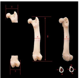
Figure 5. Measurements taken from the three-dimensional models of the femur and the humerus; 1 — diaphysis diameter of the femur (medio-lateral); 2 — distal width of the femur; 3 — maximum length of the femur; 4 — proximal width of the femur; 5 — diaphysis diam-eter of the humerus; 6 — distal width of the humerus; 7 — maxi-mum length of the humerus; 8 — proximal width of the humerus.
diagnosis, and treatment of many diseases [13, 15, 20]. In addition, bone morphometric measurements are made with the help of the digital calliper [23]. In
the present study, the humerus and femur bones of 12 adult male guinea pigs were modelled as 3D and various parameters were measured. Some morpho-metric parameters of the same bones that were meas-ured by using a digital calliper were also evaluated.
In the present study, it was determined that while a statistical difference was observed in the left and right
humeral volume parameters obtained from the data of 3D models (right: 447.70; left: 601.85, p = 0.046), no difference was observed in other parameters (max-imum length of the humerus [mm] right: 32.22; left: 32.15, maximum length of the femur [mm] right: 38.75; left: 38.61, proximal width of the humerus [mm] right: 7.72; left: 7.79, proximal width of the femur [mm] right: 10.69; left: 9.74, distal width of the humerus [mm] right: 6.28; left: 6.17, distal width of the femur [mm] right: 8.16; left: 7.80, diaphysis diameter
Table 1. Measurement results performed on the three-dimensional models of the humerus and femur in adult male guinea pigs
Parameters Right Left P
Maximum length of the humerus [mm] 32.22 ± 1.16 32.15 ± 0.85 > 0.05
Maximum length of the femur [mm] 38.75 ±1.65 38.61 ±1.83 > 0.05
Proximal width of humerus [mm] 7.72 ± 0.74 7.79 ± 0.41 > 0.05
Proximal width of the femur [mm] 10.69 ± 0.89 9.74 ± 0.61 > 0.05
Distal width of the humerus [mm] 6.28 ± 0.59 6.17 ± 0.30 > 0.05
Distal width of the femur [mm] 8.16 ± 0.38 7.80 ± 0.41 > 0.05
Diaphysis diameter of humerus (lateral-lateral) [mm] 2.89 ± 0.4 3.10 ± 0.30 > 0.05 Diaphysis diameter of the femur (cranio-caudal) [mm] 4.07 ± 0.33 4.16 ± 0.40 > 0.05 Diaphysis diameter of the femur (lateral-lateral) [mm] 5.03 ± 0.49 5.09 ± 0.55 > 0.05 Diaphysis diameter of the humerus (cranio-caudal) [mm] 3.78 ± 0.40 4.11 ± 0.30 > 0.05
Surface area of the humerus [mm2] 515.74 ± 59.63 597.49 ± 140.24 > 0.05
Surface area of the femur [mm2] 823.88 ± 84.70 802.54 ± 158.88 > 0.05
Volume of the humerus [mm3] 447.70 ± 52.70 601.85 ± 183.50 < 0.046
Volume of the femur [mm3] 770.12 ± 148.18 853.84 ± 85.88 > 0.05
Data are shown as mean ± standard deviation.
Table 2. Morphometric measurement results of the humerus and femur in adult male guinea pigs
Parameters Right Left P
Maximum length of the humerus [mm] 32.92 ± 0.83 (32.78) 32.88 ± 0.63 (32.70) > 0.05 Maximum length of the femur [mm] 39.66 ± 1.47 (39.52) 39.48 ± 1.70 (39.36) > 0.05
Proximal width of the humerus [mm] 8.23 ± 0.52 8.36 ± 0.34 > 0.05
Proximal width of the femur [mm] 10.00 ± 0.30 9.92 ± 0.17 > 0.05
Distal width of the humerus [mm] 7.01 ± 0.52 (6.92) 7.05 ± 0.66 (6.77) > 0.05 Distal width of the femur [mm] 8.40 ± 0.20 (8.34) 8.40 ± 0.40 (8.37) > 0.05 Diaphysis diameter of the humerus (lateral-lateral) [mm] 2.91 ± 0.16 2.95 ± 0.20 > 0.05 Diaphysis diameter of the humerus (cranio-caudal) [mm] 4.11 ± 0.28 4.13 ± 3.23 > 0.05 Diaphysis diameter of the femur (lateral-lateral) [mm] 5.11 ± 0.43 5.22 ± 0.35 > 0.05 Diaphysis diameter of the femur (cranio-caudal) [mm] 3.95 ± 0.30 3.91 ± 0.17 > 0.05
Cortex thickness of the humerus [mm] 0.67 ± 0.09 0.74 ± 0.15 > 0.05
Cortex thickness of the femur [mm] 0.84 ± 0.11 0.76 ± 0.10 < 0.039
Medullary cavity diameter of the humerus [mm] 2.49 ± 0.46 2.30 ± 0.36 > 0.05
Medullary cavity diameter of the femur [mm] 3.36 ± 0.25 3.23 ± 0.35 > 0.05
of the humerus (lateral-lateral) [mm] right: 2.89; left: 3.10, diaphysis diameter of the humerus (cranio-cau-dal) [mm] right: 4.07; left: 4.16, diaphysis diameter of the femur (lateral-lateral) [mm] right: 5.03; left: 5.09, diaphysis diameter of the femur (cranio-caudal) [mm] right: 3.78; left: 4.11, surface area of the humerus [mm2] right: 515.74; left: 597.49, surface area of the
femur [mm2] right: 823.88; left: 802.54, volume of
the femur [mm3] right: 770.12; left: 853.84) (Table 1,
p > 0.05). In the literature reviews, several studies in which CT was used in guinea pigs were accessed [7, 37, 42], a limited number of studies investigating the humerus and femur were accessed. The humerus and femur lengths calculated by Witkowska et al. [40] in 1-year-old guinea pigs via this model were similar to the present study. Pazvant [35] stated that the dif-ference between the maximum lengths of the right and left humerus and the anterio-posterior diameter of the diaphysis was significant in the measurement results of the CT images of the guinea pig. Pazvant [35] reported that the difference observed in results of the measurement performed between medio-lateral and anterio-posterior diameters of the diaphysis of the femur on CT images of guinea pig was statisti-cally significant; whereas, no such difference was determined in measurements performed on bone and model as a result of the study. The differences between the results of the study may be caused by the difference in methodology.
Among the measurements performed by means of the digital calliper in the present study, a statistically significant difference was found between right (0.84 mm) and left (0.76 mm) cortical thickness of the femur (p < 0.05); whereas, no difference was found in other parameters (maximum length of the humerus [mm] right: 32.92; left: 32.88, maximum length of the femur [mm] right: 39.66; left: 39.48, proximal width of the humerus [mm] right: 8.23; left: 8.36, proximal width of the femur [mm] right: 10.00; left: 9.92, distal width of the humerus [mm] right: 7.01; left: 7.05, distal width of the femur [mm] right: 8.40; left: 8.40, diaphysis diameter of the humerus (lateral-lateral) [mm] right: 2.91; left: 2.95, diaphysis diameter of the humerus (cranio-cau-dal) [mm] right: 4.11; left: 4.13, diaphysis diameter of the femur (lateral-lateral) [mm] right: 5.11; left: 5.22, diaphysis diameter of the femur (cranio-caudal) [mm] right: 3.95; left: 3.91, cortex thickness of the humerus [mm] right: 0.67; left: 0.74, medullary cavity diameter of the humerus [mm] right: 2.49; left: 2.30, medullary cavity diameter of the femur [mm] right: 3.36; left: 3.23)
(Table 2, p > 0.05). In the studies conducted between the sexes in guinea pigs, it has been specified that there is no difference in the maximum length of the right and left humerus on the direct osteometric and radiography images and the right and left values in all measurements of the femur [34, 35] and the femur and humerus lengths in both genders of nandrolone-ad-ministered rats [23].
CONCLUSIONS
In conclusion, it was determined that among the data obtained from 3D modelling, there was a statis-tical difference in the right and left humeral volume parameter and also in the right and left cortical thick-ness from the measurements performed by the digital calliper; whereas, no statistical difference was ob-served in other parameters. It can be asserted that CT can be used in the modelling of both bones; however, 3D modelling in some parameters in small bones and micro CT in measurements may be more beneficial.
Acknowledgements
A portion of this study’s abstract was presented in “1st International Health Science and Life Congress”.
This study was supported by SUBAPK (17401095).
REFERENCES
1. Alkan Z. Bilgisayarlı tomografi. Veteriner Radyoloji. Ankara. Mina Ajans Bask. ; 1999: 94–105.
2. Anonim 2017a.
http://radyoloji.blogcu.com/temel-radyolo-ji-fizigi_722957.html (Access date: 8 March 2017).
3. Anonim 2017b. http://www.4cmedikal.com.tr/mimics.
asp (Access date: 8 March 2017).
4. Bahar S, Bolat D, Dayan MO, et al. Two- and three-dimen-sional anatomy of paranasal sinuses in Arabian foals. J Vet
Med Sci. 2014; 76(1): 37–44, doi: 10.1292/jvms.13-0172,
indexed in Pubmed: 24004969.
5. Bahar S, Dayan MO. Two and three-dimensional computed tomographic anatomy of the guttural pouch in arabian foals. J Animal Vet Adv. 2014; 13: 694–70.
6. lack CR. Geometric morphometric analysis of skeletal shape variation across the pleuronectiformes, Master Thesis, University of Northern Iowa, A B D. 2014: 39–42. 7. Capello V, Cauduro A. Clinical technique: application of
computed tomography for diagnosis of dental disease in the rabbit, guinea pig, and chinchilla. J Exotic Pet Med.
2008; 17(2): 93–101, doi: 10.1053/j.jepm.2008.03.006.
8. Christensen EI, Grann B, Kristoffersen IB, et al. Three-di-mensional reconstruction of the rat nephron. Am J Physiol
Renal Physiol. 2014; 306(6): F664–F671, doi: 10.1152/
ajprenal.00522.2013, indexed in Pubmed: 24477686. 9. Dayan MO, Besoluk K. Three-dimensional reconstruction
from computed tomography images of respiratory system in New Zealand rabbits. J Vet Scien. 2011a; 27(3): 145–148. 10. Dayan MO, Besoluk K. Three-dimensional reconstruction
from computerized tomography images. Israel J Vet Med. 2011b; 86(3): 108–113.
11. Deleon VB, Smith TD. Mapping the nasal airways: using histology to enhance CT-based three-dimensional re-construction in Nycticebus. Anat Rec (Hoboken). 2014;
297(11): 2113–2120, doi: 10.1002/ar.23028, indexed in
Pubmed: 25312369.
12. Dursun N. Veteriner anatomi I, Ankara, Medisan Yayınevi. 2008: 1–280.
13. Eken E, Gezici M. The influence of stomach volume on the liver topography in cats. Anat Histol Embryol. 2002; 31(2):
99–104, indexed in Pubmed: 12047245.
14. Elad D, Einav S. Three-dimensional measurement of biological surfaces. ISPRS J Photogrammetry Remote
Sensing. 1990; 45(4): 247–266, doi:
10.1016/0924-2716(90)90047-f.
15. Freitas Ede, Yoshito P, Silv Jda. Use of Rapid Prototyping and 3D Reconstruction in Veterinary Medicine. Adv Appl Rapid Prototyping Tech Modern Engineering. 2011,
doi: 10.5772/23303.
16. Gelsvartas J, 2010. Geometric morphometrics. Availble
at: http://homepages. inf. ed. ac. uk/rbf/CVonline/LOC AL_ COPIES. AV0910/gelsvartas.pdf (Accessed: 3 March 2017). 17. González-Solá M, Al-Khayat HA, Behra M, et al. Zebrafish
cardiac muscle thick filaments: isolation technique and three-dimensional structure. Biophys J. 2014; 106(8):
1671–1680, doi: 10.1016/j.bpj.2014.01.050, indexed in
Pubmed: 24739166.
18. Guan Y, Yoganandan N, Zhang J, et al. Validation of a clin-ical finite element model of the human lumbosacral spine.
Med Biol Eng Comput. 2006; 44(8): 633–641, doi: 10.1007/
s11517-006-0066-9, indexed in Pubmed: 16937205. 19. Huang JW, Xie MK, Zhang Y, et al. Reconstruction of
penile urethra with the 3-dimensional porous bladder acellular matrix in a rabbit model. Urology. 2014; 84(6):
1499–1505, doi: 10.1016/j.urology.2014.07.044, indexed
in Pubmed: 25306480.
20. Kim M, Huh KH, Yi WJ, et al. Evaluation of accuracy of 3D re-construction images using multi-detector CT and cone-beam
CT. Imaging Sci Dent. 2012; 42(1): 25–33, doi: 10.5624/
isd.2012.42.1.25, indexed in Pubmed: 22474645. 21. Kumaş A. Radyasyon fiziği ve tıbbi uygulamaları, Ankara,
Palme Yayıncılık. 2009: 15–80.
22. Kuru M. Omurgalı hayvanlar, Ankara, Gazi Üniversitesi. 1994: 461–481.
23. Lök S. Sporda doping amaçli kullanılan nandrolonun puberta dönemindeki ratlarin femur ve humerus’u üzer-ine morfometrik etkisi, Doktora Tezi, Selcuk Üniversitesi, Konya. 2009: 19–54.
24. Lu S, Xu YQ, Zhang YZ, et al. A novel computer-assisted drill guide template for lumbar pedicle screw placement: a ca-daveric and clinical study. Int J Med Robot. 2009; 5(2): 184–
–191, doi: 10.1002/rcs.249, indexed in Pubmed: 19280584.
25. Nomina Anatomica Veterinaria. International committee on veterinary gross anatomical nomenclature. 6th ed (Rev.vers). USA, Pub. By the Ed. Com. Hanover, Columbia, Ghent, Rio de Janeiro. 2017.
26. Nickel R, Schummer A, Seiferle E, et al. The anatomy of the domestic animals, Volume I: The locomotor system. Verlag Paul Parey-Springer Verlag, Berlin. 1986: 9–168. 27. Ohlerth S, Scharf G. Computed tomography in small
ani-mals--basic principles and state of the art applications. Vet J.
2007; 173(2): 254–271, doi: 10.1016/j.tvjl.2005.12.014,
indexed in Pubmed: 16516508.
28. Onar V, Kahvecioğlu KO, Çebi V. Computed tomographic analysis of the cranial cavity and neurocranium in the German shepherd dog (Alsatian) puppies. Vet Arhiv. 2002; 72(2): 57–66.
29. Özkadif S, Eken E. Three-dimensional reconstruction of multidetector computed tomography images of paranasal sinuses of New Zealand rabbits. Turkish J Vet Animal Scien.
2013; 37: 675–681, doi: 10.3906/vet-1301-53.
30. Özkadif S, Eken E, Kalaycı I. A three-dimensional re-constructive study of pelvic cavity in the New Zealand rabbit (Oryctolagus cuniculus). Scien World J. 2014;
2014: 489854, doi: 10.1155/2014/489854, indexed in
Pubmed: 25379534.
31. Özkadif S, Eken E, Beşoluk K, et al. Three-dimensional reconstruction of New Zealand rabbit antebrachium by multidetector computed tomography. Iran J Vet Res. 2015;
16(2): 205–209, indexed in Pubmed: 27175177.
32. Özkadif S. Üç boyutlu rekonstrüksiyon kullanılarak yapılan bazı veteriner anatomik çalışmalar. Batman Üniversitesi Yaşam Bilimleri Dergisi. 2015; 5(2): 288–295.
33. Özkurt A. 2002. Üç boyutlu örneksel veriden yüzey modeli üretimi. Dokuz Eylül Ünv Mühendislik Fak Fen ve Mühendislik Dergisi. 2015; 4: 27–36.
34. Pazvant G, Kahvecioğlu KO. Kobaylarda ön ve arka bacak uzun kemiklerinin homotipik varyasyonları üzerinde araştırmalar. İstanbul Üniversitesi Veteriner Fakültesi Der-gisi. 2013; 39(1): 20–32.
35. Pazvant G. Tavşan ve kobaylarda ön ve arka bacak uzun kemiklerinin (humerus, radius, ulna, femur, tibia, fibula, metacarpus ve metatarsus) homotipik varyasyonları üzerinde araştırmalar. Doktora tezi, İstanbul Üniversitesi, İstanbul. 2003: 38–83.
36. Rohlf FJ, Marcus L. A revolution morphometrics. Trends
Ecol Evolut. 1993; 8(4): 129–132, doi:
10.1016/0169-5347(93)90024-j.
37. Souza M, Greenacre C, Avenell J, et al. Diagnosing a Tooth Root Abscess in a Guinea Pig (Cavia porcellus) Using Micro Computed Tomography Imaging. J Ex-
otic Pet Med. 2006; 15(4): 274–277, doi: 10.1053/j.
jepm.2006.09.007.
38. Topçu V. Bilgisayarlı tomografide imaj oluşumu ve göster-imi. Üç boyutlu (3B) imaj işletme ve gösterim teknikleri. İstanbul. TC Sağlık Bakanlığı Taksim Eğitim ve Araştırma Hastanesi Radyodiagnostik Kliniği. Uzmanlık tezi. 2005: 11–15.
39. Wehausen JD, Ramey RR. Cranial morphometric and evolutionary relationships in the northern range of ovis canadensis. J Mammal. 2000; 81(1): 145–161,
doi: 10.1093/jmammal/81.1.145.
40. Witkowska A, Alibhai A, Hughes C, et al. Computed tomography analysis of guinea pig bone: architecture, bone thickness and dimensions throughout development.
PeerJ. 2014; 2: e615, doi: 10.7717/peerj.615, indexed in
Pubmed: 25289194.
41. Yavru N, Yavru S. Deney hayvanları, Konya, Selçuk Üniver-sitesi Veteriner Fakültesi Yayın Ünitesi. 2000: 143–162. 42. Yin H-X, Zhao T, Liu B, et al. Visualization of guinea pig
cochleae with computed tomography of diffraction en-hanced imaging and comparison with histology. J X-Ray Scien Technol. 2007; 15(2): 73–84.