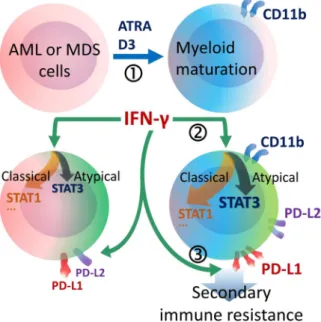
Myeloid maturation potentiates STAT3-mediated atypical IFN-gamma signaling and upregulation of PD-1 ligands in AML and MDS
Tam metin
Şekil
Benzer Belgeler
See discussions, stats, and author profiles for this publication at: https://www.researchgate.net/publication/261727268 Predicting Chemotherapy Sensitivity Profiles for Breast
Başımı çevirip de sağ cihetten arkama baktığımda üç dört karının otur duğunu ve birinin yılıştığını ve kulağıma değdirmiş olduğu tüyün de
Bunlar: Murtaza’ nın kişiliğinde işlenen ve sosyal adaletsizliğin değerlerini bünyesinde bulunduran; bulunduruyor gibi görülen Emniyet Müdürü ve komiser;
Immune checkpoint regulator PD-L1 expression on tumor cells by contacting CD11b positive bone marrow derived stromal cells. Cell
The association of clinicopathologic features and peripheral blood parameters with high PD-L1 expression in non-small cell lung cancer Introduction: Programmed death ligand 1
This article performs a narrative and aesthetic analysis of Reha Erdem’s movie, Kosmos (2009), through an engagement with Gilles Deleuze and Félix Guattari’s philosophical
However, each processor will have larger subspace (see Figure 3.1) so the pruning algorithm performs less pruning of candidate clusters at the upper level, and the number of the
In the decision-level fusion scheme with global (sensor-independent) training model, a common classification model is used for the feature vectors extracted from
