The
protective
effect of
vitamin
C,
vitamin E
and
selenium combination therapy
on
ethanol-induced duodenal mucosal injury
M Koyuturk* , S Bolkent2, S Ozdil3, S Arbak4 and R Yanardag5'Deartment
ofHistology and Embryology, Faculty ofMedicine, KadirHas University, 80810 Gayrettepe, Tur ey;2Department of
Biology,
FacultyofScience,Istanbul University, 34459 Vezneciler, Turkey;3Department of InternalMedicine, Istanbul MedicalFaculty, Istanbul University, 34390 Capa, Turkey;
4Department ofHistologyand Embryology, Marmara Medical Faculty, Marmara University, 81326
Haydarpasa, Turkey;
5Department ofChemistry, Faculty ofEngineering, Istanbul University, 34850Avcilar, Istanbul, Turkey
In this study, the effect ofa combination ofvitamin C, vitamin E and selenium on ethanol-induced duodenal mucosal damage in rats was investigated morphologi-cally and biochemically. The duodenal mucosal injury wasproducedby oraladministration of1 mLofabsolute ethanol toeachrat.Animals receivedvitamin C (250 mg/
kg), vitamin E(250mg/kg)and selenium(0.5mg/kg)for3 days and absolute ethanol 1 hour after last antioxidant administration and weresacrificed1 hourafter absolute ethanol. Extreme degeneration in intestinal mucosa of rats given ethanol was observed morphologically. In
addition, an increase in neuronal nitric oxide synthase
immunoreactive areas was observed in the rats of the group given ethanol. On the other hand, a normal
morphological appearance and a decrease in neuronal nitric oxide synthase immunoreactive areas were
de-tected in theratsgivenethanol+vitamin C+vitamin E+ selenium. In the group to which ethanol was adminis-tered,anincrease in serum cholesterol and adecrease in serum albumin levels were determined. On the other
hand,inthegrouptowhich ethanol +vitaminC+vitamin E+seleniumwere administered, serum cholesterol value decreased, and the serum albumin level increased. As a result, we can say that the combination of vitamin C, vitamin Eandselenium hasaprotective effect on ethanol-induced duodenal mucosal injury.Human & Experimen-tal Toxicology(2004)23, 391-398
Key words: duodenal injury; ethanol; nitric oxide synthase;
selenium;vitaminC;vitamin E
Introduction
Gastrointestinal mucosal lesions are caused by
various agents such as
stress,"2
ischemic-reperfu-sion,3 aspirin as nonsteroidal anti-inflammatory
drug,4'5 70% ethanol,6 80% ethanol,7 96%
etha-nol,8'9 absolute
ethanol,6'9
aceticacid,10"'1
indo-methacin andreserpin.'12"3
It is known that free radical production increased significantly in thetissue damage. It is shown that pretreatment with
vitaminCorvitaminEprior totheadministration of ethanol inhibited generation of free radicals and
DNA strand breaks in the
liver.14
It was recently reported that vitamin E might exert a protective*Correspondence:MeralKoyuturk,KadirHasUniversityMedical
Faculty, HistologyandEmbryologyDepartment,Vefa Bey S.N. 5 80810Gayrettepe, Istanbul, Turkey
E-mail:[email protected]
Received 7 November 2003; revised 26 April 2004; accepted
4 May 2004 © Arnold 2004
effect against nonsteroidal anti-inflammatory drug-induced gastric mucosal injury.5 The protective
effects of selenium seem to be primarily associated
with its presence in the glutathione peroxidases,
which are known toprotectDNAand other cellular
components from damage by oxygen radicals.'5 A
number ofinvestigations have revealed that vitamin
C, vitamin E and seleniumlevels were decreased by
exposure to ethanol.2 Selenium alone hasbeen shown to produce significant anti-ulcer activity,13 and cells adequately supplied with selenium are less susceptible to the damaging effects of endogen-ously or exogenously generated oxygen radicals.15 Vitamin Candvitamin E orvitamin E and selenium
exert a synergistic effect in the prevention of biological membranes from oxidants.'14"7
Nitric oxide synthase (NOS) is constitutively present in rat small intestine; the predominant form (90%) is neuronal nitric oxide synthase 10.1191/0960327104ht468oa
(nNOS).18 The relationship between the degree of
gut injury and nNOS activity is reported.19 Nitric oxide (NO) is a niultifunctional messenger that is involved ina wide range ofphysiological processes in many systems.20 Intracellular NO can regulate oxidants through its ability to react
rapidly
with radical oxygen species. There is clear evidence for protective effects of NO on excess production of reactive oxygen species.21AmoderateconcentrationofNO protectsthe cells against oxidative stress and plays an important role as regulatory mediator in various signalling processes.22
The presentmorphologicalandbiochemicalstudy wasundertaken to investigate the protective effect of
a combinationofvitamin C,vitaminEandselenium
onduodenal mucosalinjuryproducedbyethanol. In addition, we aimed to investigate the role of nNOS expression in ethanol-induced duodenal damage and the relationship between nNOS and antioxi-dants such as vitamin C, vitamin E and selenium.
Materials and methods
Animals
Forty, 4-5-month old, adult female
Sprague-Daw-leyrats,weighing200-250 g, obtainedfrom DETAM (Istanbul University Centre for Experimental Medi-cal Research and Application) were used in this study. The experiments were reviewed and ap-proved by the local institute's Animal Care and
Use Committees. The animals were fed with pellet
chow and tap water ad libitum before the experi-ments and fasted for 24 hours prior to the experi-ments. All rats were clinicallyhealthy.
Experimental design and treatment ofanimals The animals were randomly divided into four groups. Group I: intact animals (control). Group II: control animals receiving vitamin C (250 mg/kg/ day), vitamin E (250mg/kg/day) and sodium sele-nate (0.5mg/kg/day) for 3 days. Group III: animals receiving 1mL absolute ethanol. Group IV: animals received vitamin C, vitamin E and selenium (in the
same doses) for 3 days and absolute ethanol 1 hour after last antioxidant administration, and sacrificed
1 hour after absolute ethanol. The antioxidants and absolute ethanol were given to rats by gavage.
Animal model for duodenalmucosallesions Duodenal damage was induced by oral administra-tion at a constant volume by 1mL absolute ethanol perrat. The animals were sacrificed by ether 1 hour after treatment with absolute ethanol.
Light
microscopical
study
First
part
of duodenum was taken from animalswhich were fasted
overnight,
under etheranaesthe-sia. The tissues which were fixed in Bouin's
solu-tion and
subsequently
processed
using
traditionalparaffin embedding techniques
forpreparation
ofparaffin
sections were stained with Masson'striple dyes
and Periodic-Acid-Schiffforhistological
evaluation.Immunohistochemical
study
Same
paraffin
blocks of duodenalspecimens
that wereprepared
forlight
microscopic
assaywereused for immunohistochemical evaluation. Slides weredeparaffinized
in toluol andhydrated
in ethanol series. Slides were treated with 0.3% Triton-X 100 for 10 min and then rinsed inphosphate-buffer
saline
(10
mM,pH
7.5).
Antigen
retrieval wasperformed
in 0.01M citrate buffer(pH
6).
AHisto-statin Plus
(Zymed
Laboratories,
SanFrancisco,
USA)
broad-spectrum
kit of thestreptavidin-biotin
system
was thenapplied.
Sections were covered withblocking
serum for 20 min to preventnon-specific
binding.
They
were then incubated withnNOS
antibody
at 1:100 dilution(Transduction
Laboratories, Lexington, USA)
overnight
at 4°C. Slides were incubated for 20 min withbiotinylated
secondary antibody
then incubated with thestreptavidin-peroxidase
conjugate
for 20 min. Theenzyme
activity
wasdeveloped
usingaminoethyl-carbazole
(AEC).
The sections were counterstained withhaematoxylin.
Negative
control sectionswere
prepared
by
substituting
the nNOSantibody
withphosphate-buffer
saline.Staining
intensitywas rated asweak(+),
moderate(++)
orstrong(+++)
by
twodifferent,
blinded observers. Electronmicroscopical
study
For
scanning
electron microscopy, duodenal tissuesamples
areprefixed
for2hours ina2%phosphate-buffered
glutaraldehyde
solution(0.1
M, pH7.2),
postfixed
for 1 hour in a 1%phosphate-buffered
osmiumtetroxide solution and
passed
fromincreas-ing
alcohol andamyl
acetateseries. Afterdrying
the tissuesamples
withaBIORAD 'Critical PointDryer'
andgold
coating
with a BIORAD SC 502, tissuesamples
were examined under a JEOL 5200JSM
scanning
electronmicroscope.
Biochemical
study
Biochemical
investigations
ofcholesterol andalbu-min in serum were measured
by
means of anStatistical analysis
The results wereevaluated using an unpaired t-test and ANOVA variance analysis using the NCSS
statistical computer package.23
Results
Lightmicroscopicalresults
As light microscopic results, expansion and com-pression of the villi, ruptures and discontinuity in
the epitheliumof the end of thevilli, oedemainthe inside of villi and submucosa, hyperaemia in the capillaries, an increase in the mononuclear cell infiltration, a decrease in PAS positive reaction
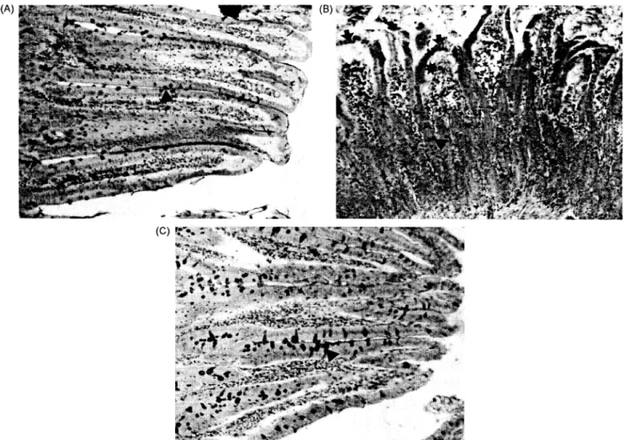
were observed induodenum of all rats of the group givenethanol,according tocontrols (Fig. 1A, B). On
the other hand, the same structures as the controls were determined in duodenum of all animals of the group given ethanol+vitamin C+vitamin E +
sele-nium. In addition, an increase in PAS positive reactionand mucus wasnoticed (Fig. 1C).
393
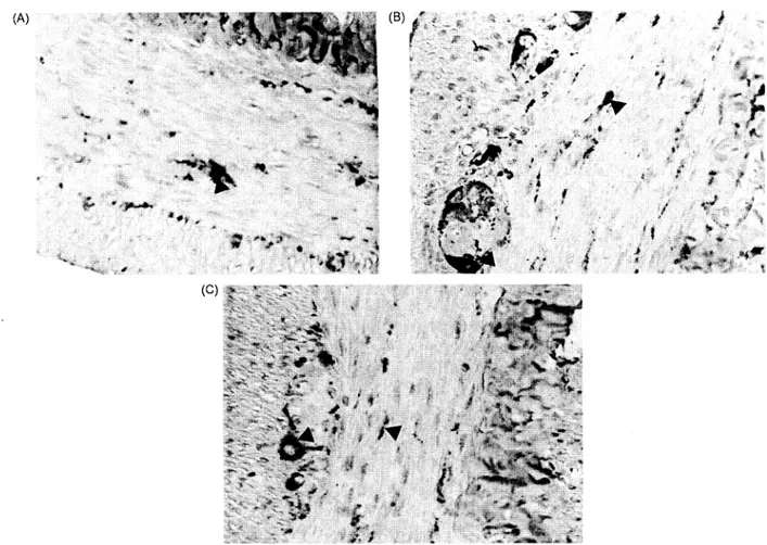
nNOSimmunoreactivity in duodenumwas deter-mined in all groups ofrats and in thesame density
(+++) (Fig. 2A-C). nNOS was localized in nerve fibres of circular muscle layer and ganglia cells of myenteric plexus. It was observed that immunor-eactive areas increasedin circularmuscle layer and ganglia cells in duodenum of all rats of the group givenethanol (Fig. 2B). Theseimmunoreactiveareas were decreased in duodenum of all animals of the group given ethanol +vitamin C+vitamin E+ sele-niumaccording to the experimental group and were almost thesame as incontrol groups(Fig. 2C). Inthe control group given antioxidants, immunoreactive areas were found the same as with intact control group.
Electron microscopical results
Scanning electron microscopical evaluation of Group I and Group II revealed good duodenal
mucosaintegritywithintactenterocytes(Fig. 3A, B). Scanning electron microscopic results of the
etha--~~~~O
(A) (B)
A~~~~~~~~~~~~~~~~~~A
(74'
Figure IA. A normal histological appearance of intestinal tissue of control rats. PAS positive reaction (A). B. The histological appearance of intestinal tissue of rats given ethanol. Expansion and compression of the villi (-*), oedema in the inside of villi and submucosa (*) and a decrease in PAS positive reaction (A*). C. The histological appearance of intestinal tissue of rats given ethanol+-i
vitamin C+vitamin E+selenium. The morphology of intestinal tissue was noticed to be nearly the same as those of the controls. An increase in PAS positive reaction ( A). PAS. x 240.
(B)
Figure 2A. nNOS immunoreactivity (A) in circular muscle layer and myenteric plexus of control rats. B. An increase in nNOS
immunoreactivity (A*)in circular musclelayerandmyenteric plexusof ratsgivenethanol.C. AdecreaseinnNOSimmunoreactivity (A)
in circularmusclelayerandmyenteric plexus ofratsgivenethanol+vitamin C+vitaminE +selenium.x520.
nol-administered group, when compared to control
groups,presented aloss ofepitheliumfrom thevilli
with exposure of underlying lamina propria.
Hae-morrhagic regions with deep erosions and fibrin deposits indicated anextreme degeneration ofvillar
surface topography (Figure 3C). Scanning electron microscopical investigation ofthe ethanol+vitamin
C+vitamin E+selenium-administered group
showed a good villar epithelial arrangement in
surface topography of villi. Closely packed enter-ocytes with mucous secretion demonstrated a
sig-nificant reduction in the severity of mucosal injury byantioxidant treatment (Fig. 3D).
Biochemical results
Serum cholesterol and albuminvaluesarepresented
in Table 1. From the obtained results, values of cholesterol in serum in the group administered
ethanol have shown no significant difference when
compared with the control group (Pt-test=0.161).
Also, a significant decrease was noted in the group
administered ethanol+vitamin C+vitaminE+
sele-nium compared to the groups administered ethanol
(Pt-test=0.0001). According to Table 1, a significant
difference in the serum cholesterol levels of four groups was observed (PANOVA=0.0001). In this
study, a statistically significant decrease was
ob-served in the serum albumin values of the group
administered ethanol, in comparison with the
con-trol group (Pvalue 0.020). Also, an insignificant
increase was detected in the group administered
ethanol+vitamin C+vitamin E+selenium, when
compared to the group administered ethanol
(Pvalue=-0.138). According to Table 1, a significant
difference in the serum albumin levels ofthe four
groups was observed (PANOVA=-0.080).
Discussion
Duodenal ulceration may heal despite
hyperchlor-hydria, suggesting that the integrity ofthe duodenal
mucosa is more crucial than the state of acid secretion in the mechanism of development ofthis ulceration. It is suggested that oxygen-derived free radicals are detrimental to the integrity of the duodenalmucosa and thatintheratoxygen-derived
(A)
(C
395 (B)
(D)
Figure 3A. Control group scanningelectronmicrograph demonstrates normal surface topography ofvillar epithelium. B. Scanning electronmicrograph ofrats receivingvitaminC+vitaminE+selenium.Regular arrangement of enterocytes suggestsanormal surface villartopography. C. Scanning electron micrograph ofintestinal mucosa ofethanol-administered ratgroup. Extreme degeneration of
surfacetopographywithdesquamated villarepithelialcells(A), deeperosionsandhaemorrhagicregions witherythrocytes (*)and fibrin
deposits.D. Scanning electronmicrograph of intestinal mucosa ofethanol+ vitamin C +vitamin E+selenium-administered ratgroup. Surfaceepithelialtopography indicatesagoodvillarepithelial arrangement,tightly packedenterocytes(0*)with mucousdischarge.Bar:
10tIm.
freeradicals are directly implicated inthe
mechan-ism of secretagogue-induced acute and chronic duodenal ulceration and that removing these radi-cals protects the duodenum against ulceration.24 In addition, it is reported that oral administration of ethanol interrupted the mucosal defence and pro-duced mucosal damage by necrosis or apoptosis, of
gastric mucosal
cells.7"5
Our study demonstrated that antioxidantsimproved theintegrity of duodenalepitheliumand reducedthedegree ofdamageinthe glandular architecture. The mucosal injury due to
ethanol administration consisted mainly of separa-tion ofthe surface epithelium from the underlying lamina propria with complete loss of epithelium.
Table1 Meanlevelsof serumcholesterol and albumin for allgroups*
Groups Control (n =10)
Cont. +vit. C+vit.E +Se(n=10)
Ethanol(n=10)
Eth. +vit.C+vit.E+Se (n=10) PANOVA
*Mean+SD.
n=Numberofanimals.
apttest =0.161 versus control groups.
bPtest=0.020versuscontrolgroups.
Cholesterol mg% 56.62+1.04 48.68+ 1.18 58.84+3.54a 51.83 + 2.02 0.0001 Pt-test 0.0001 0.0001 Albuming% 4.32 + 0.18 Pt-test 4.41 +0.58 3.99+ 0.36b 4.19 +0.19 0.080 0.649 0.138 sei.Rs S
This fact is due to the hazardous effect of ethanol which rapidly penetrates gastroduodenal mucosa
causing membrane damage.
The subsequent increase in mucosal permeability togetherwiththe release of vasoactiveproductsfrom
mast cells, macrophages and otherblood cells may lead to vascular injury, necrosis and ulcer
forma-tion.9 It is suggested that chronic ethanol
adminis-tration induces oxidative stress, mainly increasing lipid peroxidation of the cell membrane and this
leads to increased membrane fluidity, disturbances ofcalciumhomeostasis andfinallycelldeath.16 Itis reported that pharmacological antioxidants could
have beneficial effects in reducing the incidence of ethanol-induced changesin cellular
lipids,
proteins and nucleic acids. The antioxidants consideredcould act by reducing free radical production, trapping free radicals themselves, interrupting the
peroxidation process or reinforcing the natural
antioxidant defence.16 In our other study, it is
shownthat theadministration ofvitamin C,vitamin E and selenium to ethanol-induced rats eliminated accumulation of lipid peroxides in the stomach, suggesting that vitamin C, vitamin E and selenium protect againstethanol-inducedoxidativedamage.26
The prominent epithelial damage in the ethanol-administered group could be due to increased lipid peroxidation of cell membranes leading to cell death. Microscopical evaluation of gastroduodenal
mucosa of the antioxidant-administered group re-vealed a significant reduction in injury formation. We could correlate these findings with the free radical trapping activity of antioxidants. As is
known, ethanol increases membranelipid
peroxida-tionandproduction of free radicals and maycause a
number oftissue lesions.27 Free radicals are highly reactive species characterized by one or more
unpaired electrons in their outer orbital. These reactive oxygen species are highly reactive and
capable of damaging many biological macromole-cules suchasRNA, DNA, proteinsand
lipids.28
The cell membrane consists of phospholipid bilayer,cholesterol and proteins. Phospholipids and choles-terol can be modified by oxidative stress and free radicals.29 In the present study, the cause of the increase observed in the values of cholesterol and decrease in the albumin levels in the group admi-nisteredethanolcanbeexplained bydamaged tissue due tothe free radicals.
Vitamin E (tocopherol) is an important antioxi-dant in biological systems and is readily absorbed from intestine.
ox-Tocopherol
is present in the lipidbilayers ofbiologicalmembranes where itmayplay a structural role.30 c-Tocopherol very efficiently scavenges lipid peroxyl radicals and thereby
pre-vents the
lipid
peroxidation
process inan uninhib-ited chain reaction.Tocopherol deficiency
ischaracterized
by
a number of chronic healthpro-blems;
thereisdamage
tocellmembranesasaresult of increasedlipid
peroxidation.3"
Ascorbate,
themajor
water-solubleantioxidant,
has been shown toefficiently
scavengehypochloride, hydroxyl
radi-cals andperoxyl
radicals,
and to restore theanti-oxidant
properties
of fat-solubleox-tocopherol.32
Selenium is an essential part of the enzyme
glu-tathione
peroxidase,
whichfunctions as apart ofanantioxidant system to protect membranes and
es-sential
proteins
from the potentiallydamaging
effects of reactive oxygen and lipid
peroxides.33
The increase in the serum albumin levels and the decreaseinthecholesterol valuesbythe
application
of ethanol and vitamin
C,
vitamin E and selenium show that antioxidants prevent the damage causedby
ethanol.NO
regulates
acid andgastricmucussecretion and alkalineproduction,
and is involved in themain-tenance of mucosal blood flow.34 Immunohisto-chemical studies have shown that the enzyme necessary for NO synthesis is expressed in enteric
neurons.'9
It isreported
that nNOSimmunoreactiv-ity
by
immunogold
staining in small intestine waslocalized in nerve profiles in myenteric plexus and circular muscle layer.35 Ethanol intake injures the functional and structural integrity ofthe intestinal mucosa and causes loss of intestinal barrier func-tion. Abnormal intestinal barrier can allow the
penetration
ofnormally
excluded luminalsubstance across the mucosa and can lead to initiation of aninflammatory
process and mucosal damage.21 NOacts as an
endogenous
mediator modulating bothrepair and integrity of the tissues, and exhibits
gastroprotective properties against different types of aggressive agents.34 Ethanol absorption is con-trolled mainly by gastric emptying, because the
primary region of ethanol absorption is the small
intestine.36 NO was reported as a neurotransmitter
that acts at receptor protein on adjacent neuronal
membranes.37 Itwas suggested that inhibition ofNO synthesis may lead to an increase of intestinal
motility.'8 Depending on its concentration, dual roles of NO can expose protective and toxic
ef-fects.38 In this study, the increase of nNOS immu-noreactivity in the rats ofthe group given ethanol
may exhibit a role related toregulation of intestinal motility and/or membrane integrity.
Immunoreac-tion levels in the animals of the group given
ethanol-+antioxidants decreased compared to the
group given ethanol as like to control group. The decrease of endogenous nNOS expression in the
a regulatory role of antioxidants against mucosal injury.
As a result, the morphological and biochemical
evaluations reveal that the combination of vitamin C, vitamin E and selenium hasaprotective effecton ethanol-induced duodenal injury. The antioxidants supplementation may be useful in alcohol-induced oxidative stressby enhancing the antioxidant capa-city, and could play a significant protective role in
the acute stage of duodenal injury. In conclusion,
397
food supplementation with vitamin C and E, and
selenium can be used in the therapy of
ethanol-induced duodenal injury.
Acknowledgements
This study was supported by the Research Fund of Istanbul University. Project No: UDP-20/20/
04072002.
References
1 Konturek PC, Konturek SJ, Browski T, Dembinski A, Zembala M, Mytar B, Hahn EG. Gastroprotective activity ofmelatonin and its precursor, L-tryptophan,
againststress-induced and ischemia inducedlesionsis mediated by scavenge of oxygen radicals. Scand J
Gastroenterol 1997; 332: 433-38.
2 Suresh MV, Kumar S, Lal JJ, Indira M. Impact of massive ascorbic acid supplementation on alcohol induced oxidative stress in guinea pigs. Toxicol Lett 1999; 104: 221-29.
3 Smith GS,MercerDW, CrossJM,BarretoJC, MillerTA. Gastricinjuryinducedbyethanol and
ischemia-reper-fusionin therat.DigDisSci 1996; 41: 1157- 64. 4 Guidobono F, Ticozzi PC, Sibilia V. Protection
by amylin of gastric erosions induced by indometh-acin orethanolinrats.BriPharmacol 1997;120: 581-86.
5 Sugimoto N, Yoshida N, Yoshikawa T, Nakamura Y,
IchikawaH,Naito Y,KondoM. Effect ofvitamin E on
aspirin-induced gastricmucosal injuryinrats. DigDis Sci 2000; 45: 599-605.
6 BravoML,Escolar G,NavarroC,FontarnauR,Bulbena 0. Morphological study of gastric lesions developing
intherat under several damaging conditions: modifi-cationsinducedby pretreatment withzinc asexamate. ScanningMicrosc 1992; 6: 855 -64.
7 Liu ESL, Cho CH. Relationship between ethanol-induced gastritis and gastric ulcer formation in rats. Digestion 2000; 62: 232-39.
8 Szelenyi I, Brune K. Possible role of oxygen free radicals in ethanol-induced gastric mucosal damage inrats. DigDis Sci 1988; 33: 865-71.
9 Bilici D, Suileyman H, Banoglu ZN,
Kiziltung
A, Avci B,(;iftgioglu
A, Bilici S. Melatonin prevents ethanol-induced gastric mucosal damage possiblydue to its antioxidant effect. Dig Dis Sci 2002; 47: 856-61.
10 Okabe S, Pfeiffer CZ.Chronicity of acetic acid ulcerin the ratstomach. AmJDigDis 1972; 17: 619-29. 11 Ito M, Segami T, Tsukahara T, Kojima R, Suzuki Y.
Effect of cimetidine and omeprazole on gastric ulcer healing of rats with limited food intake time. EurJ
Pharmacol 1994; 263: 245-51.
12 Mozsik G, Jovar T. Biochemical and pharmacological
approach to the genesis ofulcer disease. DigDis Sci 1988; 33: 92-105.
13 Al-Moutary AR, Tariq M. Effect of vitamin E and selenium on hypothermic restraint stress and chemi-cally-induced ulcers. DigDisSci 1996;41: 1165-71.
14 Navasumrit P, Ward HT, Dodd NJV, O'Connor J. Ethanol induced free radicals andhepatic DNA strand breaksarepreventedin vivobyantioxidants: effects of acute and chronic ethanol exposure. Carcinogenesis 2000; 21: 93 -99.
15 Schrauzer GN. Anticarcinogenic effect of selenium. Cell MolLifeSci 2000; 57: 1864-73.
16 NordmannR. Alcohol and antioxidant systems. Alco-holAlcoholism 1994; 29: 513 -22.
17 Horwath PM, IpC. Synergisticeffect of vitamin E and selenium in the chemoprevention of mammary carci-nogenesis in rats. Cancer Res 1983; 43: 5335-41. 18 Hebeiss K, Kilbinger H. Cholinergic and GABAergic
regulation ofnitric oxide synthesis in the guinea pig ileum.AmJPhysiol 1999; 276: G862 -66.
19 Sanders KM, Ward SM. Nitric oxide as a mediator of
nonadrenergic noncholinergicneurotransmission. Am
JPhysiol 1992; 262:G379- 92.
20 Garcia-Victoria M, Garcia-Corch6n C, Rodrfguez JA, Garcia-Amigot F, Burrell MA. Expression ofneuronal nitric oxide synthase in several cell types of the rat gastric epithelium. J Histochem Cytochem 2000; 48: 1111-19.
21 Binion DG,FuS, Ramanujam KS,Chai YC, Dweik RA, Drazba JA, Wade JG,ZiatsNP, ErzurumSC,Wilson KT. iNOS expression in human intestinal microvascular
endothelial cells inhibits leukocyte adhesion. Am J
Physiol 1998; 275:G592-603.
22 Droge W. Freeradicalsin the physiological control of
cell function.AmJPhysiol 2002; 82:47-98.
23 Hintze JL.Copyright C. 865, East 400. North Kaysville, UT84, 037 (801), 546-0445, 1986.
24 Salim AS. Role of oxygen-derived free radicals in mechanism ofacute and chronic duodenal ulceration in the rat.DigDisSci 1990; 35: 73 -79.
25 Piotrowski J, Piotrowski E, Skrodzka D, Siomiany A, Siomiany BL. Gastric mucosal apoptosis induced by ethanol: effect of antiulcer agents. Biochem Molec Biol Int 1997;42: 247- 54.
26 Ozdil S, Yanardag R, Koyuturk M, Bolkent S, Arbak S. Protective effects of ascorbic acid, DL-cx-tocopherol acetate and sodium selenate on ethanol-induced gas-tric mucosal injury ofrats. Biol Trace Elem Res 2004; 98(in press).
27 Coudray C, Boucher F, Richard MJ, Arnaud J, Leiris J, Favier A. Zinc deficiency, ethanol, and myocardial ischemia affect lipoperoxidation in rats. Biol Trace ElemRes 1991; 30: 103- 18.
28 Tian L, Cai Q, Wei H. Alterations of antioxidant enzymes and oxidative damage to macromolecules in different organs ofrats during aging. FreeRadicBiol Med 1998; 24: 1477- 84.
29 Zima T, Fialova L, Mestek0,Janebova M, Crkovska J, Malbohan I, Stipek S, Mikulikova L, PopovP. Oxida-tive stress, metabolism of ethanol and alcohol-related diseases. IBiomedSci 2001; 8:59-70.
30 Kostner GM, Otti D, Jauhiainen M, Ehnholm C, Esterbauer H. Human plasma phospholipids transfer protein accelerates exchange/transfer of oc-tocopherol betweenlipoproteins and cells. BiochemJ 1995; 305: 659-67.
31 Yllmaz 0,
Qelik
S,Naziroglu M, $ay M, DilsizN. The effects of dietary selenium and vitamin E and their combination on the fatty acids of erythrocytes, bone marrow and spleen tissue lipids of lambs. Cell Bio-chemFunct 1997;15: 1-7.32 Skrzdlewska E, Farbiszewski R. Lipid peroxidation and antioxidant status in the liver erythrocytes, and
serum of rats after methanol intoxication. J Toxicol EnvironHealth 1998; 53:637-49.
33 Yanardag R, OrakH. Total selenium concentration in various waters ofTurkey. Environ Technol 2001; 22: 237-46.
34 Martin MJ, Jimenez MD, MotilvaV. New issuesabout nitricoxide anditseffectsonthe gastrointestinaltract.
CurrPharmDes 2001; 7: 881- 908.
35 BerezinI, Snyder SH, BredtDS,DanielEE. Ultrastruc-tural localization of nitric oxide synthase in canine small intestine and colon. Am JPhysiol 1994; 266:
C981-89.
36 Holt S, Stewart MJ, Adam RD, Heading RC. Alcohol absorption, gastric emptying and a breathalyser. BrJ
ClinPharmacol 1980; 9: 205-208.
37 Snyder SH. Nitric oxide: first in a new class of neurotransmitters. Science 1992; 257: 494-96. 38 FerrazJG, TigleyA, WallaceJL. Paradoxical effects of
L-arginine on gastricmucosal integrity. EurJ