SELF-ASSEMBLED PEPTIDE TEMPLATE DIRECTED
SYNTHESIS OF ONE-DIMENSIONAL INORGANIC
NANOSTRUCTURES and THEIR APPLICATIONS
A DISSERTATION SUBMITTED TO
MATERIALS SCIENCE AND NANOTECHNOLOGY PROGRAM OF THE GRADUATE SCHOOL OF ENGINEERING AND SCIENCE
OF BILKENT UNIVERSITY
IN PARTIAL FULFILLMENT OF THE REQUIREMENTS FOR THE DEGREE OF
DOCTOR OF PHILOSOPHY
by
HANDAN ACAR December, 2012
iii
I certify that I have read this thesis and that in my opinion it is fully fully adquate, in scope and quality, as a dissertation for the degree of doctor in philosophy.
Assist. Prof. Dr. Mustafa Özgür Güler (Advisor)
I certify that I have read this thesis and that in my opinion it is fully fully adquate, in scope and quality, as a dissertation for the degree of doctor in philosophy.
Prof. Dr. Engin Umut Akkaya
I certify that I have read this thesis and that in my opinion it is fully fully adquate, in scope and quality, as a dissertation for the degree of doctor in philosophy.
iv
I certify that I have read this thesis and that in my opinion it is fully fully adquate, in scope and quality, as a dissertation for the degree of doctor in philosophy.
Prof. Dr. Baki Emir Denkbaş
I certify that I have read this thesis and that in my opinion it is fully fully adquate, in scope and quality, as a dissertation for the degree of doctor in philosophy.
Assist. Prof. Dr. Necmi Bıyıklı
Approved for the Institute of Engineering and Science
Prof. Dr. Levent Onural Diresctor of the Graduate School
v
ABSTRACT
SELF-ASSEMBLED PEPTIDE TEMPLATE DIRECTED
SYNTHESIS OF ONE-DIMENSIONAL INORGANIC
NANOSTRUCTURES and THEIR APPLICATIONS
Handan Acar
Ph.D. in Materials Science and Nanotechnology Supervisor: Assist. Prof. Dr. Mustafa Özgür Güler
December, 2012
Engineering at the nano scale has been an active area of science and technology over the last decade. Inspired by nature, synthesis of functional inorganic materials using synthetic organic templates constitutes the theme of this thesis. Developing organic template directed synthesis approach for inorganic nanomaterial synthesis was aimed. For this purpose, an amyloid like peptide sequence which is capable of self-assembling into nanofibers in convenient conditions was designed and decorated with functional groups showing relatively high affinity to special inorganic ions, which are presented at the periphery of the one-dimensional peptide nanofiber. These chemical groups facilitated the deposition of targeted inorganic monomers onto the nanofibers yielding one-dimensional organic-inorganic core-shell nanostructures.
vi
The physical and chemical properties of the synthesized peptide nanofibers and inorganic nanostructures were characterized using both qualitative and quantitative methods.
First, silica nanotubes were obtained by silica mineralization around these peptide nanofiber templates for the construction of sensors for explosives. The fluorescence dye was used to coat the silica nanotubes to detect explosive vapor. The surface of the silica nanotubes were porous enough to adsorb more dye compared to the silica nanoparticles and silica film, and causes faster fluorescence quenching in the presence of explosives like trinitrotoluene and dinitrotoluene. The silica nanotubes which synthesized with this peptide nanofiber templates can be used in catalysis and sensors in which high surface area is advantageous. In the second part of the thesis, titanium dioxide nanotubes were obtained from titania mineralization. They are well-known with their fascinating properties as a result of the one-dimensional nanostructure, such as more efficient electron transfer and less electron-hole recombination. The sufficient photoactivity of titanium dioxide makes them suitable materials for Dye-Sensitized Solar-Cell construction. It is demonstrated that the peptide nanofiber templated titanium dioxide nanotubes have more than two times more efficiency compared to template-free synthesized titanium dioxide particles. Finally, designed peptide sequence was conducted to a multi-step seeding mediated growth method for gold mineralization around peptide nanofibers. The gold-peptide hybrid nanostructures with different packing characteristics and sizes were synthesized and fully characterized. Further, it was demonstrated that the dry film of these nanostructures showed a resistive switching dominant conductivity, due to the
vii
nanogaps in between gold nanoparticles as a result of particle alignment driven by the peptide nanofiber.
The results obtained in this thesis encourage use of a new “bottom-up” synthesis approach. Specially designed peptides with desired properties and functional groups were synthesized and peptide nanofibers formed were further used as templates for inorganic mineralization. Not only it is possible to synthesis high amount of nanostructure with this approach, but also formed one-dimensional nanostructures show advance functionalities used in several applications as a part of the thesis scope. This methodology is suitable for many metals and metal oxide based applications.
Keywords: Biomimetic mineralization, Nanomaterials, Peptide, Self-assembly, One-Dimensional Nanostructures, Template Directed Synthesis.
viii
ÖZET
KENDİLİĞİNDEN DÜZENLENEN PEPTİT KALIPLAR
YARDIMIYLA İNORGANİK TEK-BOYUTLU NANOYAPILARIN
SENTEZİ ve UYGULAMA ALANLARI
Handan AcarMalzeme Bilimi ve Nanoteknoloji Programı, Doktora
Tez Yöneticisi: Yar. Doç. Dr. Mustafa Özgür Güler Aralık, 2012
Son yıllarda, bilim ve teknoloji alanında nano düzeyde mühendislik oldukça yoğun çalışılan bir alan olmuştur. Bu tez; işlevsel inorganik malzemelerin, doğadan ilham alınarak, yapay organik kalıplar yardımıyla sentezlenmesi üzerine yeni bir yöntem sunmaktadır. Organik kalıp ile inorganik nanomalzeme sentezlenmesi yönteminin geliştirilmesi amaçlanmıştır. Bu amaçla, uygun koşullarda nanofiber haline gelebilmesi için programlanmış amiloyid benzeri peptit sekansı tasarlanmıştır. Bu tasarım sırasında, oluşan nanofiberin dış kısmında kalan ve ortamdaki belli inorganik iyonlarla bağlanma eğilimi gösteren işlevsel gruplarla döşenmiştir. Bu işlevsel kimyasal gruplar, hedeflenen inorganik monomerlerin peptit nanofiberlerin üzerlerinde birikerek, tek-boyutlu organik içerikli- inorganik kabuklu nanomalzeme oluşması için görevlidir. Sentezlenen peptit nanofiberlerin ve inoganik
ix
nanomalzemelerin fiziksel ve kimyasal özellikleri, kalitatif ve kantitatif metotlarla karakterize edilmiştir.
İlk olarak, Silisyum ile peptit nanofiber kalıpların etrafında yapılan mineralizasyon işleminden elde edilen silika nanotüpler, patlayıcı sensörü geliştirilesinde kullanılmıştır. Silika nanotüpler patlayıcıların varlığını algılaması için, floresan boya ile kaplanmıştır. Nanotüplerin yüzeylerinin bir hayli gözenekli olmaları, nanoparçacıklara ve film yüzeye göre floresan boyayı daha fazla tutmasını ve dolayısıyla trinitrotoluen ve dinitrotoluen gibi patlayıcıların olduğu ortamlarda daha hızlı floresan sönümlemesi göstermesini sağlamıştır. Bu şekilde peptit nanofiber kalıp kullanılarak sentezlenen tek boyutlu silika nanotüpler, yüksek yüzey alanları sayesinde katalizör ve sensör gibi malzemelerin hazırlanmasında kullanılabilirler. İkinci kısımda, titanyum mineralizasyonu ile titanyum dioksit nanotüpler elde edilmiştir. Tek-boyutlu yapı, elektron transferini daha verimli gerçekleşrirebilmesi ve elektron-boşluk yeniden birleşmesinin daha az meydana gelmesi gibi üstün özellikler göstermektedir. Bu özellikler ile elde edilmiş titanyum dioksit tek-boyutlu nanoyapıların gelen ışığa gösterdiği tepki artar ve bunlar boyayla uyarılan güneş pili yapımında kullanılmaya uygun hale gelir. Peptit nanofiberlerin kalıp olarak kullanılmasıyla elde edilen titanyum dioksit nanotüpler ile yapılan güneş pillerinin herhangi bir kalıp kullanılmadan sentezlenen titanyum dioksit parçacıklar ile yapılanlara göre iki katından daha fazla verimle çalıştığı gösterilmiştir. Son olarak, altın mineralizasyonu çalışmasında, çok basamaklı, tek noktadan büyütme metodu geliştirilmiştir. Bu yöntemle farklı uzunluklarda ve şekillerde birçok altın-peptit hibrit nanoyapıları elde edilmiştir. Özellikle, büyüklükleri birbirine benzer altın nanoparçacıkların peptit nanofiberler etrafında tutunması ile oluşan tek-boyutlu
x
nanoyapılar, kontrollu bir şekilde elde edilmiştir. Tek-boyutlu altın-peptit hibrit nanoyapılarındaki altın nanoparçacıkların arasındaki boşluklardan dolayı, nanoyapıların oldukça iletken olduğu gözlemlenmiştir. Bu yapıların kuru filmlerinde ise, tünellemenin baskın olduğu direnç değişimi gösteren ile bir iletkenlik gözlemlenmiştir.
Bu tezde elde edilen sonuçlarla yeni bir temelden yukarı yaklaşımı gösterilmiştir. İstenilen özelliklerde ve işlevsel gruplarla tasarlanmış peptitler sentezlenmiş ve bunlardan inorganik mineralizasyonlarda kalıp olarak kullanılabilen nanofiberler oluşması sağlanmıştır. Bu yaklaşımla, yüksek miktarda nanaomalzeme sentezi yapılması mümkün olabileceği gibi, oluşan tek-boyutlu nanoyapıların oldukça gelişmiş özellikleri ile birçok uygulama alnında kullanmak mümkün olabilir. Bu sentez yöntemi, birçok metal ve metal oksit temelli uygulamada yer bulabilir.
Anahtar Kelimeler: Biyomimetik mineralizasyon, Nanomalzemeler, Peptit, Kendiliğinden Düzenlenme, Tek-Boyutlu Nanoyapılar, Kalıp Kullanarak Sentez.
xi
xii
Acknowledgement
Dr. Mustafa Özgür Güler; you thought me to be a well-disciplined, conscientious and self-motivated researcher, and the moral values of being a fair scientist. The most valuable thing that you taught to me is the esteem that I profess to science. When I got lost, you enlightened my path with your guidance and support. I owe this thesis to your advice with your words: “I do not recommend you to do another job”. I will be always proud of being your student. The scientific advance you brought to me and the ecole of BML group will always be with me through my academic life. You sculptured me into a fair scientist.
Dr. Ayşe Begüm Tekinay; thank you for your encouragement to be curious about everything in science. You are a role model for me to be a successful female scientist. I was always welcome whenever I visited you and asked for your advice. Thank you for being so kind to me.
Dr. Aykutlu Dâna, your intelligence and your curiosity in all branches of science always evoked my admiration. I learnt a lot from you while working with you. It will be always honor for me to have worked with you. You were always very tactful to me, and I will always remember you with admiration.
I would like to thank Prof. Dr. Engin Umut Akkaya, Ass. Prof. Dr. Ali Osmay Güre and Ass. Prof. Dr. Emrah Özensoy for their fruitful discussions and guidance during my Ph.D.
Sıla Toksöz; it is a honor for me to be your friend and lab mate. I gained different aspects to science from you. You are also a sister for me. I am very lucky that you made me feel like a part of your noble and generous family. I will always remain
xiii
grateful to you and your family. I wish always the best for you which you deserve very much. I am sure I will be proud of your triumphs in the future. Ruslan Garifullin, you were with me almost from the beginning. We worked together in the lab. You have a huge work on this thesis and I owe a big thank to you. Adem Yıldırım, I learnt a lot from your sincere helps and patience. You are a very special and valuable scientist, and I will always be proud of having the chance of working with you.
Mustafa Güler, you put up my whims so much. You helped me whenever I needed you, thank you very much. Your observations always guided my research. Since we worked for hours together in a dark TEM room, you have become very close friend of mine. I will always remember you and your style in my life.
Okan Öner Ekiz, we started together in the same office. I wish I can say that we grown up together here, but we mostly got older. I am sure that, any objective aspect can see your successes and appreciate them. Hakan Ceylan, you always inspired me. Although I got some troubles in Boston, I am very grateful to that travel, since I earned a brother like you. I always appreciate your optimism (which is scientifically proved) and your excitement. Melis Şardan, Oya Ustahüseyin, Göksu Çınar; I am very lucky to be so close with you. Your friendship and intelligence helped me to get over the bad times. Being in the same research group with you will always be a honor for me. Being close friends with you will always warm my heart. I will always feel your support with me. Dr. Rükan Genç, thank you very much for enlightening my way with your not only scientific point of view, but also friendship. I learnt a lot from your optimism.
xiv
Oğuz Hanoğlu, how can I describe your place in my life; squirrels! Your determination of following your dreams always inspired me. I learnt a lot from you and your view of life, and I am sure I will continue to learn. You are very valuable and special friend of mine.
Can Ataca, talking with you always made me very self-confident. What is your secret about this feeling? I hope it is just being realistic. I feel your support with me whether you are on the other side of the world! Your sense of humor, sympathy and intelligence evoke my admiration. And Hasan Şahin, you both were very close and special friends for me. I will always remember the times with you. I owe a thank to both of you, because you colorized my life in UNAM. I always appreciate your friendship and feel very lucky to have you. I am sure that I will always be proud of your successes wherever you are.
Nilay Arkün, you are like a sister for me. I had many troubles and you were always there. The rational thoughts of you always evoked my admiration. I learnt to say “I love you” without hesitation from you. I always felt myself very comfortable with you and your big genuine family. Onurcan Özçelik, my brother, you accept me as I am. I have never listened someone else for that much long time with a full focus. The intelligence, honesty and politeness of your thoughts always amazed me. I know that, where ever you both will be in future, we will always be close. Yıldıray Kabak, you were there when I needed you most! We like each other very much, which makes it easier for me to understand your badly desire to find a passion. I am sure that you will have whatever you want in your life, with the help of your talent and ambitions. As I talked with you, we are both lucky in different points of view. As
xv
you said, sometimes leaving and starting from zero is the best. I will not start from zero, I will always have you, my very precious friends.
Kavaklıdere Rotaract Club, you are my family in Ankara. As I say from the beginning, being a part of you is a great prestige for me. It is a fortune to me to have that much big, wonderful family. I always feel very comfortable, cheerful and excited when I am with you. I always know that, I am a member of not only a club, but also a huge and warm family.
Yelloz and Tomtiş, you thought me something that I could never learn from a human but you two lovely cats. I learnt to love and be loved. I really appreciate you both because of the feeling of peace you gave to me when I needed it most.
One of the most important things in life is feeling of belonging to a home. Whatever happens to me, I know that I have a family which I belong to. Mom, Hande and Aydan, you are the most important people in my life. Where you are is home to me. We have been through a lot of things during my Ph.D., and we got over all of them together. We will always do it together, whatever it comes in life. I love you all.
My mom, although you never told us anything what you have experienced, I can understand it very deeply. You did it for us, for our future. And, I did it, for you…
xvi
Table of Contents
ABSTRACT ... iii
ÖZET ... viii
Table of Contents ... xi
Table of Figures ... xix
Table of Schemes ... xxvi
1. Introduction ... 2 2. Background ... 11 2.1. Electrostatic Interactions... 12 2.2. Metal coordination ... 13 2.3. Hydrogen bonding ... 19 2.4. π-π interactions ... 27 2.5. Solvophobic interactions... 31
xvii
2.6. Van der Waals interactions ... 33
3. Synthesis and Characterization of Peptide Nanofibers as Template for Synthesis of Inorganic One-Dimensional Nanostructures ... 36
3.1. Introduction ... 36
3.2. Experimental ... 37
3.3. Results and Discussions ... 42
4. Template-Directed Synthesis and Characterization of Silica Nanotubes and Their Application as Explosive Detection ... 58
4.1. Introduction ... 58
4.2. Experimental ... 60
4.3. Results and Discussions ... 64
5. Template Directed Synthesis and Characterization of Titania Nanotubes and Their Application as Dye-Sensitized Solar Cell ... 79
5.1. Introduction ... 79
5.2. Experimental ... 82
5.3. Results and Discusions ... 85
6. Template Directed Synthesis and Characterization One-Dimensional Au Nanocomposites and Their Conductivity Properties... 104
6.1. Introduction ... 104
6.2. Experimental Section ... 106
xviii
7. Conclusion ... 133 8. References ... 138
xix
Table of Figures
Figure 1.1. Fischer’s synthesis of glycoglycin. ... 4 Figure 2.1. Bis-biotinylated terpyridine forms a linear tetrabiotinylated connector, the
[Fe(Biot2-terpy)2]2+ complex, upon reaction with ferrous ion. The presence of streptavidin results in formation of linear coordination polymers [65]. ... 15 Figure 2.2. SEM images of MOPF bundles containing CaCl2. (a) 8x10⁻⁷ M Fe
solution containing 0.05 M CaCl2, 5 min at RT; and (b) a zoom into it. (c-e)
Biomineralized bundles of 8 x 10⁻⁶ M Fe solution, 12 hours: (c) 0.01 M CaCl2;
(d-e) 0.05 M CaCl2 [65]. ... 16
Figure 2.3. Chemical structure of the dendron-rod-coil molecules and TEM image of cadmium sulfide precipitated in a suspension of DRC nanoribbons, at an early stage growth. The inset shows the start of nucleation at different points and the organic ribbon under CdS as indicated by the arrow [71]. ... 19 Figure 2.4. Alternating D- and L-amino acids assembling into cyclic peptide
nanotubes via the antiparallel ring stacking. Extensive intersubunit hydrogen bonding can be seen in the sketches. Reproduced with permission from [77]... 21 Figure 2.5. The chemical structure and the cartoon of the peptide amphiphile
xx
aliphatic tails. (b) is the critical β-sheet hydrogen bonding portion of the peptide. (c), the peripheral peptide region, constitutes the interface between the fiber and the environment [80]. ... 23 Figure 2.6. Chemical structure of the dendron-rod-coil molecule. (a) Bright field
TEM image of unstained 0.004 wt% DRC molecules, dissolved in styrene. (b) High magnification TEM image of a thin slice of the scaffold material, containing 1 wt% DRC molecules. Arrows represent perpendicularly placed individual bimolecular ribbons [89]. ... 25 Figure 2.7. Open-ended tubular architectures: (a) aggregated helices from a hollow
helix, (b) stacked rings from a ring, (c) stacked rosettes from a rosette, (d)
monolayer-based lipid nanotubes from an unsymmetrical bolaamphiphile [92, 93]. ... 26 Figure 2.8. Oligoamide macrocycles self-assembling into transmembrane channels
through face-to-face stacking [104]. ... 29 Figure 2.9. The chemical structure of hexa-peri-hexabenzocoronene and the
self-assembly mechanism into nanotubes and helical coils: (A) A graphitic bilayer tape; each layer consists of one dimensional columns of -stacked
hexabenzocoronene units. (B) A nanotube formed by tight rolling-up of the bilayer tape. (C) A helical coil formed by loose rolling-up of the bilayer tape [111]. ... 30 Figure 2.10. The chemical structures of amylin derivatives. Both structures
assembled into helical fibrils and nanotubes. (Scale bar: 2 µm) [123]. ... 33 Figure 3.1. Ac-KFFAAK-Am Peptide ... 36
xxi
Figure 3.2. Mass spectrum of ALP molecule. MS: (m/z) calculated 751.92, [M+H] found 752.4476, [M+2H] found 376.7297. [2M+H] found 1503.8870. ... 43 Figure 3.3. Liquid Chromatogram, absorbance at 220 nm. ... 43 Figure 3.4. (a) Peptide gel (1 wt % peptide in ethanol); (b) SEM image of peptide
fibers, 3 nm Au/Pd coated, scale bar: 3 µm(c) 2 µm (d) TEM image of peptide fibers in, scale bar: 500 nm.; (e) 20 nm. ... 46 Figure 3.5. SEM images of the peptide fiber bundles in ethanol, dried with critical
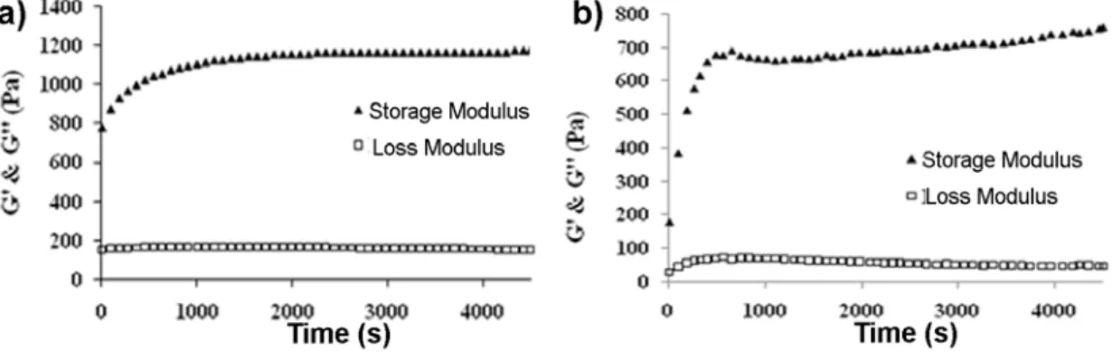
point dryer. ... 47 Figure 3.6. Time dependent oscillatory rheology measurements of gels (a) in ethanol and (b) in H2O at pH 10. ... 48
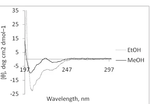
Figure 3.7. CD spectra of 0.03 wt % peptide solution in ethanol and methanol... 49 Figure 3.8. CD spectra of ALP in H2O with different concentrations a) at pH 10 and;
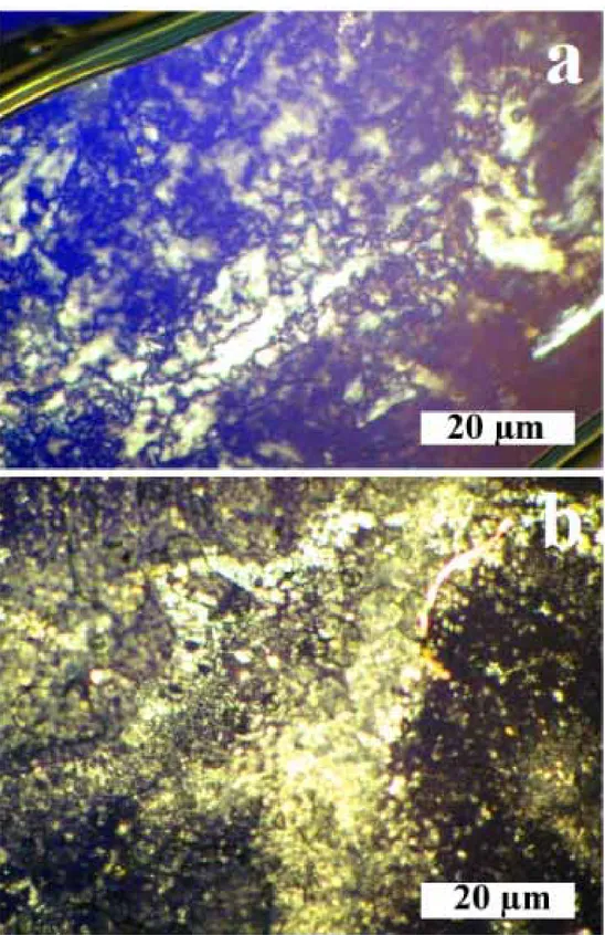
b) at pH 5. ... 50 Figure 3.9. Birefringence effect of self-assembled ALP nanostructures (a) in ethanol,
(b) in H2O at pH 10, magnification 500X. ... 51
Figure 3.10. FT-IR spectra of the ALP samples; (a) in H2O at pH 10, (b) in H2O at
pH 5, (c) in ethanol, and (d) in methanol. ... 54 Figure 3.11. UV-Vis absorption spectra of the ALP; (a) in H2O at pH 5 and pH 10,
(b) in ethanol and in methanol. ... 55 Figure 3.12. Fluorescence emission spectra of the ALP; (a) in H2O at pH 5 and 10,
(b) in ethanol and methanol. ... 55 Figure 3.13. Thermal Gravimetric Analysis of peptide powder. ... 56 Figure 4.1. TEM images of the silica nanotubes after calcination. Both scale bars: 20 nm. ... 66
xxii
Figure 4.2. SEM images of critical point dried silica nanostructures after calcination at 350 ºC scale bars; (a) 4 µm, (b) 5 µm and (c) EDX spectrum of the samples. . 66 Figure 4.3. Silica structures synthesized without peptide nanofibers as templates. .. 67 Figure 4.4. (a)-(b) STEM images and (c) EDX spectrum of the silica nanotubes after
template removal by washing. ... 68 Figure 4.5. AFM image of the fluorescent silica nanotube sensor surface. ... 70 Figure 4.6. Absorption (blue) and emission (red) spectra of TCPPH2 in ethanol
(dashed line) and in silica nanotubes (solid line). ... 71 Figure 4.7. (a) Fluorescence quenching of FSNT surface with TNT. (b) Quenching
efficiencies of the sensor upon exposure to TNT, DNT, and NB with respect to time. ... 73 Figure 4.8. SEM image of silica nanoparticles on the sensor surface... 75 Figure 4.9. Quenching efficiencies of FNST, Silica nanoparticle and silica thin film
sensors upon exposure to TNT with respect to time. ... 75 Figure 4.10. Quenching efficiencies of 7 analytes tested after 10 minutes exposure.
The FSNT sensor is found to be very selective to the TNT and DNT. NB
(nitrobenzene), BA (benzoic acid), Tol (toluene), DBA (dihydroxybenzoic acid), Xyl (xylene). ... 76 Figure 5.1. 3D network of titania nanotubes after Critical Point Dryer and calcination ... 86 Figure 5.2. TEM images of critical point dried titania nanotubes after calcination at
450⁰C. ... 86 Figure 5.3. a) TEM and b) SEM images of calcined TiO2 particles without the ALP
xxiii
Figure 5.4. XRD spectrum of titania nanotubes after calcination ... 91 Figure 5.5. XRD spectra of template-free TiO2 (blue), peptide templated TiO2
nanotubes (green), on FTO and FTO only (red). ... 91 Figure 5.6. A prototype of the constructed DSSC. ... 92 Figure 5.7. Titania nanotubes on surface after calcination and EDX spectrum of the
surface. ... 93 Figure 5.8. a) The isothermal histogram and, b) MultiPoint BET analysis based on
the histogram of template-free TiO2. ... 95
Figure 5.9. a) The isothermal histogram and, b) Multi-Point BET analysis based on the histogram of peptide-1 templated TiO2 nanotube network. ... 96
Figure 5.10. The chemical structure of N 719 dye. ... 98 Figure 5.11. UV-Vis absorption spectrum of N719 sensitizer dye. ... 98 Figure 5.12. Linear concentration calibration curve of N719. ... 99 Figure 5.13. Representative J-V spectra of devices based on template-free, peptide-1
and peptide-2 templated materials. ... 100 Figure 6.1. Representative TEM images of a) peptide nanofibers stained with uranyl acetate and b) gold nanoparticles formed on the surface of peptide nanofibers. 110 Figure 6.2. TEM images of gold nanoparticle formation on the peptide nanofibers
(13.3 mM) in ethanol at different KAuCl4 to peptide molar ratios; a) 1, b) 2 and c)
3. ... 110 Figure 6.3. Absorbance of the gold nanoparticles coated peptide nanofibers
(KAuCl4/peptide ratio is 1 molar ratio). ... 111
Figure 6.4. SEM images of a) micron sized gold aggregates formed in the absence of peptide after 2 days, and nanowire formation in the presence of peptide (13.3
xxiv
mM) captured over time, b) 10 min, c) 30 min; d) 1 h, e) 1 day, f) 2 days after the addition of equal molar of ascorbic acid to pre-seeded peptide solution with gold ions in 1:1 molar ratio. ... 112 Figure 6.5. (a) Spectrophotometric analysis of gold nanoparticle formation on the
peptide nanofibers at changing concentration of ascorbic acid at 60 min of incubation (Inset shows change in absorbance at 320 and 550 nm depending on the ascorbic acid concentration). (b) As time passes the absorption at 320 nm decreases while 550 nm increases at 13.3 mM ascorbic acid concentration (Inset shows relationship between peak maxima at 320 and 550 nm). (c) SEM image of gold nanoparticles formed on the peptide nanofiber template at optimized
incubation time and reducing agent concentration. ... 114 Figure 6.6. TEM image of 1-2 nm nanoparticles after the addition of first portion of
the gold precursor. The AuCl4−/AuCl2− ions bind to protonated amine groups and with the help of first ascorbic acid addition, gold nanoparticles were observed. ... 117 Figure 6.7. Morphological characterization of the gold nanostructures formed on the
changing concentrations of the peptide nanofiber template by STEM, and comparison of the effect of the reducing agent behavior at increased NaOH concentrations (0-0.06 mM). All scale bars are 200 nm. ... 119 Figure 6.8 STEM image of gold nanowires synthesized using multi-step seed
directed methodology a) 8.9 mM peptide concentration, b) XRD pattern, and c) the absorbance spectrum of the nanostructures ... 121
xxv
Figure 6.9. TEM images of gold nanostructures formed after 1 day of aging. a) Gold nanowires obtained in the presence of 6.6 mM peptide and b) peptide surrounded nanowires assembled in the presence of 11.9 mM peptide. ... 123 Figure 6.10. a) Dilute gold-peptide nanofiber network is dropcast on a silicon wafer
with 1µm thick oxide barrier. A voltage can be applied to the cantilever (denoted by C), or to any of the AuPd contacts (denoted by A and B). b) Simultaneous measurement of topography, and c) surface potential show the low and non-uniform conductivity of the nanofibers. Several arrows are used to denote changes in surface potential along individual nanofibers or nanofibers in contact, when a bias of VAB = 0.5 V is applied. In contrast, AuPd contacts show uniform surface
potential distribution. ... 127 Figure 6.11. a) Cyclic current versus bias measurements revealed asymmetry in the
conductance as well as hysteresis. The samples were typically high resistance initially, and switching to a high conductivity state occurs around 4.5 V bias (see inset). Several cycles were superimposed to show the extent of the repeatability of the measurements. b) The current was attributed primarily to tunneling as seen in the Fowler-Nordheim presentation, when ln(I/V2) was plotted against 1/V for increasing (red) and decreasing (blue) bias sweeps. Solid lines were guides for the eye. The transition voltage from direct to Fowler-Nordheim tunneling is a
function of tunneling gap and effective tunnel barrier height, and was seen to be modulated during cyclic voltage sweeps. ... 128 Figure 6.12. Conductivity measurements of bare peptide nanofiber was also
xxvi
Table of Schemes
Scheme 1.1. Schematic description of Solid-Phase Peptide Synthesis ... 6 Scheme 4.1. The formation of peptide nanofibers, nucleation and accumulation of
inorganic materials around it and obtaining of inorganic nanotube by calcination of the organic peptide. ... 64 Scheme 4.2. The peptide nanofibers formed, the nucleation and accumulation of
inorganic materials around the peptide nanofibers completed, the inorganic nanotubes were obtained by washing the organic part. After addition of sensitizer dye, the silica nanotubes spin coated on a surface for sensor development. ... 69 Scheme 5.1. Schematic illustration of self-assembly and mineralization of peptide
nanofibers and formation of hollow nanotubes. ... 85 Scheme 5.2. The schematic illustration of DSSC constructed with one-dimensional
xxvii
Scheme 6.1. Schematic presentation of gold nanostructure formation using peptide nanofibers as template. ... 105 Scheme 6.2. Schematic illustration of multi-step seed mediated growth methodology. ... 116 Scheme 6.3. Ascorbyl radical formed from Ascorbic Acid ... 122 Scheme 6.4. The deposition of free peptide molecules around gold nanoparticles. 125
1
Chapter 1
Introduction
2 1. Introduction
Organic materials and polymers are strongly integrated into modern technology; nevertheless, inorganic materials still preserve their status as the basic elements in engineering. However, there is an increasing awareness that the conventional methods of “heat and beat” have several limitations in fulfilling the requirements for future advanced materials. In particular the limitations are generally in the construction of complex architectures from nanoscale to macroscale. This knowledge creates a vital need for novel synthesis methods.
Among nanoscale assembly techniques, top-down approaches have attracted attention for many years. Neverthless, researchers in areas such as lithography and etching have faced difficulties related to the cost, process speed and diffraction limit of top-down devices. On the other hand, “bottom-up” approaches can offer large-scale, rapid, and low-cost production of nanostructures with a diverse range of starting materials. Self-assembly is a bottom-up approach, the spontaneous aggregation of many different subunits into larger, well-defined, functional objects with different properties.
In the self-assembly processes; atoms, molecules, particles, and other building blocks organize themselves into functional structures as driven by the energetic of the system. Molecular self-assembly is also a useful technique for material designing. It involvs non-covalent supramoleculer interactions, such as hydrogen bonding, hydrophobic, electrostatic, metal-ligand, π-π and van der Waals interactions. Althpugh these interactions are relatively weak interactions with compare to covalent bonds, sufficient number of them can yield a stable assembly. Many new
self-3
assembling material can be design to organize into ordered coplex structures by understanding the mechanism of these assembly processes [1]. Self-assembly is controlled by many factors including temperature, pH, and electrolyte concentration. Novel self-assembled materials for both biological and nonbiological applications are being developed for regenerative medicine [1-5], electronics [6-9], optics [10], and as pH detection [11]. There is no doubt that new approaches using conceptually new solutions, such as “biomimetic design” and self-assembly, should be developed and used to expand the frontiers of possibility in this field. The ability of biomimetic materials to organize inorganic “bricks” into nanoscale, microscale and macroscale materials has great potential for electronics [12, 13], catalysis [14, 15], sensors[16], molecular recognition [17], magnetism [18-20], optics [21], photonics [22] and biomedical applications [23].
One-dimensional nanostructures have found widespread use in the fabrication of nanoscale electronic, mechanic, magnetic, optical, and combinatorial devices owing to their high ratio of surface area to volume and quantum-confinement effects [24-26]. By studying one-dimensional nanostructures, the effects of reduction of size and dimensionality on mechanical, thermal and electrical properties can be investigated. An example of mechanical one-dimensional supramolecular nanostructures in nature can be seen in cytoskeletons. Actin filaments, intermediate filaments and microtubules are the three main classes of protein filaments that form the cytoskeleton; actin and microtubules act in concert during cell movement and morphogenesis [27]. Although nanoscience is still far away from being able to mimic such intricacy, biology continues to inspire the design of nanodevices. Amyloid plaques are another natural example of one-dimensional nanostructures formed by
4
very stable self-assembling peptides, and are known to play a role in Alzheimer’s disease [28]. As a result of the biological relevance of one-dimensional aggregates in neurodegenerative diseases, the self-assembly mechanisms of such one-dimensional nanostructures are of interest to researchers.
Amino acids are the building blocks of proteins, and of the living organisms. They are also utterly important for the formation of inorganic structure of the living organism. Synthesis of peptide in desired sequences attracts the scientists for many years, due to the such importance of proteins. The first peptide synthesis published by Emil Fischer (9 October 1852 – 15 July 1919) and Fourneau [29]. Fischer won Nobel Prize in Chemistry in 1902 by the synthesis of Diketopiperazine hydrolysis glycoglycin (Figure 1.1). This reaction opened tremendous fields in many branches of science and although it has been more than 100 years, synthesis of an artificial peptide with desired properties is still a hot topic.
Figure 1.1. Fischer’s synthesis of glycoglycin.
After Fischer’s innovation, longer sequences intended to synthesis by wet chemistry. Solid Phase Peptide Synthesis (SPPS) pioneered by Robert Bruce Merrifield (July 15, 1921 – May 14, 2006), who won the Nobel prize in 1984 [30]. SPPS allows the synthesis of both natural and unnatural peptides with many functional groups. In this method, amino acids bind each other via amide (peptide)
5
bonds. The peptide synthesis occurs by coupling the C-terminus of one amino acid to the N-terminus of another. To prevent unintended reactions, protecting groups are used.
As can be seen in scheme 1.1 this synthesis strategy is based on the usage of solid insoluble beads modified with linkers. The coupling reaction starts on this linkers. The free N-terminal amine of a solid-phase attached amino acid is coupled to a single C-terminus activated and N-terminus protected amino acid unit. The N-terminus of the amino acid is protected due to avoid unintended reactions, with Fluorenylmethyloxycarbonyl (Fmoc) group. After deprotection of N-terminus of the new attached amino acid, it coupled to the other amino acid. This strategy is based on repeated cycles of coupling- washing- deprotection- washing processes. At the end of coupling of all predetermined sequence of amino acids, the linker between solid-phase and peptide cleaved by a trifluoroacetic acid (TFA). After the filtration of remained solid-phase, synthesized peptide-TFA solution would be separated.
6
7
Proteins and peptides can assist forward the synthesis of inorganic materials in an environmental-friendly strategy via a process known as biomineralization. Nature inspired synthetic peptide networks have wide applications including bioactive tissue scaffolds [31, 32], carrier agents [33, 34], and templated synthesis of inorganic materials [35, 36]. Recently, the high mechanical stability of peptide nanofiber networks was shown [37, 38].
The design of the amino acid sequence could be chosen to implement any functionality to the peptide. To determine the functional groups of a peptide, the affinity of binding with intended mineral ions in the medium should be considered. The ions of different minerals have some particular properties such as being Lewis acid or base.
The design of the peptide used in this thesis was achieved with the consideration of properties of functional groups. The electrostatic interactions between functional groups and mineral ions in the medium designed by taking the Ralph Pearson’s Hard Soft Acid Base (HSAB) Theory [39]. According to this theory; soft acids react faster and form stronger bonds with soft bases, whereas hard acids react faster and form stronger bonds with hard bases. Here 'hard' implies to species which are small, have high charge states and 'soft' implies to species which are big, have low charge states.
In this thesis, we described the design and synthesis of a peptide molecule with special functional groups. The peptide molecule is able to self-assemble into one-dimensional nanostructure under appropriate conditions. The functional groups have affinity to bind particular mineral ions in the medium, by which these peptide nanofibers act as templates. The structures formed by accumulation of minerals
8
around these peptide nanofibers are uniformed and well-defined nanostructures and have many application areas such as sensors, dye-sensitized solar cells and electronics as described in this thesis.
In Chapter 4, the synthesis of fluorescent silica nanotubes forming a porous network by using biomineralization process through self-assembled peptidic nanostructures are described. Fluorescent porous organic-inorganic thin films are interest of explosive detection because of their vapor phase fluorescence quenching property. The amine groups on the peptide nanofibrous scaffold were used for silicon ions seeding and accumulation. Silica nanotubes were used to prepare highly porous surfaces and they were doped with a florescent dye by physical adsorption for explosive sensing. These porous surfaces exhibited fast, sensitive and highly selective fluorescence quenching against nitro-explosive vapors. The materials developed in this work have a vast potential in sensing applications benefiting from the elevated surface area.
The synthesis of one-dimensional anatase titanium (IV) oxide nanostructures was explained in Chapter 5 by using amine functionalized self-assembled amyloid-like peptide nanofibers as templates. Mineralization of peptide templates leads to hybrid organic-inorganic material, where surface of nanofibers is coated with a titanium oxide precursor. Further formulation of this material as a paste and its direct application to transparent conductive oxide (TCO) glass surface, followed by calcination, leads to a fully functional electrode with a nanostructured anatase TiO2
layer. Photovoltaic performance of the obtained layer is assessed in dye sensitized solar cell efficiency measurements.
9
The amyloid like peptide molecule self-assembling into one-dimensional nanofibers has affinity to gold ions. These molecules were used as templates for growth of one-dimensional gold nanostructures as described in Chapter 6 in the presence of ethanol as a seeding agent and ascorbic acid as a reducing agent. We performed multi-step seed mediated synthesis of gold nanowires by evaluating several parameters including the molar ratios of peptide/ gold precursor, peptide and ascorbic acid concentration as well as the effect of the reduction kinetics of the ascorbic acid. Gold nanostructures with wide range of morphologies (e.g. smooth nanowires, noodle-like one-dimensional nanostructures and also this homogenous aggregates of spherical nanoparticles) were synthesized by use of green chemistry method and they have potential use in several applications including electronics and optics.
10
Chapter 2
Background
This work is partially described in the following publication:
Toksoz, S., H. Acar, and M.O. Guler, "Self-assembled one-dimensional soft nanostructures", Soft Matter, 6(23),p.,5839-5849, 2010.
11
2. Background
Supramolecular interactions are driving forces of self-assembly in simple systems. Understanding interactions in the assembly mechanisms of biological molecules has become a crucial factor in designing nanoscale materials. It is important for appreciate the high complexity of self-assembly process of nature.
The interactions that coordinate the amino acids in natural one-dimensional systems are highly dynamic and often delicate, due to their relatively weak nature in comparison to that of covalent bonds [40]. However, the sufficient number of these weak interactions can yield strong and stable aggregations. Biological one-dimensional entities including viruses [41] and fungi [42] have been found to be used as templates for nanostructure synthesis, such as wires [43, 44].
In order to understand and control the self-assembly of supramolecular structures, the non-covalent interactions taking part in this process must be studied in detail [45, 46]. This background informaiton focuses on one-dimensional self-assembled organic nanostructures classified according to the forces acting on their formation ranked, in order from the strongest interaction to the weakest. (Table 2.1).
12
Type of Interaction Strength (kJ/mol) Properties
Electrostatic 50-300 Non-selective
Coordination binding 50-200 Directional
Hydrogen Bonding 5-120 Selective, directional
π-π Stacking 0-50 Directional
Solvophobic Depends on solvent type Little directional constraint
van der Waals < 5 Non-directional,
non-selective
Covalent 350 Irreversible
Table 2.1. Strength and properties of non-covalent interactions. Adapted from [40, 47].
2.1. Electrostatic Interactions
Electrostatic interactions are based on Coulombic attraction between opposite charges. In host/guest chemistry, many receptors for anions and cations use electrostatic interactions to hold the guest in place [48]. The principles of formation of nanostructures through electrostatic interactions can be read in Faul and Antonietti’s review [47].
Zhang et al. developed ionic self-complementary peptides, one of which is named RADA16, a peptide which forms nanofibers in aqueous solutions by using β-sheet structures [49]. RADA16 contains negatively charged aspartic acids and positively charged arginines. Forming hydrogels in physiological media, the gels promoted the growth of neural cells in an integrated network that showed synaptic activity [50]. In this case, the charged properties of the peptide nanostructures served not only for their self-assembly but also for guidance of the cells. These hydrogels have actually been shown to improve the attachment and
13
differentiation of a variety of cell types, including stem [51] and endothelial cells [52].
Peptide amphiphile molecules [53] exploit a number of non-covalent interactions to self-assemble [54]. Two oppositely charged peptide amphiphiles have been shown to self-assemble into one-dimensional nanofibers by electrostatic interactions in aqueous solution at neutral pH [55]. The use of oppositely charged biomacromolecules to induce self-assembly has also been demonstrated, where heparin molecules screened the positive charge of a peptide sequence [56]. Size of the macromolecules plays an important role in formation of nanofibers as well [57], as larger molecules including DNA and chondrotin sulfate result in gelation of positively-charged peptides, whereas bovine serum albumin, a smaller molecule, cannot induce self-assembly.
Tetrapyrroles are large macrocyclic molecules containing four pyrrole rings. They self-assemble into light-harvesting and energy-transferring nanostructures in biological systems. Porphyrins from tetrapyrrole family are attractive building blocks to synthesize photocatalytic and light-responsive nanotubes; two oppositely charged porphyrins were employed to self-assemble through electrostatic interactions [58]. It is possible to change the function and the structure of the nanotubes by modifying the porphyrin building blocks, suggesting a high degree of control over the nanotubes.
2.2. Metal coordination
Over the past few decades, many studies have been carried out regarding coordination polymers and crystal engineering of metal complexes; selective
14
metal ion binding has been found to be a promising approach to control the fabrication of nanoscale self-assembled structures. In metal coordinated structures, metal ion functioning had been used along with some other non-covalent interactions [59-62] including hydrogen bonding, π–π stacking interactions and van der Waals forces. Potential applications of these nanostructures lie in the development of new materials, including metal-organic frameworks [63, 64], with magnetic, non-linear optical and photoluminescent properties.
Metal centers interact with ligands via medium strength directional metal-ligand bonding. A broad knowledge of coordination chemistry contributes to the selection of appropriate metal-ligand pairs and binding modes for the assembly of supramolecular structures with differing shapes. A coordination system consists of a central metal atom, called the coordination center, ligated to other atoms, called ligands, where the coordination bonds are delocalized over the ligands, thus reducing the Coulomb repulsion between the electrons. The d-orbital occupation changes the symmetry of the metal coordination sites, thereby changing the supramolecular shape. To obtain the desired structure, the metal coordination environment and binding mode of the linkers must be carefully designed, where the shape is encoded both in the ligands and the metal ions. For the metal center, transition metal ions can be useful as they not only stabilize the structures but also interact with other elements, allowing construction of more complex structures. To form complex structures, the metal centers should be available for further coordination; bulky ligands can be used to hinder the attachment of same or other ligands to the metal centers, thereby preventing
15 saturation of the metal centers.
Figure 2.1. Bis-biotinylated terpyridine forms a linear tetrabiotinylated connector, the [Fe(Biot2-terpy)2]2+ complex, upon reaction with ferrous ion. The presence of streptavidin results in formation of linear coordination polymers [65].
16
Figure 2.2. SEM images of MOPF bundles containing CaCl2. (a) 8x10⁻⁷ M Fe
solution containing 0.05 M CaCl2, 5 min at RT; and (b) a zoom into it. (c-e)
Biomineralized bundles of 8 x 10⁻⁶ M Fe solution, 12 hours: (c) 0.01 M CaCl2; (d-e)
0.05 M CaCl2 [65].
The transition-metal connectors in conjunction with proteins are used to create one-dimensional metal-organic protein frameworks [63]. It was demonstrated that streptavidin, a homotetrameric protein with four biotin binding sites, combined with a linear tetrabiotinylated connector, the [Fe(Biot2-terpy)2]2+ complex bearing four
biotin groups, with the aim of producing a collagen mimetic material for calcite biomineralization. Self-assembly took place through non-covalent interactions of coordination polymers and protein aggregates into a one-dimensional metal–organic protein framework (MOPF) to yield millimeter-sized one-dimensional matrices. Ferrous ions were used in the synthesis of [Fe(Biot2-terpy)2]2+, as terpyridine readily
17
biomineralization of calcite, carbondioxide vapor was streamed over MOPF bundles for nucleation, growth and assembly of calcite microcrystals on the bundles (Figure 2.2). The negatively charged glutamic acid and aspartic acid side chains are thought to have favored chelation of calcium [65].
Self-assembly of de novo designed peptides into well-designed structures can be achieved through an appropriately chosen metal ion [66-68]. Selective recognition and binding of the metal ion to the peptide might induce a conformational change. Self-assembly of a fibril forming peptide, a structural variant of a three-stranded helical bundle forming a trimeric leucine zipper, was triggered by silver(I) ion binding to proximal histidine residues [66]. The trigonal planar geometry in the metal ion binding site favored binding of silver(I) ions, which in the presence of soft-donor nitrogen ligands can adopt a trigonal planar coordination in the presence of soft-donor nitrogen ligands. Alternative coordination geometry using ions like copper, zinc and nickel failed to induce self-assembly. In an earlier study, same peptide was shown to self-assemble via a pH-dependent route [69]. Although, the final fiber morphologies were similar for two assembly routes, the lateral association between fibrils to form fibers larger in diameter was less extensive for silver ion induced fibrils. This might be related to the Columbic repulsion due to the partial co-localization of positive charges by counter ions: metal ion induced fibers were positively charged due to the presence of metal ions, in this case one silver(I) ion per peptide; whereas pH-induced fibers have a negative charge at neutral pH.
Cd(II) centers are known for their use as metal coordination, because the d10 configuration of cadmium complexes results in formation of various coordination
18
geometries. Kong et al. reported that an imidazole-containing tripodal ligand reacted with Cd(II) complexes of bromide, tetrafluoroborate, and nitrate, and three new coordination polymers were obtained [70]. In the presence of [CdCl4]2- as a counter
anion, one of the complexes adopted a one-dimensional zigzag chain structure. The other two complexes adopted branched chain structures. The Cd(II) centers of the three complexes had different geometries such as distorted square pyramide, tetrahedron, and distorted octahedron. Tetrafluoroborate and nitrate anions were not found in the distorted geometries, but they had an impact on the self-assembly process so that on the final framework. The obtained coordination polymers demonstrated photoluminescence, as all complexes exhibited a blue fluorescence at room temperature in the solid state and could serve as a good candidate for photoactive materials.
Dendron-rod-coils, having unique triblock architecture, self-assemble into one dimensional nanoribbons which have found use as a template for the synthesis of cadmium sulfide nanohelices [71, 72]. Cadmium sulfide semiconductors have potential photovoltaic applications. Cadmium ions have an affinity towards the hydroxy containing dendron region of the ribbons over the organic solvent, which was tetrahydrofuran in this study [71]. After exposure to hydrogen sulfide gas, the localized cadmium ions started to nucleate and grow as cadmium sulfide on one face of the twisted ribbon to form nanohelices with a pitch of 40-50 nm, nearly twice that of ribbons. Nucleation happened in many points at the same time, as revealed by transmission electron microscopy image (Figure 2.3) [73]. In some cases, double coils were encountered, possibly due to the mineralization occured at both faces of the ribbon.
19
Figure 2.3. Chemical structure of the dendron-rod-coil molecules and TEM image of cadmium sulfide precipitated in a suspension of DRC nanoribbons, at an early stage growth. The inset shows the start of nucleation at different points and the organic ribbon under CdS as indicated by the arrow [71].
2.3. Hydrogen bonding
Arguably the most important noncovalent interaction in the self-assembly process is hydrogen bonding, due to its directionality and strength. The hydrogen atoms act as a bridge between two electronegative atoms; the hydrogen bond donor group consists of an electronegative atom bound to a hydrogen atom that
20
has a small positive charge due to dipole formation, and the hydrogen bond acceptor group consists of a dipole where the interacting atom of the acceptor group has a source of electrons.
The enthalpy of the hydrogen bond-based self-assembled systems must be balanced in consideration of the enthalpic loss and entropic gains due to hydrogen bonding and stacking interactions/hydrophobic effects, respectively [74]. Generally, hydrogen bonding is not the sole interaction in the self-assembly process; it is usually accompanied by other non-covalent interactions which have lower energies. Although hydrogen bonds are mostly used for constructing 2-D and 3-D nanostructures due to their selectivity and high directionality [75], there are also one-dimensional nanostructures which are self-assembled through hydrogen bonding interactions [76].
Macrocycles containing an even number of alternating D- and L-amino acids, developed by Ghadiri and co-workers, are assembled into nanotubes by orthogonal hydrogen bonding, with the hydrogen bonds of the tubular structures perpendicular to the plane of the individual molecules (Figure 2.4) [77]. In the design of the four cyclic peptides, four non-polar amino acids and one polar amino acid were employed. Non-polar residues were chosen to study the effects of increasing hydrophobic surface contact; the polar residue, glutamine, was selected because of its hydrogen-bonding donor/acceptor capability and its possible participation in intra- and intertubular hydrogen bonding interactions, thus contributing to the structural stability of the system. Similar designs have been shown to form trans-membrane channels for ion transport [78].
21
Figure 2.4. Alternating D- and L-amino acids assembling into cyclic peptide nanotubes via the antiparallel ring stacking. Extensive intersubunit hydrogen bonding can be seen in the sketches. Reproduced with permission from [77].
Peptide amphiphile design of Hartgerink et al. was first presented in a biomineralization study [53], where the amphiphilic molecule consisted of a hydrophilic peptide headgroup, consecutive cysteine residues for formation of disulfide bonds, a flexible linker region, a phosphorylated serine residue for inducing biomineralization and a hydrophobic alkyl tail. A recent study of the self-assembly mechanism of peptide amphiphiles by Velichko et al. revealed that hydrogen bonding is the main interaction contributing to the final one-dimensional shape [79]. In their model, the head groups were designed as
22
electroneutral in order not to calculate the effects of electrostatics in the self-assembly process. This design enabled them to perform the Monte Carlo-stochastic dynamics simulations in a reasonable time and still capture various aspects of the process. Their coarse-grained model states that transitions from random molecules in solution to different micellar structures are based on the interaction between hydrophobic interactions and hydrogen bonding. Paramonov
et al. designed 26 different peptide amphiphiles to study a number of parameters
in self-assembly such as amino acid choice, directionality of hydrogen bonding and the reasons that the nanofiber structure is favored over other structures [80]. They found that the amino acids near the alkyl tail of the peptide amphiphiles were the main contributors to the β-sheet structure, formed along the Z-axis of the fiber. There, the disruption of the hydrogen bonds, which occurred by methylating the glycine amino acids forming the hydrogen bonds, resulted in a transition from nanofiber structure to nanovesicle structure after a specific number of methylations, due to the fact that the remaining energy was not enough to hold the supramolecular structure of a nanofiber together (Figure 2.5). Guler et
al. developed peptide amphiphile molecules conjugated to nucleic acids, resulting
in thermally more stable duplexes of the conjugate molecule-DNA/RNA in comparison to nucleic acid-nucleic acid duplexes [81]. Such a molecule could be useful in RNA interference studies.
23
Figure 2.5. The chemical structure and the cartoon of the peptide amphiphile nanofiber: The inner most region, (a), is the hydrophobic core composed of aliphatic tails. (b) is the critical β-sheet hydrogen bonding portion of the peptide. (c), the peripheral peptide region, constitutes the interface between the fiber and the environment [80].
While studying the ability of very short aromatic peptides to form well-ordered amyloid fibrils, Reches et al. observed β-amyloid diphenyalanine structural motif of Alzheimer’s disease self-assembling into one-dimensional nanotubes [82]. The nanotubes worked well as a mold for casting silver metal nanowires, first by metal ion reduction and then enzymatic degradation of the mold. By using diphenylalanine structural motif of Alzheimer’s disease self-assembling into one-dimensional nanotubes [82]. The nanotubes worked well as a mold for casting silver metal nanowires, first by metal ion reduction and then enzymatic degradation of the mold. By using D-phenylalanine in the core structure of the peptide, the researchers achieved the construction of proteolitically stable nanotubes, which might have applications as nanotube-based biosensors. In another study, “teslian” (metal-insulator-metal) coaxial nanocables were developed by using the diphenyalanine peptide nanotubes as templates [83]. Silver ions were reduced in the nanotubes which were then chemically modified
24
with linker peptides consisting of a diphenylalanine motif and cysteine amino acid. The diphenylalanine motif was devised for interaction with the nanotube surface and cysteine in order to facilitate the imaging process of the structures, aided by gold ions that interact with the thiol group of the amino acid.
Hong et al. produced silver nanowires [84] by reducing the silver ions in their organic nanotubes, which consisted of a reduced form of calix[4]quinone with four hydroquinone moieties [85]. Of the eight hydroxyl groups, four outer groups led to self-assembled structures with intermolecular hydrogen bonding in the presence of water molecules. Repeating tubular calix[4]hydroquinone octamers formed short hydrogen bonds between themselves to stabilize a linear tubular polymeric structure, where intertubular π-π stacking interactions contributed to the stability as well.
Hydrogen bonding is a powerful strategy in the self-assembly of one-dimensional structures formed by dendrimeric molecules [86]. Some dendritic dipeptides were shown to self-assemble into helical porous one-dimensional nanostructures [87]. The characterization of the helical nanostructures indicated that the controlled design of periodic non-biological porous structures in bulk and in solution was achieved by dendrimer chemistry. The molecular recognition and self-assembly process are strong enough to tolerate a range of modifications to the amphiphilic structure.
Dendron-rod-coils, which assemble into one dimensional ribbons, form gels in certain solvents and at certain concentrations via hydrogen bonding in the hydroxyl rich regions and π-π stacking of the conjugated segments [88-90]. Zubarev et al. designed dendron rod-coil molecules as additives to modify the
25
properties of polystyrene [90]. The molecules were dissolved in organic solvents to form one-dimensional birefringent ribbon-like nanostructures with a width of 10 nm and a thickness of 2 nm, even at an extremely dilute concentration (Figure 2.6). When the gels were heated to a temperature above the boiling point of the organic solvent (e.g. styrene, dichloromethane, or acrylate derivatives), the gels did not melt revealing the irreversibility of gel structure by temperature. To disassemble the gels, hydrogen bonding must have been disrupted by polar solvents. At 1 wt% concentration, ribbons aggregated into bundles of 5 to 10 flat ribbons, where excess organic solvent use made the relatively isolated ribbons twist and gain a 20 nm pitch. The twisting probably protects the hydroxyl groups in the center of the ribbon from the organic solvent.
Figure 2.6. Chemical structure of the dendron-rod-coil molecule. (a) Bright field TEM image of unstained 0.004 wt% DRC molecules, dissolved in styrene. (b) High magnification TEM image of a thin slice of the scaffold material, containing 1 wt% DRC molecules. Arrows represent perpendicularly placed individual bimolecular ribbons [89].
26
Lipid nanotubes are promising templates for producing one-dimensional nanostructures that provide organic, discrete, tubular structures composed of a very high number of identical lipid molecules, as Young’s modulus of a single lipid nanotube has been determined to be around 700 MPa [91]. Various nanotube architectures are available with reference to the type of building block, as can be seen in Figure 2.7 [92, 93].
Helices can intertwine and produce aggregated helices; when they reduce into rings, face-to-face non-covalent interactions result in the formation of tubes; instead of rings, rosettes can arrange into stacked rosettes; amphiphilic molecules might roll into sheets to produce nanotubes [93].
Figure 2.7. Open-ended tubular architectures: (a) aggregated helices from a hollow helix, (b) stacked rings from a ring, (c) stacked rosettes from a rosette, (d) monolayer-based lipid nanotubes from an unsymmetrical bolaamphiphile [92, 93].
27
functionalization of both sides are important in order to obtain well-defined and tailor-made architectures for many different purposes. Kameta et al. self-assembled fluorescent lipid nanotubes from an unsymmetrical bolaamphiphile to encapsulate and track the passage of guest molecules through the hollow nanotubes, in order to better understand how the molecules act while entering and exiting the cell through the channels of the lipid bilayer [94]. The same group also developed cardanyl-β-D-glucopyranoside lipid nanotubes to reveal the role of water confined in a lipid nanotube by incorporating 8-anilinonaphthalene-1-sulfonate as a probe, with the ultimate aim of guest encapsulation [92]. Hydroxyl groups of glucopyranoside covering the inner surfaces interacted with water molecules, lowering the solvent polarity to a value similar to that of propanol rather than bulk water.
2.4. π-π interactions
Even though much weaker and less directional when compared to hydrogen bonds, π stacking interactions also drive the self-assembly process for π-conjugated systems. The nature of π-π interactions is not very clear, but it is suggested that the geometrical arrangement of the fragments, as well as π electrons, contribute to these interactions [95]. In order to obtain one-dimensional structures as end products, it is necessary to favor growth along the stacking direction rather than lateral growth alongside chains. With the progression of efficient and high yield synthesis methodologies of bulk molecules [96] like dendritic molecules and macrocycles, it has become possible to build nanoscale architectures. Arylene ethynylene macrocycles [97, 98], molecules with large planar surfaces, minimal ring
28
strain and highly tunable ring sizes of 0.5−5 nm, have been employed mainly for the synthesis of 2-D and 3-D structures [99, 100], as simple self-assembly methods [101] resulted in the formation of agglomerates, aided by solvophobic interactions between the alkyl side chains. Balakrishnan et al. achieved the self-assembly of arylene ethylene macrocycles into one-dimensional nanofibers by implementing the sol-gel process [102]. Cooling a homogenous solution results in the gelation of the molecules, which decreases molecular mobility, thus minimizing the lateral growth due to side chain association.
Another type of macrocycles, constituted from oligoamides, is known to form fibrillar structures [103]. The self-assembly mechanism is suggested to rely on face-to-face stacking, aligning the macrocycles into nanotubes containing a large channel (Figure 2.8) [104]. Such nanotubes are proposed to be used as transmembrane channels by modifying the side groups to tune the solubility and membrane compatibility of the macrocycles. Employing different functional groups resulted in differing pore diameters, thus affecting the conductance of the transmembrane channels, showing that it is possible to mimic the ionic channels in biological membranes.
29
Figure 2.8. Oligoamide macrocycles self-assembling into transmembrane channels through face-to-face stacking [104].
Engelkamp et al. demonstrated that the tuning of the supramolecular chirality of one-dimensional objects can be done in a controlled way by simply controlling the strength and directionality of non-covalent interactions [105]. Disk molecules, derived from a phthalocyanine ring and covered with crown ether moieties, formed fibers with right-handed helicity through π-π stacking, which eventually yielded superhelices with left-handed helicity. The advantages of using π-π stacking in the design of nanostructures are its relatively less complex method,and high electron mobility through the nanostructure. High mobility is generally attributed to strong overlapping in a stack of neighboring molecules’ electronic wave functions [106] which increases bandwidth and consequently electrical conductivity [107].
In the molecular electronics field, nanowires and nanocables self-assembled via π-π stacking are drawing attention [108-110]. This type of stacking is commonly observed in aromatic compounds with extended π systems. To give an example, hexabenzocoronene is a PAH (Polycyclic Aromatic Hydrocarbon) compound consisting of 13 fused benzene rings, and it was shown to self-assemble into nanocables; researchers also devised a method for putting the cables into organic
30
field effect transistors by use of elastomer stamps [111]. In another study, a derivative of hexabenzocoronene self-assembled into nanotubes in tetrahydrofuran, where the walls of the tubes consist of helical arrays of π-stacked coronenes covered by hydrophilic glycol chains (Figure 2.9) [112]. The final structures have been found to be electroconductive and have a resistivity comparable to that of gallium nitride semiconductors.
Figure 2.9. The chemical structure of hexa-peri-hexabenzocoronene and the self-assembly mechanism into nanotubes and helical coils: (A) A graphitic bilayer tape; each layer consists of one dimensional columns of -stacked hexabenzocoronene units. (B) A nanotube formed by tight rolling-up of the bilayer tape. (C) A helical coil formed by loose rolling-up of the bilayer tape [111].
Hexabenzocoronene had also been used for producing discotic liquid crystals, called mesogens, by chemically modifying the periphery of hexabenzocoronene, whose liquid crystalline phase showed a rapid switching process within the applied electrical field [113]. π- π stacking and hydrogen bonding are the two
31
synergistic intermolecular forces controlling the assembly of the central aromatic subunits of the liquid crystals.
As a very well established study to understand the driving forces of self-assembly, Wang et al study on supra-amphiphiles can be given as an example [114]. In this study, many interactions introduced to the peptide structure to achieve rigid self-assembled nanostructures. They used mesogenic groups, strong π-π stacking groups, hydrophobic interactions in water, electrostatic interactions, and so on, to self-assembly of building blocks.
2.5. Solvophobic interactions
Solvophobic interactions, which have little directional constraint, differ from the other non-covalent interactions in terms of inducing self-assembly that hydrophobic interactions are stabilized due to favorable entropy rather than favorable enthalpy, which might even be unfavorable as long as entropy is favored. The solvophobic parts of the molecules tend to associate to minimize their surface area contacting the solvent, whereas the solvophilic parts try to remain in contact with the solvent. The two opposing forces compete with each other, one tending to decrease the interfacial area per molecule, the other tending to increase.
Solvophobic interaction is observed in solvents with a spatial hydrogen-bond network. It is thought to consist of two stages, namely solvation and interaction. According to Rodnikova et al., the solvation is related to the lability of the hydrogen-bond network, whereas elasticity of the network determines the interaction part [115].
![Table 2.1. Strength and properties of non-covalent interactions. Adapted from [40, 47]](https://thumb-eu.123doks.com/thumbv2/9libnet/6011829.126697/39.892.174.780.146.467/table-strength-properties-non-covalent-interactions-adapted.webp)

![Figure 2.8. Oligoamide macrocycles self-assembling into transmembrane channels through face-to-face stacking [104]](https://thumb-eu.123doks.com/thumbv2/9libnet/6011829.126697/56.892.182.720.149.392/figure-oligoamide-macrocycles-self-assembling-transmembrane-channels-stacking.webp)