Signal Processing: Image Communication 7 (1995) 225-230
SIGNAL PROCESSING:
lilMAGE
COMMUNICATIONMotion-compensated prediction based algorithm for medical image
sequence compression*
Seyfullah H. O&z, 6mer N. Gerek*, A. Enis Cetin
Department of Electrical and Electronics Engineering, Bilkent University Bilkent, Ankara 06533, Turkey
Received 15 November 1993
Abstract
A method for irreversible compression of medical image sequences is described. The method relies on discrete cosine transform and motion-compensated prediction to reduce intra- and inter-frame redundancies in medical image se- quences. Simulation examples are presented.
1. Introduction
Digital medical images are produced by a num- ber of medical imaging devices including computed
tomography (CT), magnetic resonance imaging
(MRI), ultrasound, digital radiography, and radio- graphic film digitizers. Due to the large amount of data per image there is a storage problem in medi- cal Picture Archiving and Communication Systems (PACS) [S, 123. Image compression is an efficient way of handling this problem [S, 12,2,4,7,9,13]. Reversible (lossless) image compression techniques usually achieve a degree of compression in the order of 3 : 1, whereas irreversible (lossy) techniques may achieve compression ratios in the order of
*This work is supported by TUBITAK - Scientific and Tech- nical Research Council of Turkey, and it was presented in the “Canadian Conference on Electrical and Computer Engineer- ing”, September 1992.
*Corresponding author.
5: 1 to 20: 1. Irreversible techniques which do not introduce any visual degradation can be used in a PACS [S].
Most of the irreversible image coding techniques use transform domain methods. For example, dis- crete cosine transform (DCT) based techniques have been successfully employed for still picture coding including medical image compression, and video coding in practice [ 12,4,2,7,9,13,10,14].
Many medical imaging devices, including the CT and MRI machines, produce image sequences. In a typical radiology department 20-30% of all data created are image sequences. Regular medical image sequences contain images of slices of the human body. Therefore, neighboring images in a CT or an MRI sequence are highly correlated with each other. In order to compress such a medi- cal image sequence, one should not only consider intra-frame coding but also exploit the high cor- relation in the third dimension to achieve further compression.
0923-5965/95/%9.50 0 1995 Elsevier Science B.V. All rights reserved SSDIO923-5965(95)00027-5
This paper presents a three-dimensional (3-D) image compression algorithm which takes advant- age of the correlation among the neighboring images. The algorithm relies on DCT-based com- pression within a medical image and ‘motion-com- pensated’ prediction [6] to remove redundancies between images in the sequence.
dom access points are determined by the GOP layer header. These headers may also contain in- formation such as the characteristics of the in- cluded slice images. The corresponding body places of the slice images contained in the GOP can also be written in the GOP header.
2. Image sequence coding algorithm
The third layer is the picture layer. In the proposed method, the coded medical image sequence consists of I-type pictures (intra-frame coded) and P-type pictures (inter-frame coded) as described in [l].
One of the key features of our algorithm is the use of ‘motion compensation’ which is utilized to remove the redundancies between image frames. Although there may not be real motion in most medical image sequences, one can also consider the changes occurring in consecutive images of the se- quence as motion of the blocks and treat accordingly.
The slice layer concept of the MPEG-I standard may or may not be used in this application. In medical applications, when an error occurs in im- age transmission, usually the whole image should be retransmitted. By removing the slice layer headers from the coded bit stream, the compression ratio improves by 0.5% in CT images with CR of approximately 10 : 1.
In essence, our algorithm is similar to the
MPEG-I standard [a]. However, the MPEG-I
standard is not suitable for coding medical image sequences directly. This standard is commonly used for digital video with 8 bits per pixel luminance and chrominance components. On the other hand, our algorithm is aimed to compress gray level medical image sequences with 10 and 12 bits per pixel resolutions. Moreover, the features of medical im- ages are different from the regular video sequences. Based on the statistical analysis of the quantized transform coefficients, we designed a new variable- length codeword (VLC) tables for coding (run, level) pairs.
The syntax of our algorithm consists of a five- layered hierarchical structure for the coded bit stream. These layers are also present in the MPEG-I standard, but with a different syntax and for different information, Each layer supports a set of functions which are essential for our coding purposes.
The ‘macroblock’ and ‘block’ layers exist in our algorithm similar to the MPEG-I standard. All the coding methods are kept the same; however new VLC codebooks are generated and used. The well- known D-type pictures [l] which are low-resolu- tion images that enable fast decompression for fast forward and reverse display purposes are not suit- able for our purposes. The aim of this work is to show the use of predictive and motion-compen- sated compression in the medical applications only. The use of bidirectionally predicted B-type pictures may increase the compression gain; however it does not contribute to the results that we obtain for this work. Furthermore, there are works [ll, l] which describe some methods discarding the B-type frames without much compression loss. As a result, B and D-type pictures are not used in our imple- mentation.
The first layer in this hierarchical structure is the sequence layer. Just like in the standard MPEG-I, the sequence header contains information to distinguish the image set of one patient from another one. This header contains data regarding the patient character- istics together with the necessary coding informations such as horizontal and vertical picture resolutions, pixel aspect ratios, and quantization.
In the block coding, we also modified the coding of rarely occurring (run, level) pairs to improve coding efficiency. Since the dynamic range of the medical pictures are 10 or 12 bits, the statistics of rarely occurring (run, level) pairs changes from those of 8-bit video images.
3. Simulation examples and conclusion
The second layer is the group of pictures (GOP) We used both objective and subjective perfor- layer which is essential for our algorithm. The ran- mance measures for the evaluation of our
S.H. O&z et al. / Signal Processing: Image Communication 7 (1995) 225-230 227 compression method. We made comparisons with
a single frame image compression method (using I-type frames only) which is basically a modified implementation of the JPEG algorithm [7].
The given test sequence contains 17 512 x 512 sized abdominal CT images with resolution 10 bits per pixel. We used the PSNR,
,
(1)
where
1
“’
(2)
to evaluate the quality of the compressed pictures.In this paper, results for two compression effici- ency levels (compression ratios (CR) of approxim- ately 10: 1 and 20: 1) are presented in Tables 1 and 2. In the last rows of Tables 1 and 2, we gave the intra-frame only coding results. By comparing these rows with the rest of the tables, we observed that better compression ratios can be achieved by using more P-type pictures for a given PSNR value for a GOP with more than one P-type picture. We also note that the average PSNR tends to decrease as the number of consecutive P-type pictures in- crease in a GOP. This imposes a trade-off between the average CR and the PSNR in choosing the GOP structure to be used. Based on our results the GOP structures IPPIPP... and IPPPIPPP. . . are the ones which balance these two features in a good way. Fixing the CR or PSNR values to an exact prescribed value is quite difficult for this scheme because the quantizer scale index (QSI) parameter modulating the quantizer step size assumes only integer values as in MPEG-I standard. However, in case of both Tables 1 and 2, the results for GOP structures IPP and IPPP show that for a given PSNR level using P-type pictures in the GOP structure enables a better compression ratio as compared to using only I-type coding. The gain in the compressed file is approximately 20 Kbytes per compressed CT image sequence consisting of 20 images.
The image shown in Fig. 1 is an original image from the test sequence. Fig. 2 shows its I-type coded version (CR = 10.55: 1). In Fig. 3, another original
Table 1
Coding results for N 10: 1 CR level in our algorithm. Compres- sion ratios and PSNR values are determined by using the en- samble of pictures in the GOP. The last row is the result of intra-frame only coding
GOP structure Average PSNR Average CR
IP 59.17 9.89 IPP 59.03 10.21 IPPP 58.93 10.27 IPPPP 58.80 10.23 IPPPPP 58.56 10.12 IPPPPPP 58.44 10.00 II . . . I 58.90 9.96 Table 2
Coding results for 5 20: 1 CR level in our algorithm. Compres- sion ratios and PSNR values are determined by using the en- samble of pictures in the GOP. The last row is the result of intra-frame only coding
GOP structure Average PSNR Average CR
IP 55.32 21.00 IPP 55.00 24.36 IPPP 54.85 23.20 IPPPP 54.54 22.56 IPPPPP 54.01 22.15 IPPPPPP 53.81 21.86 II I 54.59 21.40
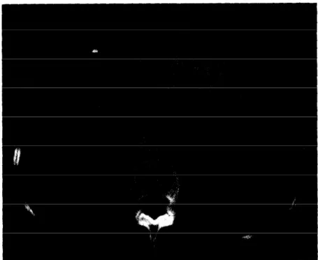
image from the same sequence is shown. The P-type coded version of this image at the CR =10.35:1 is shown in Fig. 4. The difference image produced by subtracting the original image from its P-type coded version (CR =22.56: 1) is shown in Fig. 5. In all the P-type coded pictures shown here, the GOP is in the form IPPPP and the picture is the last P-type picture.
As a subjective measure we resorted to the opin- ions of a group of medical doctors. This group contained one radiologist, one internal medicine expert and a practitioner. They evaluated the orig- inal and the compressed images in terms of(i) the overall picture quality, and (ii) the picture quality based on diagnostic features. The group stated that the compressed pictures with approximately 10: 1 compression ratios cannot be distinguished from
Fig. 1. An original image from the test sequence
S.H. O&z et al. / Signal Processing: Image Communication 7 (1995) 225-230 229
Fig. 3. Another original image from the test sequence.
Fig. 5. The difference image (12 x [original - reconstructed]) for P-type coding at CR =22.56 (0.44 bits/pel). The darker the points get, the higher the difference is.
their originals. This is valid for both I- and P-type pictures in all GOP-types tested. However, there are visible degradations in approximately 20: 1 compressed pictures which may still be used in a PACS for archiving purposes, if a radiologist’s report accompanies the image sequence. Similar results can be obtained for other medical image sequences, too.
Three-dimensional coding of medical image se- quences was first considered by Huang et al. [3]. In [3], a 3-D DCT-based coding method was de- veloped. However, this method does not use ‘motion-compensation’ techniques which are widely utilized in video-sequence coding including MPEG-I [7]. In this paper, we experimentally ob- served that the use of predictive coding and motion-compensated coding in a medical image
sequence compression increases the coding effici- ency without compromising objective and subjec- tive picture quality.
References Cl1 121 II31 c41 PI PI c71 C81 c91 Cl01 II111 Cl21 Cl31 Cl41
G. Bjentegaard, Proposal 06, VADIS/COST Forward Prediction Coding, MPEG91/270, November 1991. A.E. Cetin, “Subband coding of DSA images”, in: H.K. Huang, 0. Ratib, A.R. Bakker and G. Witte, eds., Picture Archioing and Communication Systems (PALS) in Medicine,
Springer, Berlin, 1991, pp. 361-363.
K.K. Chan, C.C. Lau, S.L. Lou, A. Hayrapetian, B.K.T. Ho and H.K. Huang, “Three-dimensional transform com- pression of images from dynamic studies”, The UCLA PACS modules and related projects - A progress report, Los Angeles, CA, 1990, pp. 60-64.
P.S. Cho, K.K. Chan and K.T. Ho, “Data storage and compression”, in: H.K. Huang, 0. Ratib, A.R. Bakker and G. Witte, eds., Picture Archiving and Communication Sys- tems (PALS) in Medicine, Springer, Berlin, 1991, pp. 71-82. H.K. Huang, 0. Ratib, A.R. Bakker and G. Witte, eds.,
Picture Archiving and Communication Systems (PACS) in Medicine, Springer, Berlin, 199 1.
N.S. Jayant and P. Nell, Digital Coding of Waveforms, Prentice-Hall, Englewood Cliffs, NJ, 1984, pp. 320, 321. D. Le Gall, “Digital multimedia systems: Digital image and video standards”, Commun. ACM, Vol. 34, 1991, pp. 47-58.
MPEG-I standard ISO/IEC IS1 1172.
M. Rabbani and P.W. Jones, Digital image Compression Techniques, SPIE Press, USA, 1991.
T.V. Ramabadran and K. Chen, “The use of contextual information in the reversible compression of medical images”, IEEE Trans. Medical Imaging, Vol. 11, No. 2, June 1992, pp. 185-195.
H. Sandgrind, NTR proposal to MPEG-S/Video tests, Kurihama, Japan, 1992.
W.S. Weinberg, M. Loloyan, R.K. Taira, K.K. Chan and H.K. Huang, “Automatic acquisition of CT, MR, and US images for PACS”, in: H.K. Huang, 0. Ratib, A.R. Bakker and G. Witte, eds., Picture Archiving and Communication Systems (PALS) in Medicine, Springer, Berlin, 1991, pp. 43-50.
P. Wilhelm, D.R. Haynor, Y. Kim and E.A. Riskin, “Lossy image compression for digital medical imaging systems”, SPlE Opt. Engrg., Vol. 30, October 1991, pp. 1479-1485.
Y.Q. Zhang, M.H. Loew and R.L. Pickholtz,“A combined - transform coding (CTC) scheme for medical images”,
IEEE Trans. Medical Imaging, Vol. 11, No. 2, June 1992, pp. 196-202.
![Fig. 5. The difference image (12 x [original - reconstructed]) for P-type coding at CR =22.56 (0.44 bits/pel)](https://thumb-eu.123doks.com/thumbv2/9libnet/5889026.121704/6.810.73.383.119.559/fig-difference-image-original-reconstructed-type-coding-bits.webp)
