1073 Ann Med Res 2020;27(4):1073-6
Annals of Medical Research
DOI: 10.5455/annalsmedres.2019.11.728
Original Article
Correlation of pulse oximetry oxygen saturation with blood
gas arterial oxygen saturation in patients with heart failure
reduced ejection fraction: Prospective cohort study
Gulay Gok1, Tufan Cinar2, Zekeriya Nurkalem1, Dursun Duman1 1Medipol University, Faculty of Medicine, Department of Cardiology, Istanbul, Turkey
2Sultan 2.Abdülhamid Han Training and Research Hospital, Clinic of Cardiology, Istanbul, Turkey Copyright © 2020 by authors and Annals of Medical Research Publishing Inc.
Abstract
Aim: An estimation of accurate oxygen saturation is a critical in the management of patients with heart failure (HF). However,
obtaining peripheral arterial blood samples may be technically difficult in some cases. The purpose of this study was to evaluate the correlation of pulse oximetry SO2 with arterial SO2 in patients with HF reduced ejection fraction (HFrEF).
Material and Methods: In total, 29 consecutive patients who were admitted to cardiology clinics with HFrEF were prospectively
enrolled in this study. We enrolled all patients if the chief physician ordered an atrial blood gas analysis. Simultaneous atrial blood gas sample and pulse oximetry measurements were collected and compared. The strength of association between atrial blood gas and pulse oximetry measurements was determined by Pearson correlation and Bland and Altman analysis.
Results: We evaluated 29 heart failure patients (the mean age was; 70.7±10.9, 44.8 % of patients were male) with a mean EF of 29.6 ±
5.5. We observed that the mean pulse oximetry SO2 and arterial SO2 in the study were 92.8 ± 4.4 and 93 ± 5.2, respectively. We found
significantly positive linear correlation between two methods according to Pearson analysis (r = 0.683, p < 0.001). A low bias was found between two methods according to Bland and Altman method (-0.2296, p = 0.23).
Conclusion: The present data may suggest that pulse oximetry oxygen saturation is an acceptable substitute for the arterial oxygen
saturation in patients with HFrEF.
Keywords: Arterial oxygen saturation; correlation; heart failure; pulse oximetry oxygen
Received: 13.11.2019 Accepted: 17.12.2019 Available online: 02.04.2020
Corresponding Author: Tufan Cinar, Sultan 2.Abdülhamid Han Training and Research Hospital, Clinic of Cardiology, Istanbul, Turkey E-mail: [email protected]
INTRODUCTION
Arterial blood gas analysis is indisputably more accurate than pulse oximetry. However, it is more difficult to obtain, more painful and expensive in addition to lasting longer time to obtain result. Some clinical studies have investigated the accuracy of pulse oximetry SO2 with
arterial SO2 in patients with uremic acidosis, diabetic
ketoacidosis, acute pulmonary edema, and living in high altitudes (1-5) However, there are few studies which mainly focused on the accuracy of pulse oximetry saturation in patients with heart failure (HF). Although pulse oximetry oxygen measurement is frequently used in daily clinical practice, it is not known whether there is a correlation between pulse oximetry oxygen saturation and arterial blood oxygen saturation in patients with heart failure with reduced ejection fraction (HFrEF). In this study, we therefore evaluated the reliability of oxygen saturation measurements obtaining by pulse oximetry and arterial
oxygen analysis in patients with HFrEF. Pulse oximetry measurements were compared with a more accurate method of arterial oxygen saturation.
MATERIAL and METHODS
Patient’s selection
This study was conducted in a high volume tertiary heart center. We enrolled all patients in the study if the chief physician ordered an atrial blood gas. Included in this prospective study was 34 consecutive patients who were admitted with the diagnosis of HFrEF. The inclusion criteria were as following; being older than 18 years, providing an informed consent, and without having significant comorbidities. The exclusion criteria included the existence of impalpable radial arteries or inconvenient vascular structures for arterial blood sampling, having hemodynamic compromise, the presence of inotrope use, anemia, infection, acute renal failure, pneumonia, and chronic obstructive pulmonary disease. The baseline
1074 Ann Med Res 2020;27(4):1073-6
demographics, clinical characteristics, New York Heart Association functional class, medication usages, echocardiographic data, and laboratory findings of patients were collected from electronic database of our hospital. All patients in the study received the standard medical treatment according to the latest HF guideline of European Society of Cardiology. The study was approved by ethics committee of the Medipol University School of Medicine (approval number: 10840098-604.01.01-E.61050-880).
Echocardiographic and laboratory examination
All blood samples were obtained in the first morning following admission. Biochemical parameters were measured by using Roche Cobas 6000 biochemistry auto-analyzer device. Arterial samples were withdrawn from the radial artery through a heparinized syringe and were carried to the laboratory. Simultaneously, fingertip pulse oximetry measurements were obtained on the same side with the standard finger sensors. G-life pulse Fs10E oximetry type was used after calibrating with the normal patient findings. All arterial blood sample results were obtained within 15 minutes. Multiple measurements were obtained from some patients to calibrate the device accuracy. The mean value recorded for patients whom had multiple results.
Standard echocardiographic evaluation was performed by an experienced cardiologist within 24-48 h following admission. The left ventricle ejection fraction was estimated using the modified Simpson method. Systolic pulmonary artery pressure was calculated according to the Bernoulli equation.
Statistical analysis
SPSS (SPSS Inc., Chicago, IL, USA) version 20.0 was used to analyze the data. Continuous variables were presented as mean ± standard deviation (mean±SD) and the categorical variables were expressed as number and percentage (%). The agreement between pulse oximetry O2 saturation and arterial O2 saturation was assessed by Bland Altman. The strength of association was determined by Pearson correlation. A p value < 0.05 was considered to be statistically significant.
RESULTS
In total, 34 patients (the mean age was 70.7 ± 10.9, 44.8 % of all cases were male) were firstly included in the study. We excluded 5 patients from the study (2 of them had diagnosis of acute renal failure, 1 patient had a missing data, and the remaining patients had diagnosis of pneumonia). The final cohort was consisted of 29 HFrEF patients.
The clinical characteristics, echocardiographic, and laboratory data of all patients were represented in Table 1. In the present research, 26 patients had ischemic HF and 3 patients had non-ischemic HF. The mean EF of the study was 29.6 ± 5.5. We observed that the mean pulse oximetry O2 saturation and arterial O2 saturation in the
Table 1.Baseline clinical, laboratory and echocardiographic features of the study population
Age, years 70.7±10.9
Gender, male (n) 44.8% (13)
Clinical characteristics
Body mass index, kg/m2 28.2±4.5
NHYA class 3-4, (n) 60.7% (17) NHYA class 1-2, (n) 39.3% (11) Ischemic heart failure, (n) 89.7% (26) Non-ischemic heart failure, (n) 10.3% (3) Hypertension, (n) 27.6% (8) Diabetes mellitus, (n) 25.9% (7) Chronic renal failure, (n) 35.7% (10)
Obesity, (n) 10.3% (3) Atrial fibrillation,(n) 30% (12) Heart rate, bpm 86.4±17.4 BP, mmHg Systolic 114.7±19.5 Diastolic 70.6±11.9 Pulse oximetry SO2 92.8±4.4 Arterial SO2 93±5.2 Vpo2 34.2±5.9 Vpco2 60.7±13.7 VHCO3 26.3±4.6 Laboratory findings
Blood urea nitrogen, mg/dL 75.4±40.5 Creatinine, mg/dL 1.6±0.6 Hemoglobin, mg/dL 12.3±2.1
Hematocrit, % 38±6
Aspartate aminotransferase, U/L 35±39 Alanine aminotransferase, U/L 46±43 Brain natriuretic peptide, ng/mL 5920±1580
Echocardiographic findings
Left ventricle diastolic diameter, mm 61.8±5.8 Left ventricle systolic diameter, mm 50.4±7.3 Ejection fraction, % 29.6±5.5 Pulmonary artery systolic pressure, mmHg 21±14.9 Left atrium diameter, mm 51.2±7.8 Moderate-severe MR, (n) 24.1% (7) Moderate-severe TR, (n) 20.7% (6)
Continuous variables are presented as mean ± SD or median, nominal variables presented as frequency (%).
Abbreviations: MR; mitral regurgitation, TR; tricuspid regurgitation
1075 Ann Med Res 2020;27(4):1073-6
study were 92.8 ± 4.4 and 93 ± 5.2, respectively. The mean Vpo2, Vpco2, and VHCO3 were 34.2±5.9, 60.7±13.7, and
26.3±4.6 respectively.
Figure 1 shows a 95% limits of agreement between the methods. According to the Bland Altman method, the range of agreement limits were from 7.4025 to -7.8617, and the t bias (mean difference) between the pulse oximetry SO2 saturation and arterial SO2 saturation was
-0.2296. As for the plot of unit values, this bias was not significant (p = 0.23).
Figure 1. Bland and Altman plot, with the representation of the
limits of agreement (dotted line) from -1.96s to +1.96
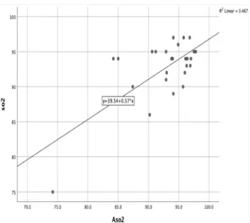
The correlation was significant between the methods (p < 0.001). According to Pearson analysis as shown Figure 2, a linear correlation was detected and the correlation coefficient was r = 0.683.
Figure 2. Pearson correlation of pulse oximetry SO2 and arterial
SO2
DISCUSSION
Arterial blood gas sampling is quintessential in guiding clinical treatment such as hospitalization, mechanical ventilator support or diuretic therapy in HF patients. However, obtaining arterial blood gas sample can be difficult, and it sometimes cause some complications, including local hematoma or infection on vascular puncture site. In some studies, pulse oximetry has been shown to be a useful for substitute of arterial blood oxygen saturation in intensive care units or emergency departments. Therefore, pulse oximetry is recommended to decrease amount of complications on vascular puncture site and to decrease economic cost (5-8).
The presence of anemia, peripheral vasoconstriction, hypotension or inotrope use may affect pulse oximetry values in patients with HF. In particular, pulse oximetry measurements may be unreliable due to low cardiac output and decreased tissue perfusion among these patients. Moreover, circulatory failure may effect peripheral oxygenation which may cause inaccurate fingertip pulse oximetry measurements. Based on these assumptions, some studies have suggested that pulse oximetry would not be an accurate method to obtain oxygen saturation in patients with impaired hemodynamics or using vasoactive agents (9). Therefore, our study focused on the specific group of HF patients without any known secondary co-morbidities including severe hemodynamic compromise, inotrope use or infection.
A previous study, which included patients with acute pulmonary edema, demonstrated that the Bland and Altman plot for arterial SO2 and pulse oximetry SO2 showed
a mean bias 0.6 and a low accuracy in comparison between SO2 and aSO2 was found (5). However, the average bias
(-0.209) for our study was close to zero, which means the two methods were systematically producing close results. This finding could be related to inclusion of stabilized patients with HFrEF in our study. In most of the studies, higher accuracy was found in pulse oximetry SO2 values
when the arterial SO2 is not extremely low, which was
similar with our findings (10,11). A significant bias and a lower sensitivity was found when arterial SO2 < 90% (5,12).
Some studies have revealed that a greater bias exists in patients using vasoactive agents, with septic shock or with chronic obstructive lung disease (9,13,14). In addition to this, in critically ill patients, venous SO2 in addition to
pulse oximetry is recommended for accurately predicting arterial oxygen saturation (15). However, in our study, such patients were not present and a linear, positive, and strong relation was detected by Pearson correlation analysis. Based on the study findings, we may suggest that pulse oximetry SO2 is an acceptable substitute for the arterial
SO2 in patients with HFrEF. However, as our data was
relatively small, prospective studies with a large groups of patients are needed to confirm our results.
1076 Ann Med Res 2020;27(4):1073-6
LIMITATION
Although some studies showed that the types of pulse oximeters may affect the agreement with the arterial blood oxygen saturation, we did not evaluate the results with different types of oximetry. In addition to this, our study was conducted in a small groups of patient population, which may also have an effect on the results. The study results might be not true for all patients with HF because we selected only patients with HFrEF.
CONCLUSION
This study has proved that the pulse oximetry SO2 is
statistically acceptable substitute for the arterial SO2 in
patients with HFrEF. Based on our results, we may suggest that pulse oximetry SO2 may be very useful in HFrEF
patients who are having inconvenient arterial structure or are under treatment of anticoagulants.
Competing interests: The authors declare that they have no competing interest.
Financial Disclosure: There are no financial supports.
Ethical approval: The study was approved by ethics committee of the Medipol University School of Medicine (approval number: 10840098-604.01.01-E.61050-880).
Gulay Gok ORCID: 0000-0003-0205-1138 Tufan Cinar ORCID: 0000-0001-8188-5020 Zekeriya Nurkalem ORCID: 0000-0001-6515-7309 Dursun Duman ORCID: 0000-0003-2542-3467
REFERENCES
1. Brandenburg MA, Dire DJ. Comparison of arterial and venous blood gas values in the initial emergency department evaluation of patients with diabetic ketoacidosis. Ann Emerg Med 1998;31:459−65.
2. Gokel Y, Paydas S, Koseoglu Z, et al. Comparison of blood gas and acid−base measurements in arterial and venous blood samples in patients with uremic acidosis and diabetic ketoacidosis in the emergency room. Am J Nephrol 2000;20:319−23.
3. J M Lozano, O R Duque, T Buitrago, et al. Pulse oximetry reference values at high altitude. Arch Dis Child 1992;67:299-301.
4. Lim BL, Kelly AM. A meta-analysis on the utility of peripheral venous blood gas analyses in exacerbations of chronic obstructive pulmonary disease in the emergency department. Eur J Emerg Med 2010; 17:246-8.
5. Masip J, De Mendoza D, Planas K, et al. Peripheral venous blood gases and pulse-oximetry in acute cardiogenic pulmonary oedema. Eur Heart J Acute Cardiovasc Care 2012;1:275-80.
6. Van de Louw A, Cracco C, Cerf C, et al. Accuracy of pulse oximetry in the intensive care unit. Intensive Care Med 2001;27:1606-13.
7. Holburn CJ, Allen MJ. Pulse oximetry in the accident and emergency department. Arch Emerg Med 1989; 6:137-42.
8. Bierman MI, Stein KL, Snyder JV. Pulse oximetry in the postoperative care of cardiac surgical patients. A randomized controlled trail. Chest 1992;102:1367-70. 9. Ibanez J, Velasco J, Raurich JM. The accuracy of
the Biox 3700 pulse oximeter in patients receiving vasoactive therapy. Intensive Care Med 1991;17:484-6 10. Jensen LA, Onyskiw JE, Prasad NG. Meta-analysis
of arterial oxygen saturation monitoring by pulse oximetry in adults. Heart Lung 1998;27:387−408. 11. Perkins GD, McAuley DF, Giles F, et al. Do changes in
pulse oxymeter oxygen saturation predict equivalent changes in arterial oxygen saturation? Crit Care 2003; 7: 67.
12. Seguin P, Le Rouzo A, Tanguy M, et al. Evidence for the need of bedside accuracy of pulse oximetry in an intensive care unit. Crit Care Med 2000;28:703-6. 13. Hannhart B, Michalski H, Delorme N, et al. Reliability of
six pulse oximeters in chronic obstructive pulmonary disease. Chest 1991;99:842-6.
14. Secker C, Spiers P. Accuracy of pulse oximetry in patients with low systemic vascular resistance. Anaesthesia 1997;52:127-30.
15. Zeserson E, Goodgame B, Hess JD, et al. Correlation of Venous Blood Gas and Pulse Oximetry with Arterial Blood Gas in the Undifferentiated Critically Ill Patient. J Intensive Care Med 2018; 33:176-81.