Scientific paper
A Simple and Highly Sensitive Turn-on
Schiff Base Type Naked-eye Fluorescent Sensor
for Aluminum Ion in Living Cells
Sedat Keskin and Mevlut Bayrakci*
Karamanoglu Mehmetbey University, Faculty of Engineering, Department of Bioengineering 70200, Karaman/TURKEY
* Corresponding author: E-mail: [email protected] Tel.: +90 338 2262200 fax: +90 338 2262214
Received: 09-28-2018
Abstract
Six different Schiff bases to be used as turn-on fluorescent probes based on photoinduced electron transfer (PET) mech-anism for the recognition of aluminum ions were successfully synthesized and characterized. The binding abilities of synthesized compounds with different metal cations were investigated by absorption and emission spectra. From the spectrophotometric experiments, it were seen that compound SK-1 displayed an excellent fluorescence response towards targeted aluminum ions probably due to its suitable chelating structure. Furthermore, such compound SK-1 also showed high sensitivity and selectivity response towards aluminum ions over other competing ions. In addition, the potential biological applications of SK-1 to detect aluminum ions in living cells were also investigated and results showed that flu-orescence sensor SK-1 could be a promising probe for determining and/or monitoring aluminum ions in both biological and/or chemical samples.
Keywords: Schiff Base, fluorescent probe, cell imaging, aluminum, PET.
1. Introduction
Aluminum is one of the most abundant metal ele-ments in the Earth’s crust and has an important place in our live. 1 Due to different reasons such as both ecological
system and human activities, high quantities of aluminum are found in the environment.2 The presence of excess
amount of aluminum in nature life affects the living be-ings. Consequently, some natural products containing large amount of aluminum in food chain are slowly con-sumed by human beings and this consumption causes many toxic effects towards human health and this toxicity leads to different diseases such as cancer, neurotoxicity, di-alysis disease, Alzheimer’s and Parkinson’s diseases.3-6
With respect to the World Health Organization, desired concentration of aluminum in drinking water must be limited to 7.4 μM.7 Therefore, it is important topic to
de-sign and develop effective analytical methods or instru-ments for detection of aluminum ions in environmental and/or biological systems. Although many different and sophisticated analytical techniques including inductively coupled plasma emission (ICP-OES) or mass
spectrome-try (ICP-MS), and atomic absorption spectromespectrome-try have been used extensively for the detection of aluminum ions,8,9 most of these techniques have some disadvantages
such as time consuming, qualified personal and high cost.10 But among them, fluorescence spectroscopy is most
popular analytical instrument for the detection of metal ions and it is preferred intensively by scientists in analyti-cal applications owing to its easy operation, comparatively low cost, and high sensitivity, etc.11 Thus, many different
fluorescence based chemosensors specific for metal ions have been designed and developed. Compared to these metal ions, just a few fluorescent probes have been report-ed for detection of trace amount of aluminum ions. Some limitations such as poor coordination ability and lack of spectroscopic characteristics have always been problemat-ic for the detection of aluminum ions.12 In addition to
these limitations, both complicated synthesis and solubili-ty properties of new fluorescent probes are also other re-strictions in the point of design of aluminum sensors.13
Therefore, it is necessary and important to design and syn-thesis of aluminum sensors that can be easily prepared and dissolved. Many sensitive and selective fluorescent sensors
for metal cations have been reported based on fluores-cence resonance energy transfer (FRET),14 chelation-
en-hanced fluorescence (CHEF),15 internal charge transfer
(ICT),16 photoinduced electron transfer (PET),17 and
exci-mer/exciplex formation mechanisms.18 However, although
photoinduced electron transfer (PET)-based fluorogenic sensors have many advantages,19 their synthesis and
appli-cation are not common in the literature. Although there are very interesting literature reports about photoinduced electron transfer (PET)-based fluorogenic sensors for dif-ferent analytes,20–22 synthesis of Schiff base type probes are
very inspirational owing to their easily preparation, faster response and high selectivity towards specific analytes of interest. Schiff bases are the most popular class of synthet-ic compounds in organsynthet-ic, medsynthet-icinal and pharmaceutsynthet-ical chemistry due to their unique biochemical properties such as antitumor, anti-HIV, antibacterial, antioxidant, in-flammatory, antifungal, pesticidal, anthelmintic and anti-hypertensive, activitie.23–25 Although, there are
appropri-ate literature reports showing their biological applications of Schiff base compounds, recently, limited number of lit-erature results about using of Schiff base derivatives as flu-orescent probe for the detection of metal ions in living cell have been existed.26–30 In the light of these literature, here,
we presented the design, synthesis and biological applica-tions of a series of Schiff base based fluorescent sensors containing ortho, meta and para hydroxy units which could detect aluminum ions by the ‘naked eye’.
2. Experimental
2. 1. General
2,3-dihydroxybenzaldehyde, 3,4-dihydroxybenzal-dehyde, ortho, meta and para aminophenol and all metal
salts were of analytical grade and purchased from Sig-ma-Aldrich or Merck and was further used without any purification. 1H NMR spectra was recorded on Agilent
Premium Compact spectrometer operating at 600 MHz. Chemical shifts were reported as δ values (ppm). Peak multiplicities were expressed as follows: s, singlet; bs, broad singlet; d, doublet; t, triplet and m, multiplet. Bruker Vertex FT-IR spectrometer (ATR) was used for FT-IR spectra. UV-vis absorbance spectra were collected by a Shimadzu UV-1800 and the fluorescence measurements were obtained by Hitachi F-7100.
2. 2. General Procedure for the Synthesis
of Schiff Base Probes
To a stirred solution of corresponding ortho, meta or para aminophenol compounds (1.5 mmol) in 20 mL abso-lute ethanol was added 1.5 mmol of 2,3-dihydroxybenzal-dehyde (for SK-1, SK-2 and SK-3, respectively) or 1.5 mmol of 3,4-dihydroxybenzaldehyde (for 2, 3 and
MK-4, respectively); the reaction mixture was stirred under
re-flux for 18 h. After completion of the reaction, excess amount of solvent was removed under reduced pressure and the solid residue was washed with 1 N HCl, brine and excess amount of water. The crude product was crystallized from CH2Cl2-C2H5OH (1:1) solvent system (Scheme 1).
SK-1: Red solid with 68% yields, FT-IR (ATR cm–1): 1616
(C=N stretching). 1H NMR (600 MHz DMSO): δ 14.21
(bs, 1H, OH) 9.88 (bs, 1H, OH), 9.02 (bs, 1H, OH), 8.91 (s, 1H, CH=N), 7.38 (d, J= 8.3 Hz, 1H, Ar-H), 7.10 (m, 1H, Ar-H), 7.02 (m, 1H, Ar-H), 6.95 (m, 1H, Ar-H), 6.87 (m, 1H, Ar-H), 6.77 (m, 1H, Ar-H), 6.69 (m, 1H, Ar-H). Anal. calcd. For C13H11O3N: C, 68.11; H, 4.84; N, 6.11. Found: C,
68.09; H, 4.90; N, 6.19%.
SK-2: Dark red solid with 70% yields, FT-IR (ATR cm–1):
1623 (C=N stretching). 1H NMR (600 MHz DMSO): δ
14.18 (bs, 1H, OH) 9.87 (bs, 1H, OH), 9.03 (bs, 1H, OH), 8.89 (s, 1H, CH=N), 7.35 (d, J= 8.4 Hz, 1H, Ar-H), 7.13 (m, 1H, Ar-H), 7.03 (m, 1H, Ar-H), 6.92 (m, 1H, Ar-H), 6.85 (m, 1H, H), 6.74 (m, 1H, H), 6.65 (s, 1H, Ar-H). Anal. calcd. For C13H11O3N: C, 68.11; H, 4.84; N, 6.11.
Found: C, 68.03; H, 4.80; N, 6.07%.
SK-3: Dark red solid with 70% yields, FT-IR (ATR cm–1):
1621 (C=N stretching). 1H NMR (600 MHz DMSO): δ
14.17 (bs, 1H, OH) 9.88 (bs, 1H, OH), 9.06 (bs, 1H, OH), 8.91 (s, 1H, CH=N), 7.34 (d, J= 8.4 Hz, 1H, Ar-H), 7.17– 7.11 (m, 3H, H), 6.83 (m, 1H, H), 6.34 (m, 1H, Ar-H), 6.21 (m, 1H, Ar-H). Anal. calcd. For C13H11O3N: C,
68.11; H, 4.84; N, 6.11. Found: C, 68.03; H, 4.88; N, 6.15%.
MK-2: Red solid with 71% yields, FT-IR (ATR cm–1): 1635
(C=N stretching). 1H NMR (600 MHz DMSO): δ 9.84 (bs,
1H, OH) 9.48 (bs, 1H, OH), 9.03 (bs, 1H, OH), 8.87 (s, 1H, CH=N), 7.27 (m, 2H, Ar-H), 7.18 (m, 2H, Ar-H), 7.10– 7.04 (m, 3H, Ar-H). Anal. calcd. For C13H11O3N: C, 68.11;
H, 4.84; N, 6.11. Found: C, 68.09; H, 4.90; N, 6.19%.
MK-3: Red solid with 65% yields, FT-IR (ATR cm–1): 1634
(C=N stretching). 1H NMR (600 MHz DMSO): δ 9.81 (bs,
1H, OH) 9.52 (bs, 1H, OH), 9.11 (bs, 1H, OH), 8.91 (s, 1H, CH=N), 7.33 (m, 2H, Ar-H), 7.25 (m, 1H, Ar-H), 7.09 (m, 1H, Ar-H), 6.88 (m, 2H, Ar-H), 6.57 (m, 1H, Ar-H). Anal. calcd. For C13H11O3N: C, 68.11; H, 4.84; N, 6.11. Found: C,
68.03; H, 4.80; N, 6.07%.
MK-4: Dark red solid with 58% yields, FT-IR (ATR cm–1):
1620 (C=N stretching). 1H NMR (600 MHz DMSO): δ
9.81 (bs, 1H, OH) 9.47 (bs, 1H, OH), 9.16 (bs, 1H, OH), 8.90 (s, 1H, CH=N), 7.34–7.29 (m, 2H, Ar-H), 7.17 (m, 2H, Ar-H), 7.03 (m, 3H, Ar-H). Anal. calcd. For C13H11O3N: C, 68.11; H, 4.84; N, 6.11. Found: C, 68.03; H,
4.88; N, 6.15%.
2. 3. Uv–Vis and Fluorescence Studies
The stock solutions of SK-1, SK-2, SK-3, MK-2,
MK-3 and MK-4 (1 mM), the guest nitrate salts of metal
cations (Li+, Na+, Ag+, Ca2+, Ba2+, Co2+, Cs+, Cu2+, Mg2+,
Hg2+, Mn2+, Pb2+, Ni2+, Sr2+, Zn2+ and Al3+) (1 mM) in
DMF were prepared. In absorption and emission experi-ments, the volume of studied solutions was adjusted as 2.0 mL. Titration experiments were performed by addition of corresponding amount of metal cation solutions to a DMF solution of targeted fluorescent probe (SK-1). The absorp-tion spectra of SK-1, SK-2, SK-3, MK-2, MK-3 and MK-4 in the presence and absence of metal cations were record-ed in the range of 200–600 nm. All emission spectra were obtained at room temperature under the excitation of 400–430 nm. The solutions were scanned (1200 nm/min) with 400 watt of PMT voltage in a spectrofluorometer with the range of 400–750 nm. The widths of the slit for the both excitation and emission were adjusted at 5 nm. The best fluorescence intensity at 530 nm was determined un-der the excitation at the wavelength of 430 nm.
2. 4. Biological Applications
The living MCF7 cells were provided by ATCC (American Type Culture Collection, Rockville, MD, USA). MCF7 cells were incubated with 10 μM of Al3+ ions in the
culture medium at 37 οC for 1 h and washed with
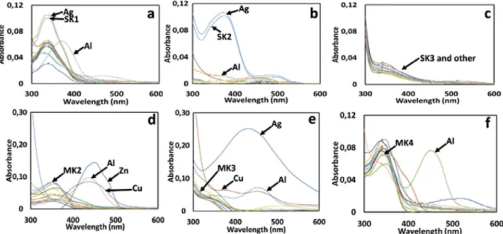
phos-Fig. 1. UV–vis absorption spectra of (1µM); (a) SK-1, (b) SK-2, (c) SK-3, (d) MK-2, (e) MK-3, and (f) MK-4 in the presence of several metal ions such as Li+, Na+, Ag+, Ca2+, Ba2+, Co2+, Cs+, Cu2+, Mg2+, Hg2+, Mn2+, Pb2+, Ni2+, Sr2+, Zn2+ and Al3+ (10 eq. for each metal ion).
phate buffered saline (PBS) followed by the addition of 10 μM of SK-1. Bright field and fluorescent images were tak-en from Leica DM3000 fluoresctak-ence microscopy.
3. Results and Discussion
3. 1. UV–vis Absorption Studies
The absorption spectrum of the Schiff bases SK-1,
SK-2, SK-3, MK-2, MK-3 and MK-4 was investigated by
the absence and/or presence of 10 equiv. of metal cations such as Li+, Na+, Ag+, Ca2+, Ba2+, Co2+, Cs+, Cu2+, Mg2+,
Hg2+, Mn2+, Pb2+, Ni2+, Sr2+, Zn2+ and Al3+. As seen in Fig.
1, the absorption spectrum of all Schiff bases, exhibited a broad absorption band attributable to π–π* transition of the imine moiety at around 342 nm. The absorption band positions generally remained unchanged over the various metal ions except Al3+ ions. After the addition of Al3+, the
appreciable bathochromic or hypochromic changes at around 342 nm for SK-1, SK-2, MK-2, MK-3 and MK-4 was observed owing to the imine nitrogen (CH=N) was involved in coordination with Al3+ ion. However,
consid-erable changes in the absorption spectra of the SK-3 was not observed over the various metal ions (Fig. 1c). Fur-thermore, new absorption bands at around 470 nm (for
SK-1, and SK-2) and 450 nm (for 2, 3 and MK-4) were seen probably due to the complexation capabilities
of these molecules with Al3+ ions. Since absorption
spec-troscopy is a complementary part of emission spectrosco-py, fluorescence emission studies were also applied for the getting more information about the spectrophotometric results.
3. 2. Fluorescence Emission Analysis
High selectivity is necessary to define the excellent chemosensor. Therefore, to evidence the usability of the synthesized Schiff bases as a selective sensor, the fluores-cence behavior of Schiff bases SK-1, SK-2, SK-3, MK-2,
MK-3 and MK-4 was investigated by Hitachi F-7100
Spec-trofluorometer upon addition of selected metal ions such as Li+, Na+, Ag+, Ca2+, Ba2+, Co2+, Cs+, Cu2+, Mg2+, Hg2+,
Mn2+, Pb2+, Ni2+, Sr2+, Zn2+ and Al3+. The reported Schiff
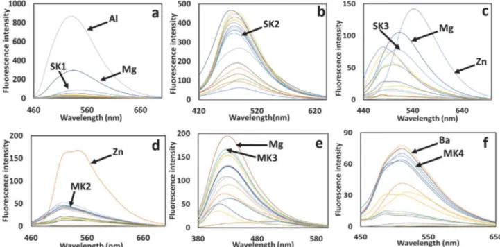
base probes (1 µM) showed a weak fluorescence emission spectrum at around 530 nm (for 1), 480 nm (for
SK-2), 508 nm (for SK-3), 518 nm (for MK-SK-2), 430 nm (for MK-3) and 518 nm (for MK-4) with an excitation of 430
(for SK-1), 400 nm (for SK-2 and SK-3,), 380 nm (for
MK-3,) and 430 nm (for MK-2 and MK-4). Other metal
ions (10 µM) such as Li+, Na+, Ag+, Ca2+, Ba2+, Co2+, Cs+,
Cu2+, Mg2+, Hg2+, Mn2+, Pb2+, Ni2+, Sr2+, and Zn2+ were
added to the solution of Schiff base probes, considerable decrease or increase in fluorescent intensity of probes were not observed in Fig. 2. Whereas, upon addition of Al3+ (10 µM) remarkable fluorescence increase
accompa-nied by a red shift of 24 nm from 530 nm to 554 nm was only noticed for the Schiff base probe SK-1 (Fig. 2a). Schiff base probe SK-1 exhibited a more than 37-fold fluorescent enhancement alone in the presence of Al3+ ions. This
in-crease in fluorescence intensity is such that the Schiff base probe SK-1 shows “OFF-ON” mode of high sensitivity for Al3+ ions. Furthermore, the Schiff base probe SK-1
indi-cated considerable color change from colorless to brilliant turquoise fluorescence in the presence of Al3+ ions under
UV light, and this color change was also easily detected by
Fig. 2. Fluorescent emission spectra of (1µM); (a) SK-1, (b) SK-2, (c) SK-3, (d) MK-2, (e) MK-3, and (f) MK-4 in the presence of several metal ions such as Li+, Na+, Ag+, Ca2+, Ba2+, Co2+, Cs+, Cu2+, Mg2+, Hg2+, Mn2+, Pb2+, Ni2+, Sr2+, Zn2+ and Al3+ (10 eq. for each metal ion).
the naked eye (Fig. 3). As a results, this Schiff base probe
SK-1 can be evaluated to determine Al3+ ions in solution
visually.
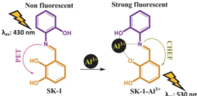
Photoinduced electron transfer (PET) mechanism includes the deactivation of the excited-state of fluorescent compounds by adding an electron to its frontier orbital. This electron addition to one of frontier orbital of excit-ed-state causes a non-emissive state for fluorophore struc-tures. For instance, the presence of one or more functional groups having free pair of electrons attached to the fluores-cent molecule may quench its fluoresfluores-cent intensity intra-molecularly due to photoinduced electron transfer (PET) mechanism. However, a possible interaction of these elec-tron donor groups with elecelec-tron acceptor metal ions re-duces efficient electron donor capabilities of these groups, thereby disconnecting the photoinduced electron transfer (PET) mechanism and increase the fluorescence output via chelation-enhanced fluorescence (CHEF).31,32 Herein,
the emission intensity of SK-1 was very low because of the quenching by the lone pair electrons of imine group through a PET mechanism. However, with increasing of Al3+ (0–10 equiv.), the fluorescence emission intensity of
SK-1 (1.0 µM) at 530 nm increased gradually (Fig. 4b). The
complexation of the imine group (-C=N) with Al3+ ion
given rise to the PET mechanism was suppressed, the fluo-rescence of the complex structure was restored.33,34
3. 3. Titration and Competition Studies
The binding properties of SK-1 with Al3+ ions were
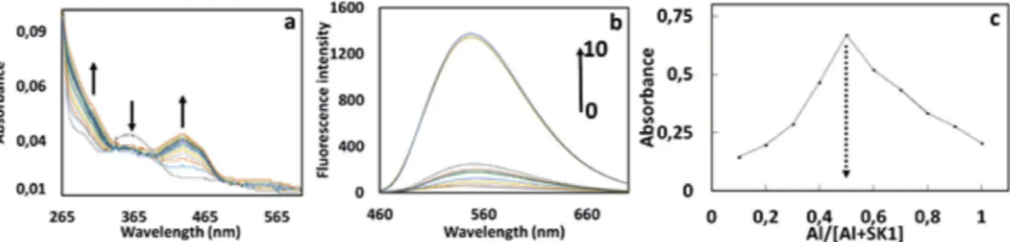
studied by both UV–vis and fluorescent titration experi-ments (Fig. 4a and 4b). Firstly, we explored the UV-vis ti-tration spectra of SK-1 with increasing concenti-trations of Al3+ in DMF. As shown in Fig. 4a, upon addition of
in-creasing amounts of Al3+ (0.0 to 2.0 equiv.), absorption
bands of SK-1 appeared at around 360 nm was gradually decreased with increasing amount of Al3+, while the
inten-sities of absorption SK-1 at around 434 nm increased. Fur-thermore, the absorbance at around 434 nm reached max-imum in the presence of 1.0 equiv. of Al3+ and showed
nearly no change with further addition of metal ion. The titration configuration of SK-1 with Al3+ in Fig. 4a
indicat-ed 1 equiv. of Al3+ reacting with same equiv. of SK-1 could
quickly reached an equilibrium, showing complex forma-tion between SK-1 and Al3+ with 1:1 stoichiometry. To
fur-ther examine the sensing properties of SK-1, sensitivity of
SK-1 as a probe toward Al3+ ions was investigated by the
fluorescence titration experiments by increasing concen-tration of Al3+ ions (0-10 equiv.) at 530 nm (Fig. 4b). Upon
excitation at 430 nm, SK-1 in the absence of any Al3+
showed practically no emission signal between the range of 460 and 700 nm which was probably due to PET pro-cess.35 However, a clear enhancement in fluorescence
in-Fig. 3. Images showing the corresponding (a) visible color and (b) fluorescence color changes of SK-1 with and without metal cations (10 equiv. of Li+, Na+, Ag+, Ca2+, Ba2+, Co2+, Cs+, Cu2+, Mg2+, Hg2+, Mn2+, Pb2+, Ni2+, Sr2+, Zn2+ and Al3+) under day light and UV light.
Fig. 4. (a) UV-Vis, (b) fluorescence titration spectra of compound SK-1 (1µM) respectively, upon addition of Al3+ (from 0 to 10 equiv.) at room
tensity of SK-1 was observed gradually at around 530 nm with increasing concentrations of Al3+ as shown in Fig 4b.
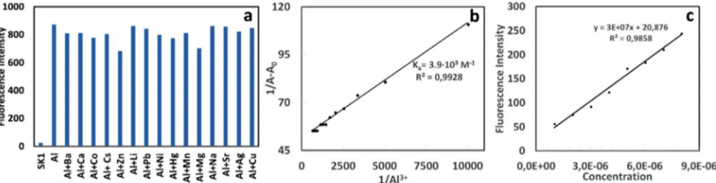
This increase in fluorescent intensity was probably due to the chelation-enhanced fluorescence (CHEF) effect that inhibiting the PET process by complexation of SK-1 with Al3+.36 Furthermore, the detection limit of Al3+ was
esti-mated based on the fluorescence titration profile (Fig. 5c). The detection limit of SK-1 in recognizing Al3+ was found
to be 4.85 · 10−7 M which was lower than some reported
literature results regarding Al3+ selective
chemosen-sors.37,38 This result was shown that this sensor could be
used for both detection and monitoring of sub-micromo-lar concentration of aluminum ions in biological and envi-ronmental systems. To verify the practical application of
SK-1 as an Al3+ selective and sensitive fluorescent sensor,
competition experiments were also performed by adding of Al3+ into SK-1 solution mixed with other coexisting
metal ions such as Li+, Na+, Ag+, Ca2+, Ba2+, Co2+, Cs+,
Cu2+, Mg2+, Hg2+, Mn2+, Pb2+, Ni2+, Sr2+, and Zn2+. As
de-picted in Fig. 5a, relatively low interference was seen for the detection of Al3+ in the presence of other competing
metal ions. Although, the slightly decreasing in emission intensity of SK-1 at around 530 nm was observed in the presence of Zn2+, and Mg2+, fluorescent response was
rela-tively detectable. However, upon addition of other com-peting metal ions under same conditions, it was seen that the fluorescence emission intensity at around 530 nm did not change considerably and SK-1 still have an efficient “turn-on” rate for the detection of Al3+. Consequently, it
was concluded that SK-1 could be a promising selective and sensitive fluorescent sensor for the detection of Al3+ in
the presence of competing metal ions.
3. 4. Binding Studies
To determine the binding stoichiometry of SK-1 with Al3+, the method of continuous variations known as Job’s
plot was used.39 In this method, each experiment
per-Fig. 5. Fluorescent selectivity of SK-1 (1µM) at 530 nm upon addition of various metal ions (10µM) Li+, Na+, Ag+, Ca2+, Ba2+, Co2+, Cs+, Cu2+, Mg2+,
Hg2+, Mn2+, Pb2+, Ni2+, Sr2+, and Zn2+, λex = 430 nm. (b) Calculation of binding constant between SK-1 and Al3+. A0 is the absorbance of free SK-1
solution (1µM); A is the absorbance of compound SK-1 solution (1µM) upon addition of different amounts of Al3, (c) Calculation of detection
limit of SK-1 for Al3+ with excitation at 430 nm and emission at 530 nm by addition of different amounts of Al3+ to SK-1 solution (1µM)
formed with different concentrations of SK-1 and Al3+ with
maintaining the total concentration at 10 μM. The plot ob-tained by measuring the fluorescence intensity at 530 nm for nine experiments with molar fraction of SK-1 (0.1 to 0.9). In this experiment, the maximum absorbance value was observed when the molar fraction was 0.5 (Fig. 4c) and it was consistent well with the UV-vis titration spectra (Fig. 4a). This data showed that 1 mole of SK-1 and Al3+
partici-pated in the complex formation and binding mode was de-termined as 1:1 stoichiometry. Furthermore, the binding constant of the probe SK-1 with Al3+ were calculated by the
Benesi–Hildebrand method.40 From curve fitting of
absor-bance values of probe SK-1 against the reciprocal of the Al3+ concentration, this plot yielded a linear fit as seen in
Fig. 5b. The value of the binding constant was calculated as 3.9 · 103 M–1 which was within the range of those (103–109
M–1) previously reported Al3+ sensors.41 In addition, the
linear plot also proved the 1:1 complexation behavior of
SK-1 to Al3+. Because, if a 1:1 metal-probe complex is
formed between receptor and metal ions, Benesi-Hildeb-rand plot should be linear.42 In related to stoichiometry, the
binding site participated in complexation was clarified by FT-IR (ATR) and 1H NMR experiments along with
stoichi-ometry confirmation as presented in Fig. 6 and 7.
The IR spectra of free SK-1 and SK-1-Al3+ complex structure showed that the characteristics frequencies of
SK-1 with 1.0 equiv. of Al3+ exhibited significant changes
as compared with those of free the SK-1 (Fig. 6). The IR spectra of the free SK-1 showed the absence of bands at around 1735 and 3300 cm–1 attributable to the carbonyl
ν(C=O) and ν(NH2) stretching vibrations and a clear
strong new band at around 1616 cm–1 due to azomethine
ν(HC=N) linkage.43–45 All these existing and disappearing
signals in IR indicated that amino and aldehyde groups in starting reactants (Scheme 1) were converted into the
SK-1 and synthesis of the SK-1 was successfully carried
out. The comparison of IR spectra of free SK-1 and its Al3+
complex (Fig. 6) demonstrated that SK-1 probe was prin-cipally coordinated to the Al3+ ion. The strong band
ap-pearing at around 1616 cm–1 due to azomethine group
shifted to a higher frequency at 1629 cm–1 in Al3+ complex,
indicating participation of azomethine group in the com-plexation with the Al3+ ion. On the other hand, the free
OH group at 3378 cm−1 was completely disappeared at 1
equiv. of Al3+. In addition, disappearing of strong band at
around 3378 cm−1 indicated that phenolic hydroxy group
of SK-1 participated in the complex formation with Al3+.
To better understand the complexation between the probe SK-1 and Al3+, 1H NMR experiment of SK-1 in
DMSO-d6 were examined by addition of 1 equiv. of Al3+.
As seen in Fig. 7, three phenolic -OH signals belonging to
SK-1 was observed at around 9.88, 9.02 and 14.21 ppm.
Compared the phenolic -OH signals, appearing signal at around 14.21 ppm attributed the ortho position of SK-1 is probably due to the intramolecular hydrogen bonding (Fig. 7). While the phenolic OH proton at 14.21 ppm dis-appeared when added of 1.0 equiv. of Al3+ to SK-1
solu-tion, the other signal at around 9.88 and 9.02 ppm shifted to downfield. Also, it was seen that the imine (CH=N) pro-ton of SK-1 at 8.91 ppm was slightly shifted to some extent. This shift for the imine proton was probably due to com-plexation ability of the azomethine group after coordina-tion of SK-1 with Al3+ · 44 All these shifting and/or
disap-pearing of signals showed that both phenolic OH group located in ortho position and imine group of SK-1 were efficient on complex formation between SK-1 and Al3+. In
the light of obtained spectroscopic data, possible complex formation mechanism was given in Fig. 8.
Fig. 7. 1H NMR spectra in DMSO-d
6 of SK-1 with (1.0 equiv of Al3+) and without Al3+.
3. 5. Biological Applications
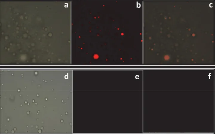
SK-1 was successfully applied for imaging of Al3+
ions in human breast cancer cells, MCF7 under fluores-cence microscope. Cells treated with free SK-1 were used as controls. When MCF7 cells were incubated with
SK-1 (10 µM), it was not seen any fluorescence response
(Fig. 9e). However, after addition of Al3+ ions, a brilliant
red fluorescence was sighted in the MCF7 cells (Fig. 9b). Merged images of fluorescence and bright-field showed that fluorescence signals were detected in the
intra-cel-Fig. 8. Proposed binding mechanism between the compound SK-1 and Al3+.
lular zone, showing the distribution of Al3+ and cell
membrane permeabilities of SK-1 molecules (Fig 9c). On the other hand, Fig. 9 indicated that SK-1 could stain Al3+ ions in living cells without any harm (cells remain
alive even after several hours of exposure to 10 µM of
SK-1), making it useful to monitor Al3+ in biological
sys-tems.
4. Conclusion
In conclusion, visual detection of highly selective and sensitive Al3+ ions by a very simple and low-cost
fluo-rescence sensor (SK-1) based on the blocking PET process was carried out successfully. SK-1 showed high sensitivity with the detection limit at around 4.8 × 10⁻⁷ M in the
mi-cromolar scale and selectivity response towards Al3+ over
other metal ions with 37-fold fluorescence enhancement. The predicted configuration of the SK-1–Al3+ complex formation was well-characterized to be 1:1 by spectroscop-ic analyses. Beyond that, SK-1 was utilized to detect sensi-tively the Al3+ ions in living cells by emitting visible
fluo-rescence. Cell applications indicated that SK-1 could be used as an excellent fluorescence probe for visualizing of Al3+ ions in cell lines.
5. Acknowledgments
The authors declare that there is no conflict of interest. This study is part of master thesis of Sedat Keskin and au-thors of this paper gratefully would like to thank Karamano-glu Mehmetbey University Research Foundation (BAP) and The Scientific and Technological Research Application Cen-ter (BILTEM) for the financial and technical supports.
6. References
1. J. Zhu, Y. Zhang, L. Wang, T. Sun, M. Wang, Y. Wang, D. Ma, Q. Yang, Y. Tang. Tetrahedron Lett., 2016, 57, 3535–3539. DOI:10.1016/j.tetlet.2016.06.112
2. X. Yan, H. Yang, F. Tian. RSC Adv., 2015, 107012–107019. DOI:10.1039/C5RA17557G
3. V. K. Gupta, S. K. Shoora, L. K. Kumawat, A. K. Jain. Sensors
Actuators, B Chem., 2015, 209, 15–24.
DOI:10.1016/j.snb.2014.10.143
4. K. Kaur, V. K. Bhardwaj, N. Kaur, N. Singh. Inorg. Chem.
Com-mun., 2012, 18, 79–82. DOI:10.1016/j.inoche.2012.01.018
5. Y. Xu, S. Mao, H. Peng, F. Wang, H. Zhang, S.O. Aderinto, H. Wu. J. Lumin., 2017, 192, 56–63.
DOI:10.1016/j.jlumin.2017.06.023
Fig. 9. Fluorescence images of Al3+ using probe SK-1 in MCF7: (a) bright field image of MCF7 cells treated with probe SK-1; (b) fluorescence image
of MCF7 cells treated with probe SK-1; (c) merged image of (a) and (b); (d) bright field image of MCF7 cells treated with probe SK-1 without Al3+;
6. L. Kang, Y. T. Liu, N.N. Li, Q. X. Dang, Z. Y. Xing, J. L. Li, Y. Zhang. J. Lumin., 2017, 186, 48–52.
DOI:10.1016/j.jlumin.2016.12.056
7. T. Han, X. Feng, B. Tong, J. Shi, L. Chen, J. Zhi, Y. Dong. Chem.
Commun., 2012, 48, 416–418. DOI:10.1039/C1CC15681K
8. M. H. Nagaoka, T. Maitani. Analyst, 2000, 125, 1962–1965.
DOI:10.1039/b006590k
9. M. Shellaiah, Y. H. Wu, H. C. Lin. Analyst, 2013, 138, 2931– 2942. DOI:10.1039/c3an36840h
10. S. Mukherjee, P. Mal, H. Stoeckli-Evans. J. Lumin., 2016, 172, 124–130. DOI:10.1016/j.jlumin.2015.11.014
11. S. Das, D. Karak, S. Lohar, A. Banerjee, A. Sahana, D. Das.
Anal. Methods., 2012, 4, 3620–3624.
DOI:10.1039/c2ay25825k
12. X. Sun, Y.-W. Wang, Y. Peng. Org. Lett., 2012, 14, 3420–3423.
DOI:10.1021/ol301390g
13. C.-Y. Huang, Y. Jhong, J.-L. Chir, A.-T. Wu. J. Fluoresc., 2014,
24, 991–994. DOI:10.1007/s10895-014-1404-1
14. H. Cheng, Y. Qian. RSC Adv., 2015, 5, 82887–82893. DOI:10.1039/C5RA15546K
15. M. Shellaiah, Y.C. Rajan, P. Balu, A. Murugan. New J. Chem.,
2015, 39, 2523–2531. DOI:10.1039/C4NJ02367F
16. S. Jana, S. Dalapati, N. Guchhait. J. Phys. Chem. A, 2013, 117, 4367–4376. DOI:10.1021/jp3120463
17. G. Q. Wang, J. C. Qin, L. Fan, C. R. Li, Z. Y. Yang. J.
Photo-chem. Photobiol. A Chem., 2016, 314, 29–34.
DOI:10.1016/j.jphotochem.2015.08.005
18. L. Wang, W. Qin, X. Tang, W. Dou, W. Liu. J. Phys. Chem. A.,
2011, 115, 1609–1616. DOI:10.1021/jp110305k
19. J. Shao, H. Lin, Z.-S. Cai, H. Lin. J. Photochem. Photobiol. B
Biol., 2009, 95, 1–5. DOI:10.1016/j.jphotobiol.2008.12.007
20. J. Kumar, M.J. Sarma, P. Phukan, D. K. Das. Dalt. Trans., 2015,
44, 4576–4581. DOI:10.1039/C4DT03932G
21. J. Wu, W. Liu, J. Ge, H. Zhang, P. Wang. Chem. Soc. Rev., 2011,
40, 3483–3495. DOI:10.1039/c0cs00224k
22. T. Gunnlaugsson, H. D. P. Ali, M. Glynn, P. E. Kruger, G. M. Hussey, F. M. Pfeffer, C. M. G. Dos Santos, J. Tierney. J.
Fluo-resc., 2005, 15, 287–299.
DOI:10.1007/s10895-005-2627-y
23. M. H. Yan, T. R. Li, Z. Y. Yang. Inorg. Chem. Commun., 2011,
14, 463–465. DOI:10.1016/j.inoche.2010.12.027
24. S. Sen, T. Mukherjee, B. Chattopadhyay, A. Moirangthem, A. Basu, J. Marek, P. Chattopadhyay. Analyst, 2012, 137, 3975– 3981. DOI:10.1039/c2an35560d
25. C. Dohno, A. Okamoto, I. Saito. J. Am. Chem. Soc., 2005, 127, 16681–16684. DOI:10.1021/ja054618q
26. L. Fan, T. R. Li, B. D. Wang, Z. Y. Yang, C. J. Liu. Spectrochim.
Acta - Part A Mol. Biomol. Spectrosc., 2014, 118, 760–764.
DOI:10.1016/j.saa.2013.09.062
27. Y. J. Lee, C. Lim, H. Suh, E. J. Song, C. Kim. Sensors Actuators B
Chem., 2014, 201, 535–544. DOI:10.1016/j.snb.2014.05.035
28. S. Guha, S. Lohar, A. Sahana, A. Banerjee, D. A. Safin, M. G. Babashkina, M.P. Mitoraj, M. Bolte, Y. Garcia, S.K. Muk-hopadhyay, D. Das. Dalt. Trans., 2013, 42, 10198-10207.
DOI:10.1039/c3dt51045j
29. S. K. Shoora, A. K. Jain, V. K. Gupta. Sensors Actuators, B
Chem., 2015, 216, 86–104. DOI:10.1016/j.snb.2015.04.038
30. M. Tajbakhsh, G. B. Chalmardi, A. Bekhradnia, R. Hossein-zadeh, N. Hasani, M. A. Amiri. Spectrochim. Acta Part A Mol.
Biomol. Spectrosc., 2018, 189, 22–31.
DOI:10.1016/j.saa.2017.08.007
31. P. Alaei, S. Rouhani, K. Gharanjig, J. Ghasemi.
Spectro-chim. Acta - Part A Mol. Biomol. Spectrosc., 2012, 90, 85–92.
DOI:10.1016/j.saa.2012.01.008
32. N. C. Lim, S. V. Pavlova, C. Brückner. Inorg. Chem., 2009, 48, 1173–1182. DOI:10.1021/ic801322x
33. J. M. An, Z. Y. Yang, M. H. Yan, T. R. Li. J. Lumin., 2013, 139, 79–83. DOI:10.1016/j.jlumin.2013.02.019
34. T. Gunnlaugsson, A. P. Davis, J. E. O’Brien, M. Glynn. Org.
Lett., 2002, 4, 2449–2452. DOI:10.1021/ol026004l
35. G. Zhu, Y. Wang, H. Fu, X. Xu, Z. Cui, X. Ji, G. Wu.
Spectro-chim. Acta Part A Mol. Biomol. Spectrosc., 2015, 137, 148–
153. DOI:10.1016/j.saa.2014.08.021
36. F. Karagöz, O. Güney, M. Kandaz, A.T. Bilgiçli. J. Lumin.,
2012, 132, 2736–2740. DOI:10.1016/j.jlumin.2012.05.005
37. D. Zhou, C. Sun, C. Chen, X. Cui, W. Li. J. Mol. Struct., 2015,
1079, 315–320. DOI:10.1016/j.molstruc.2014.09.050
38. W.-H. Ding, D. Wang, X.-J. Zheng, W.-J. Ding, J.-Q. Zheng, W.-H. Mu, W. Cao, L.-P. Jin. Sensors Actuators B Chem., 2015,
209, 359–367. DOI:10.1016/j.snb.2014.11.144
39. J. S. Renny, L. L. Tomasevich, E. H. Tallmadge, D. B. Collum.
Angew. Chem. Int. Ed. Engl., 2013, 52, 11998–2013.
DOI:10.1002/anie.201304157
40. H. A. Benesi, J. H. Hildebrand. J. Am. Chem. Soc., 1949, 71, 2703–2707. DOI:10.1021/ja01176a030
41. C. Kim, T. G. Jo, J. Lee, E. Nam, K. H. Bok, M.H. Lim. New
J. Chem., 2016, 40, 8918–8927. DOI:10.1039/C6NJ01544A
42. H. M. Kim, C. Jung, B. R. Kim, S.-Y. Jung, J. H. Hong, Y.-G. Ko, K. J. Lee, B. R. Cho. Angew. Chemie Int. Ed., 2007, 46, 3460–3463. DOI:10.1002/anie.200700169
43. İ. Kaya, E. Kartal, D. Şenol. Des. Monomers Polym., 2015, 18, 524–535. DOI:10.1080/15685551.2015.1041084
44. V. K. Gupta, A. Kumar, S. Lokesh, K.Kumawat. Sensors
Actu-ators, B Chem., 2015, 195, 98-108.
DOI:10.1016/j.snb.2013.12.092
45. V. K. Gupta, B. Sethi, R. A. Sharma, S. Agarwal, A. Bhartia. j. Mol. Liq., 2013, 177, 114-118.
Povzetek
Za detekcijo aluminijevih ionov na osnovi fotoinduciranega prenosa elektronov smo sintetizirali in okarakterizirali šest Schiffovih baz. Z absorpcijskimi in emisijskimi spektri smo raziskovali možnost vezave sintetiziranih spojin z različnimi kovinskimi kationi. S spektrofotometrijo smo ugotovili, da daje spojina SK-1 odličen fluorescenčni odziv na ciljne alumi-nijeve ione. Razlog je verjetno njena kelatna struktura. Spojina SK-1 je za alumialumi-nijeve ione pokazala večjo občutljivost in selektivnost kot za ostale ione. Rezultati raziskav možnosti uporabe SK-1 za detekcijo aluminijevih ionov v celicah so pokazali, da je lahko fluorescenčni senzor SK-1 obetavna sonda za določanje in/ali monitoring aluminijevih ionov v bioloških in/ali kemijskih vzorcih.
Except when otherwise noted, articles in this journal are published under the terms and conditions of the Creative Commons Attribution 4.0 International License