RESEARCH
CT evaluation of the bony nasal pyramid dimensions in Anatolian
people
D Karadag*
,1, NC Ozdol
1, K Beriat
2and T Akinci
11
Ufuk University, Faculty of Medicine, Department of Radiology;2Ufuk University, Faculty of Medicine, Department of Ear Nose Throat, Ankara, Turkey
Objectives: The aim of this study was to evaluate the nasal bone and bony nasal pyramid in adult Anatolian people.
Method: A total of 80 patients (48 males, 32 females, mean age of 40.03 years) were all evaluated using CT. Upper, intermediate and inferior thickness of the nasal bone on each side and on the lateral and medial osteotomy line were measured. In addition, nasal bone length and pyriform aperture width were determined.
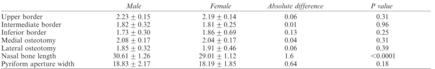
Results: The bone thickness was 2.23 mm ¡ 0.15 mm in males and 2.19 mm ¡ 0.14 mm in females at the level of upper border of the nasal bone; 1.82 mm ¡ 0.32 mm in males and 1.81 mm ¡ 0.25 mm in females at the intermediate level; and 1.73 mm ¡ 0.30 mm in males and 1.86 mm ¡ 0.69 mm in females at the lower border of the nasal bone. The mean thickness on the lateral osteotomy line was 1.85 mm ¡ 0.32 mm in males and 1.91 mm ¡ 0.46 mm in females. The mean thickness of the medial osteotomy line was 2.08 ¡ 0.17 mm in males and 2.04 mm ¡ 0.17 mm in females. The mean length of the nasal bone was 30.61 mm ¡ 1.26 mm in males and 29.01 mm ¡ 1.12 mm in females. The mean width of the pyriform aperture was 18.83 mm ¡ 2.17 mm in males and 18.19 mm ¡ 1.85 in females.
Conclusion: The dimensions of the nasal pyramid are known to be important in the selection of appropriate osteotome. Our results can be used for pre-operative evaluation of Anatolian people undergoing nasal surgery.
Dentomaxillofacial Radiology (2011) 40, 160–164. doi: 10.1259/dmfr/35578628
Keywords: nasal bone; pyriform aperture; computerized tomography; Anatolian people
Introduction
The nasal bones consist of two pieces that are situated side by side at the middle and upper part of the face. Each bone has two surfaces and four borders. The superior border has a narrow, thick surface which articulates to the frontal bone. The inferior border is thin and is attached to the lateral cartilage of the nose. The lateral border articulates to the frontal process of the maxilla. The medial border articulates to the spine of the frontal bone, the perpendicular plate of the ethmoid and the septal cartilage of the nose.
The nasal bone structure can be examined by physical examination or radiologic methods. Physical examination and radiographic data may not provide a concrete and objective result as experts’ opinions may
vary or examination methods may differ. Therefore, three dimensional (3D) CT is a perfect technique for obtaining objective results. This technique provides an advanced examination of the craniofacial anatomic anomalies, paranasal sinuses and nasal cavity, as well as dental structures for orthodontic therapy. It also facilitates a maxillofacial and facial plastic surgery or transnasal approach.
The shape and the size of the nasal bone varies in different races, ethnic groups, genders and ages.1
Choosing the appropriate size of osteotome pre-opera-tively or having the option to change it intraoperapre-opera-tively increases the success of the surgery. Furthermore, pre-operative CT assessment of the bony nasal pyramid can be a valuable tool, providing satisfactory accuracy and reliability for planning craniomaxillofacial reconstruc-tive procedures. The normareconstruc-tive data for the bony nasal pyramid dimensions among ethnic and gender groups
*Correspondence to: Demet Karadag, Ufuk Universitesi Tip Fakultesi Radyoloji ABD. Ankara/Turkey; E-mail: [email protected]
Received 15 December 2009; revised 30 March 2010; accepted 31 March 2010 http://dmfr.birjournals.org
could provide credible and objective reference for the estimation of optimal thickness of nasal augmentation and determining the ideal sites for fixation device placement.2 For these reasons, we undertook a study
to better understand the nasal morphology. In this study, we have analysed the bony nasal pyramid of the Anatolian population by using CT. Unlike other CT studies, we also analysed nasal bone thickness at three different levels on both sides.
Materials and method
This study was approved by the local research ethics committee. A total of 80 patients who underwent 3D maxillofacial CT examination between January 2008 and February 2009 were retrospectively examined. All CT examinations were performed at our institution with a 16-channel multislice CT scanner (LightSpeed, Gene-ral Electric Medical Systems, Milwaukee, WI). The parameters were 120 kV, 160 mAs, 512 6 512 matrix, 0.6 mm slice thickness, 0.5 mm pitch. Reformatted images in the standard coronal and sagittal planes were made at a slice thickness of 0.625 mm.
Patients with a history of nasal surgery or significant facial trauma were excluded from the study. 48 men (median age 39.2 years; age range 16–65 years) and 32 women (median age 41.2 years; age range 19–72 years) were examined during the study.
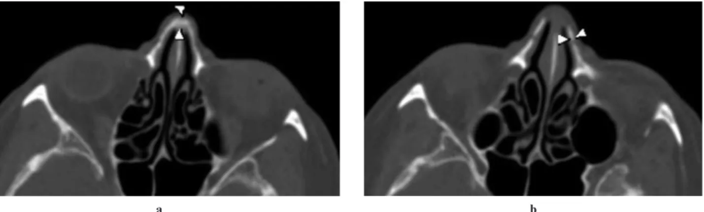
We measured upper, intermediate and inferior thick-ness of the nasal bone on both sides. In addition, nasal bone thickness at three points (low, intermediate and high) was measured on the lateral and medial osteot-omy line (Figure 1). The low level point (just cephalad to the pyriform aperture), the intermediate level point (just caudal to infraorbital rim) and the high level point (just cephalic to the infraorbital rim) were the reference points where the measurements were taken. The length of nasal bone and the upper width of pyriform aperture were measured in sagittal and coronal reformatted images. The width of pyriform aperture was measured between the inferior borders of the nasal bones
(Figure 2). All these measurements were done by 3 radiologists, who had between 3 and 12 years of experience of reporting maxillofacial CT findings, using an Advantage Workstation version 4.2 P (General Electric Medical System, Milwaukee, WI). We used an Active Matrix Liquid Crystal Display (AMLCD) monitor (size 21 inch, resolution 2048 6 1536, 3 MP (MFGD-3420)) which is regularly quality tested and calibrated. The light conditions were 240 (Nom.400)/ 0 (Lmaks/Lmin)(Lux).
We performed t-test using SPSS 17.0 software for Windows (SPSS, Inc, Chicago, IL) for statistical ana-lyses. Interobserver variability was measured by com-paring the results of measurements of three radiologists, blinded to each others’ measurements.
Results
The mean nasal bone thickness was 2.23 mm ¡ 0.15 mm in males and 2.19 mm ¡ 0.14 mm in females at the level of upper border of the nasal bone; 1.82 mm ¡ 0.32 mm in males and 1.81 mm ¡ 0.25 mm in females at the intermediate level; and 1.73 mm ¡ 0.30 mm in males and 1.86 mm ¡ 0.69 mm in females at the lower border of the nasal bone. The mean thickness on the lateral osteotomy line was 1.85 mm ¡ 0.32 mm in males and 1.91 mm ¡ 0.46 mm in females. The mean thickness of the medial osteotomy line was 2.08 mm ¡ 0.17 mm in males and 2.04 mm ¡ 0.17 mm in females.
The mean length of the nasal bone was 30.61 mm ¡ 1.26 mm in males and 29.01 mm ¡ 1.12 mm in females. Nasal bone length was significantly longer in males compared with females. The mean width of the pyriform aperture was 18.83 mm ¡ 2.17 mm in males and 18.19 mm ¡ 1.85 mm in females (Table 1).
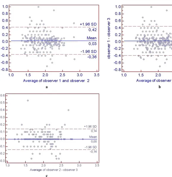
The Bland and Altman model, coefficient of varia-tion, was used to measure interobserver measurement variability. Bland and Altman plots were constructed for comparison of measurements between observers (Figure 3a,b,c). These plots indicate a standard deviation
a b
Figure 1 Axial CT image demonstrating the measurements of the nasal bone thickness. The arrowheads show the medial (a) and lateral (b) osteotomy points
(SD) of ¡0.4 between observer 1 and 2 (DK and NCO); a SD of ¡0.4 between observer 1 and 3 (DK and TA); and a SD of ¡0.14 between observer 2 and 3 (NCO and TA) (Figure 3, Table 2).
The mean absolute differences of nasal bone thick-ness width between both genders lie within the 95% confidence interval (CI) bounds.
The plots suggest that there is neither a falling nor a rising precision with rising measurement values.
Discussion
Although the most frequent surgical procedures per-formed by facial plastic surgeons are nasal bone reconstruction and rhinoplasty, standard osteotomies are still performed blindly, generally with only physical or radiographic examination. Therefore, to ensure confidence and better performance during nasal sur-gery, pre-operative evaluation of the 3D anatomy and the measurements of the nasal bone is advised.
There is increasing interest in understanding racial differences in nasal morphology. A few studies have been published on race differences of the nasal bones
and pyriform aperture. Lee et al measured the bone thickness at the sites of the lateral and intermediate osteotomy line and evaluated nasal bone shape in 75 Korean subjects by using 3D CT.1 In this study,
there was no significant difference in the thickness of the bone at the lateral and intermediate osteotomy point. Citardi et al measured the nasal bone thickness of 8 patients and found out that the lateral osteotomy thickness was 2.39 mm ¡ 0.68 mm and the intermedi-ate osteotomy thickness was 1.8 mm ¡ 0.3 mm.2
Ac-cording to this study, there was a difference between the lateral and intermediate osteotomy thickness. Lee et al measured the thickness of the bone along the track of a medial, intermediate and lateral osteotomy3and found
that the average bone thickness along the path of a lateral osteotomy was 2.61 mm ¡ 0.66 mm at the low level, 2.75 mm ¡ 0.76 mm at the middle level and 2.72 mm ¡ 0.53 mm at the high level. The mean bone thickness at the level of medial osteotomy at the low and high levels was 2.54 mm ¡ 0.31 mm and 2.77 mm ¡ 0.30 mm, respectively. Hwang et al studied 88 dried skulls from Korean adults and measured the height and width of nasal bone and pyriform aperture.4
According to Hwang et al, the nose shape of Korean people was different from the Germans; the width of
a b
d c
Figure 2 (a) Midline sagittal CT view and the related 3D image of the nasal bone shows the measurement of the nasal bone length (b). Representative measurement of the pyriform aperture width on coronal reformatted CT image (c) and the related 3D image (d)
Table 1 Nasal bone thickness, nasal bone length and pyriform aperture width (mm)
Male Female Absolute difference P value
Upper border 2.23 ¡ 0.15 2.19 ¡ 0.14 0.06 0.31
Intermediate border 1.82 ¡ 0.32 1.81 ¡ 0.25 0.01 0.96
Inferior border 1.73 ¡ 0.30 1.86 ¡ 0.69 0.13 0.25
Medial osteotomy 2.08 ¡ 0.17 2.04 ¡ 0.17 0.04 0.31
Lateral osteotomy 1.85 ¡ 0.32 1.91 ¡ 0.46 0.06 0.39
Nasal bone length 30.61 ¡ 1.26 29.01 ¡ 1.12 1.6 ,0.0001
the nasal bone in Koreans is shorter than that of Germans and Type B nasal bone was the most common type in Koreans.
In our study, the mean thickness of the nasal bone on the lateral osteotomy line was 1.85 mm ¡ 0.32 mm in males and 1.91 mm ¡ 0.46 mm in females. The mean thickness of the medial osteotomy line was 2.08 mm ¡ 0.17 mm in males and 2.04 mm ¡ 0.17 mm in females.
In the second step of our study, we measured the nasal bone height and pyriform aperture width as, according to opinion, these can be related to the climate. It has been suggested that in dry and cold air, the inhalant air can be better moisturized and heated if
the nasal bone is long and narrow.5 Ofodile has
reported that the mean nasal bone length of the Austrian was 30.2 mm while the black American was 27.9 mm, and the width of the pyriform aperture of the Austrian was 2.16 mm while the black American was 2.34 mm.5Lang and Baumeister reported that German
nasal bone length was 24.9 mm ¡ 3.2 mm and the width of the pyriform aperture was 23.6 mm ¡ 1.8 mm.6 Hwang reported that Korean nasal bone
length was 25.9 mm ¡ 3.8 mm in males and 24.5 mm ¡ 3.7 mm in females while the width of the pyriform aperture was 25.7 mm ¡ 1.7 mm in males and 25.4 mm ¡ 2.1 mm in females.4According to our
results, the mean length of the nasal bone is longer a
c
b
Figure 3 a,b,c: Bland Altman plots show interobserver variability. SD, standard deviation
Table 2 Interobserver variability
Inter-observer variation N Alpha R CV(%) Bland Altman plot, 95%CI limit
Observer DK-NCO 400 0.88 0.79 3.8 0.74 – 0.82
Observer DK-TA 400 0.79 0.67 4.2 0.61 – 0.72
Observer NCO-TA 400 0.92 0.86 1.9 0.96 – 0.98
and the width of the pyriform aperture is smaller in Anatolian people than in Koreans, Austrians and Germans.
Previously, direct nasal inspection and X-ray cephalo-metric analysis were used for nasal bone measurements.7,8
Cadaver studies that are used for measurements are objective but are not useful to determine normal ranges because of the small sample size. CT can be used to evaluate the bony nasal pyramid objectively with a larger sample size and to provide detailed anatomical informa-tion valuable for surgical planning.
The primary limitation of our study was that the measurements were manually obtained by using the mouse cursor. In order to reduce the measurement error, repeated measurements were done on the same subjects by three observers who were blind to each
other’s results during data collection, and all measure-ments were performed on bone window scans. The second limitation of the study was the small sample size. Because we excluded patients with any history of nasal surgery or significant facial trauma from the study, we did not use a control group.
In conclusion, the results of the present study demonstrated that the nasal bone thickness and the lengths of Anatolian people were quite different from other races. Furthermore, the results of this quantita-tive anatomical study highlight the comprehensive pre-operative evaluation in patients who will be under-going nasal surgery. Additional multicentric studies are necessary to create a data bank of normal subjects of nasal bone and pyriform aperture among different races.
References
1. Lee SH, Yang TY, Han GS, Kim YH, Jang TY. Analysis of the nasal bone and nasal pyramid by three-dimensional computed tomography. Eur Arch Otorhinolaryngol, 2008; 265: 421–424.
2. Citardi MJ, Hardeman S, Hollenbeak C, Kokoska M. Computer-aided assessment of bony nasal pyramid dimensions. Arch Otolaryngol Head Neck Surg 2000; 126: 979–984.
3. Lee H, Kang HJ, Choi JH, Chae SW, Lee SH, Hwang SJ. Rationale for osteotome selection in rhinoplasty. J Laryngol Otol 2002; 116: 1005–1008.
4. Hwang TS, Song J, Yoon H, Cho BP, Kang HS. Morphometry of the nasal bones and pyriform apertures in Koreans. Ann Anat 2005; 187: 411–414. 5. Ofodile FA. Nasal bones and pyriform apertures in blacks. Ann
Plast Surg 1994; 32: 21–26.
6. J. Lang, R. Baumeister. U¨ ber, das postnatale Wachstum der Nasenho¨hle, Gegenbaurs Morphol. Jahrb 1982; 128: 354–393. 7. Begg RJ, Harkness M. A lateral cephalometric analysis of the
adult nose. J Oral Maxillofac Surg. 1995; 53: 1268–1274. 8. Nakagawa K,Matsumoto N, Takatsuka S. New cephalometric images