Light and electron microscopic studies on the oviduct epithelium
of the Pekin duck (Anas platyrhynchos)
Asuman ÖZEN1, Emel ERGÜN2, Aytül KÜRÜM2
1 Department of Histology and Embryology, Faculty of Veterinary Medicine, Ankara University, Dışkapı, Ankara, Turkey. 2 Department of Histology and Embryology, Faculty of Veterinary Medicine, Kırıkkale University, Kırıkkale, Turkey.
Summary: The present study was undertaken to investigate the histochemical properties and light and electron microscopic
morphology of the oviduct of laying Pekin ducks and Pekin ducks in the quiescent phase of the reproductive cycle. The oviducts of five healthy, two-year-old adult laying Pekin ducks and five five-month-old Pekin ducks in the quiescent phase of the reproductive cycle, obtained from the Farm of Ankara University Faculty of Veterinary Medicine, constituted the material of the present study. The oviduct of the Pekin duck was segmented into five regions, namely, the infundibulum, magnum, isthmus, uterus and vagina. Along the oviduct, the tunica mucosa forms convolutions protruding into the lumen, and the height of these convolutions were determined to increase in the regions of the magnum and uterus. The lamina epithelialis was ascertained to be composed of ciliated and secretory cells. The lamina propria was filled with glands in the magnum, isthmus and uterus. In laying ducks, the PAS-positive reaction in the isthmus and the performic acid/Alcian blue pH 2.5 reaction in the magnum were determined to be very strong. On the other hand, due to the cells forming the lamina epithelialis and the proprial glands not having developed fully, these reactions were observed to be weak in ducks in the quiescent phase of the reproductive cycle. In electron microscopic examinations, a single type of secretion granule, of medium electron density and varying size, was observed in the secretory cells of the lamina epithelialis. Electron dense secretion granules were present in the proprial glands of the magnum and isthmus, whereas proprial glands of the uterus contained electron light granules.
Key words: Histochemistry, oviduct, Pekin duck.
Pekin ördeği (Anas platyrhynchos) ovidukt epitelinde ışık ve elektron mikroskobik çalışmalar
Özet: Bu çalışma, yumurtlayan ve yumurtlamaya başlamamış Pekin ördeği ovidukt’unun histokimyasal özelliklerini, ışık ve
elektron mikroskobik morfolojisini incelemek amacıyla yapıldı. Çalışmada materyal olarak Ankara Üniversitesi Veteriner Fakültesi çiftliğinden sağlanan 5’er adet sağlıklı, yumurtlayan (erişkin, 2 yaşında ) ve yumurtlamaya başlamamış (5 aylık) Pekin ördeği oviduktu kullanıldı. Pekin ördeği oviduktu; infundibulum, magnum, istmus, uterus ve vagina olmak üzere beş bölgeden oluşmuştu. Oviduktta tunika mukozanın lümene doğru kıvrımlar yaptığı ve bu kıvrımların yüksekliğinin magnum ve uterusda arttığı gözlendi. Lamina epitelyalisin silyumlu ve sekretorik hücrelerden oluştuğu görüldü. Magnum, istmus ve uterusda lamina propriya bezlerle doluydu. Yumurtlayan ördeklerde istmus bölgesinde PAS(+) reaksiyonun, magnum bölgesinde performik asit/Alcian blue pH 2.5 reaksiyonunun çok kuvvetli olduğu görüldü. Yumurtlamayanlarda ise lamina epitelyalisi oluşturan hücrelerin ve lamina propriyadaki bezlerin henüz gelişmediği ve buna bağlı olarak reaksiyonların zayıf olduğu izlendi. Elektron mikroskopik incelemelerde, lamina epitelyalisdeki sekretorik hücrelerde değişik büyüklükte tek tip, orta yoğunlukta salgı granülleri gözlendi. Magnum ve istmus bölgelerinin lamina propriyasındaki bezlerde elektron koyu salgı granülleri görülürken, uterus bölgesindeki bezlerde ise elektron açık granüller görüldü.
Anahtar sözcükler: Histokimya, ovidukt, Pekin ördeği.
Introduction
The avian oviduct is divisible into five anatomically distinguishable regions. These five regions include the infundibulum, which forms a strong perivitellin membrane around the egg yolk, the magnum, responsible for the synthesis and secretion of albumin, the isthmus, which forms a fibrous membrane around the egg white, the uterus, which forms the eggshell, and finally the vagina, which connects the uterus to the cloaca (1,3,4,9,18). The wall of these regions are made up of three layers, namely the tunica mucosa, tunica muscularis and tunica serosa (1,2,9).
In all regions of the oviduct, the lamina epithelialis of the tunica mucosa is composed of ciliated and nonciliated secretory cells (1,3,5,6,19). The lamina propria is filled with compactly arranged glands of which properties vary between the regions of the magnum, isthmus and uterus (9,11,20). The tunica mucosa forms convolutions which protrude into the lumen. These mucosal convolutions, which bulge towards the lumen in the form of primary, secondary and tertiary branches, are lined with mostly ciliated cells along the luminal surface and are mainly composed of secretory cells in the basal region. The number of ciliated cells is reported to be
greater than secretory cells in the infundibulum and vagina (4,5,14,16).
The isthmus is the region where the eggshell membrane is formed. In this region, the proprial glands are reported to be rich in neutral mucopolysaccharides and sulphur-containing proteins (1,3,8).
The uterus, which is also known as the shell gland, is the part of the oviduct in which the eggshell is fabricated. Although the ciliated and secretory cells which line the uterus epithelium are made up of a single layer, the nuclei being positioned apically in ciliated cells and basally in secretory cells results in a pseudostratified appearance which conveys an erroneous impression as if there is more than one layer of cells (1,12).
Primary, secondary and tertiary mucosal convolutions also exist in the region of the vagina, which is responsible for the selection, storage and transport of sperms. This region, in which the lamina propria does not contain exocrine tubular glands, is reported to comprise a great number of ciliated cells (1,5,10,17).
The uterovaginal region which connects the uterus to the vagina, and the distal part of the infundibulum are where sperm is stored. These regions are reported to contain a glycogen-rich secretion (1,5,13).
In studies conducted using the SEM (4) nonciliated secretory cells were reported to dominate the base of crypts. In a study conducted on the oviduct epithelium of the Pekin duck using the TEM (6), secretion granules containing electron dense structures were observed to be present in secretory cells.
The present study was aimed at the investigation of the light and electron microscopic structure of the oviduct epithelium and the histochemical localisation of the mucus content of the oviduct in the Pekin duck.
Materials and Methods
The oviducts of five healthy, two-year-old adult laying Pekin ducks and five five-month-old Pekin ducks in the quiescent phase of the reproductive cycle, obtained from the Farm of Ankara University Faculty of Veterinary Medicine, constituted the material of the present study. The animals were decapitated for the extraction of the oviduct, and subsequently, tissue samples were taken from the five regions of the oviduct, namely, the infundibulum, magnum, isthmus, uterus and vagina.
For light microscopic examinations: The tissue
samples taken were fixed in 10% neutral formol, washed and passed through graded alcohol, methyl benzoate and benzol solutions prior to being blocked in paraplast. Five-micron-thick sections were cut and applied Mallory’s triple staining technique modified by Crossmon for general histological examination (7), Periodic acid-Schiff (PAS) reaction for neutral
mucosubstance (7), Alcian blue (Ab) pH 2.5 reaction for acidic mucosubstance (7), PAS/Ab pH 2.5 reaction for the demonstration of both neutral and acidic mucosubstance (7), Best’s carmine technique for glycogen (7) and the Performic acid/Alcian blue method for the demonstration of sulphur-containing proteins (7).
For electron microscopic examinations: The tissue
samples taken were pre-fixed in glutaraldehyde paraformaldehyde (pH 7.4) for 24 hours, in compliance with the method described by Karnovsky (15). After being washed in cacodylate buffer for 3 hours, the samples were fixed for a second time in 1% osmic acid for 2 hours. Later, the samples were left in 0.5% uranyl acetate for 2 hours and passed through graded alcohols and propylene oxide before being embedded in araldite M. 300-400 Å-thick-sections cut from these blocks were contrasted as described by Veneable and Coggeshall (21) and were examined under a Carl Zeiss EM 9S-2 model transmission electron microscope.
Results
Light microscopic examinations revealed the oviduct of the Pekin duck to be divided into five regions, and these regions to be composed of the tunica mucosa, tunica muscularis and tunica serosa. The tunica mucosa was determined to form convolutions protruding into the lumen, and the lamina epithelialis was ascertained to be composed of ciliated and nonciliated secretory cells (Figure.1). The nucleus was demonstrated to be positioned basally in secretory cells and apically in ciliated cells. The organisation of the monolayered ciliated and secretory cells forming the lamina epithelialis, particularly in the regions of the uterus and vagina, created an illusion of cellular stratification. For, the nuclei of these cells are disposed at different levels, the nuclei of ciliated cells being positioned apically, and the nuclei of secretory cells being positioned basally due to secretion.
In laying ducks, the epithelium and cilia were determined to be well-developed and the lamina propria was filled with glands in the regions of the magnum, isthmus and uterus (Figure.2A). In ducks which were in the quiescent phase of the reproductive cycle, the ciliated and secretory cells lining the epithelium and the proprial glands were determined not to have fully developed (Figure 2B).
Particularly in the region of the isthmus, secretory cells were ascertained to be rich in PAS-positive material. Proprial glands of the isthmus also contained PAS-positive granules, and while the glands positioned near the epithelium stained lightly, those situated more deeply were observed to stain darker (Figure 3). This reaction was determined to be very weak in ducks which were in the quiescent phase of the reproductive cycle.
Figure 1A: Epithelium of the vagina in laying Pekin duck, triple, 1B: Epithelium of the uterus in laying Pekin duck, triple, ciliated cells (arrows), proprial glands of the uterus (arrow heads) triple, x 900.
Şekil 1A: Yumurtlayan pekin ördeğinde vagina epiteli, triple, 1B: Yumurtlayan pekin ördeğinde uterus epiteli, silyumlu hücreler (oklar), uterus bezleri (ok başı) triple, x 900.
Figure 2A: Epithelium of the isthmus in laying Pekin duck, ciliated cell (Ci), nonciliated secretory cell (Se), proprial glands of the isthmus (arrow heads), triple, 2B: Epithelium of the isthmus during quiescent phases of the reproductive cycle, triple, x 900.
Şekil 2A: Yumurtlayan Pekin ördeğinde istmus epiteli, silyumlu hücre (Ci), sekretorik hücre (Se), istmus bezleri (ok başı) triple, 2B: Yumurtlamaya başlamamış hayvanda istmus epiteli, triple, x 900.
Figure 3: Region of the isthmus in laying Pekin duck, weakly stained corpus glandule (arrows), strongly stained corpus glandule (arrow heads), PAS/Ab pH 2.5, x 550.
Şekil 3: Yumurtlayan Pekin ördeğinde istmus bölgesi, zayıf boyanan korpus glandulalar (oklar), kuvvetli boyanan korpus glandulalar (ok başları), PAS/Ab pH 2.5, x 550.
Figure 4: Ciliated and nonciliated secretory cells in the epithelium of the magnum. Transverse section of the cilium (arrow heads), microvillus (m), nucleus (N), ciliated cell (Ci), nonciliated secretory cell (Se), secretion granules (arrows), x 22000.
Şekil 4: Magnum epitelinde silyumlu ve sekretorik hücre, silyumların enine kesiti (ok başları), mikrovillus (m), nukleus (N), silyumlu hücre (Ci), sekretorik hücre (Se), salgı granülleri (oklar), x 22000.
Figure 5: Ciliated and nonciliated secretory cells in the epithelium of the vagina, ciliated cell (Ci), nonciliated secretory cell (Se), secretion granules (Sg), x 15000.
Şekil 5: Vagina epitelinde silyumlu ve sekretorik hücre, silyumlu hücre (Ci), sekretorik hücre (Se), salgı granülleri (Sg), x 15000.
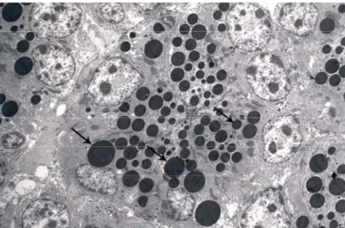
Figure 6: Proprial glands of the magnum, electron dense granules (arrows), x 2200.
Şekil 6: Magnum bezleri, elektron koyu granüller (oklar), x 2200.
Figure 7: Proprial glands of the uterus, electron light granules (arrows), x 11500.
Şekil 7: Uterus bezleri, elektron açık granüller (oklar), x 11500.
PAS-positive material did not exist in the secretory cells of the infundibulum, uterus, magnum or vagina. Staining with Best’s carmine gave a positive reaction in only the epithelial cells of the region of the isthmus.
The localization of neutral and acidic mucosubstance was determined using the PAS/Ab pH 2.5 combined staining technique. While epithelial cells in the region of the isthmus contained both acidic and neutral mucosubstance (Figure 3), the epithelium of the region of the magnum contained only acidic mucosubstance. No reaction occurred for any of the two stains in the vagina. In ducks which were in the quiescent phase of the reproductive cycle, the epithelium and glands were not fully developed and the reaction was very weak.
The performic acid/Ab pH 2.5 staining method produced a positive reaction in only the epithelium of the magnum in laying ducks, and neither the remaining regions in laying ducks nor the oviduct epithelium of ducks which were in the quiescent phase of the reproductive cycle stained.
Electron microscopic examination also revealed the lamina epithelialis to be composed of monolayered prismatic ciliated and secretory cells, microtubules to be arranged in the 9+2 formation in the transverse sections of the apically located cilia of ciliated cells, and short microvilli to be present in the apical region of secretory cells (Figure 4). A single type of secretion granule, of varying size, was observed to be present in secretory cells (Figure 5). The lamina propria was determined to be filled with glands in the regions of the magnum, isthmus and uterus. These glands were demonstrated to contain electron dense secretion granules in the magnum (Figure 6) and isthmus, and electron light secretion granules in the uterus (Figure 7)
Discussion and Conclusion
In previously conducted studies, the avian oviduct is reported to be segmented into five regions, and the lamina epithelialis is indicated to be composed of monolayered prismatic epithelium comprising ciliated and secretory cells (1,3,4,6,9). It is noted that the infundibulum and vagina do not contain proprial glands, whereas that the lamina propria of the magnum, isthmus and uterus is filled with glands (1,3,9,11). The findings obtained in the present study are in compliance with these reports.
Artan et al. (3) reported the PAS-positive reaction to produce lighter staining in the glands situated nearer to the lumen and darker staining in the more deeply localised glands of the magnum region in the hen. In the present study, PAS-positive reaction did not occur in the glands of the magnum region in the Pekin duck. In compliance with the reports of the above mentioned researchers, in the region of the isthmus, superficially located glands were determined to stain lighter and deeply located glands were demonstrated to stain darker.
Davidson et al. (8) and Özen (18) reported acidic mucopolysaccharides to be found in the regions of the magnum and vagina, and neutral mucopolysaccharides to be found in the isthmus in the hen. Findings obtained from the magnum and isthmus in the present study, are in compliance with the results of the indicated researchers. However, neutral and acidic mucopolysaccharides were determined not to exist in the vagina of the Pekin duck. In a study conducted by Fertuck and Newstead in the quail and hen (11), the properties of the proprial glands of the region of the magnum were determined to change with the phase of the reproductive cycle. In the present study, proprial glands were determined not to have developed in the region of the magnum in Pekin ducks which were in the quiescent phase of the reproductive cycle.
In the quail, three types of secretion granules, namely electron dense, electron light and medium
electron density are reported to exist in the proprial glands of the magnum (11). In the hen, proprial glands of the magnum (9) and isthmus (1) are reported to contain electron dense material, and the proprial glands of the uterus (1) are indicated to stain palely. In the present study, the proprial glands of the regions of the magnum and isthmus were determined to contain electron dense secretion granules, and the proprial glands of the uterus were demonstrated to contain electron light granules.
Balachandran et al. (6) reported secretory cells to contain secretion granules of medium electron density in the Pekin duck. Findings obtained in the present study are in compliance with the report of these researchers, for, a single type of secretion granule of medium electron density was demonstrated to be found in secretory cells.
In conclusion, the oviduct is divided into five regions in the Pekin duck, the infundibulum and vagina do not contain any glands, the remaining three regions are rich in proprial glands, and the epithelium is composed of ciliated and secretory cells. Light microscopic examination revealed the mucosubstance content of the proprial glands of the isthmus to be PAS-positive, and the epithelium of the magnum to be Ab-positive. Furthermore, electron microscopic examination revealed the presence of a single type of secretion granule, of medium electron density, in secretory cells.
References
1. Aitken RNL (1971): The Ovidukt. 1237-1352. In: DJ Bell, BM Freeman (Ed), Physiology and Biochemistry of the Domestic Fowl.Academic Pres Inc, London, Newyork. 2. Applegate TJ, Harper D, Lilburn MS (1998): Effect of
hen production age on egg composition and embryo development in commercial Pekin ducks. Poult Sci, 77,
1608-1612.
3. Artan ME, Dağlıoğlu S (1984): Tavuk, keklik ve
bıldırcında yumurta yolunun mikroskopik yapısı üzerinde karşılaştırmalı bir çalışma. İstanbul Univ Vet Fak Derg,
10, 17-28.
4. Bakst M, Howarth B (1975): SEM preparation and
observations of the hen’s oviduct. Anat Rec, 181, 211-226.
5. Bakst MR (1998): Structure of the avian oviduct with
emphasis on sperm storage in poultry. J Exp Zool, 282,
618-626.
6. Balachandran A, Bhatnagar MK, Geissinger HD (1985): Scanning and transmission electron microscopic
studies on the oviduct of Pekin ducks fed methyl mercury containing diets. Scanning Microsc, 1, 311-322.
7. Culling CFA, Allison RT, Barr WT (1985): Cellular
Pathology Technique. Fourth edition, Butterworth,
Wellington.
8. Davidson MF, Draper MH, Leonard EM (1968):
Structure and function of the oviduct of the laying hen. J
Physiolo, 196, 9-10.
9. Draper MH, Johnston HS (1968): The fine structure of
the oviduct of the laying hen. J Physiolo, 196, 7.
10. Eroschenko VP (1979): Morphological alterations in the
cells of the developing quail oviduct as influenced by estradiol-17b and the insecticide Kepone:I.observations by light and scanning electron microscopy. Biol Reprod, 21,
625-638.
11. Fertuck HC, Newstead JD (1970): Fine structural
observations on magnum mucosa in quail and hen oviduct.
Z Zellforsch, 103, 447-459.
12. Friedenbach DJ, Davison KL (1977): Scanning and
transmission electron microscopic changes associated with duck and chicken shell gland cilia after p,p’-DDT administration. Toxicol Appl Pharmacol, 40, 291-297.
13. Gilbert AB, Lake PE, Wood-Gush DG (1966): Aspects
of the physiology of the transport of the ovum through the oviduct of the domestic hen. 11-13. In: C. Horton-Smith,
EAmoroso (Ed), Physiology of the Domestic Fowl. Oliver&Body, Edinburg, London.
14. Gounon P, Laine MC,Sandoz D (1987): Cytokeratin
filament organization in the ciliated cells of the quail oviduct . Eur J Cell Biol, 44, 229-237.
15. Karnovsky MJ (1965): A formaldehyde-glutaraldehyde
fixative of high osmolality for use in electron microscopy. J
Cell Biol, 27, 137A-138A.
16. Lemullois M, Chantal Marty M (1990):
Immunocytochemical study of the formation of striated rootlets during ciliogenesis in quail oviduct. J Cell Sci, 95,
423-432.
17. Lemullois M, Klotz C, Sandoz D (1987):
Immunocytochemical localizations of myosin during ciliogenesis of quail oviduct. Eur J Cell Biol, 43, 429-437.
18. Özen A (2002): Tavuklarda ovidukt üzerinde ışık
mik-roskopik çalışmalar. Turk J Vet Anim Sci, 26, 1283-1288.
19. Sharma RK, Duda PL (1989): Histomorphological
changes in the oviduct of the mallard (Aves:Anatidae).
Acta Morphol Neerl Scand, 27, 183-192.
20. Solomon SE, Fryer JR, Baird T (1975): The
ultrastructural localization of calcium in the avian shell gland. J Microsc, 105, 215-222.
21. Veneable J H, Coggeshall R (1965): A simplified lead citrate stain for use in electron microscopy. J Cell Biol, 25, 407-408.
Geliş tarihi: 20.05.2008 / Kabul tarihi: 03.06.2008
Address for correspondence: Doç. Dr. Asuman Özen
Ankara University, Veterinary Faculty, Histology and Embryology Department 06110 Dışkapı-Ankara, Turkey. e-mail :[email protected]