KEY WORDS: alginate, flurbiprofen, insert, nanoparticle, nanostructured lipid carrier, ocular. * Author to whom correspondence should be addressed. E-mail: [email protected]
Lat. Am. J. Pharm. 35 (5): 972-9 (2016)
Revised version: January 6, 2016 Accepted: January 7, 2016
Novel Nanostructured Lipid Carrier Based Flurbiprofen
Loaded Sodium Alginate Inserts for Ocular Drug Delivery
Evren H. GÖKÇE 1& Neslihan ÜSTÜNDAG OKUR 2 1Department of Pharmaceutical Technology, Faculty of Pharmacy,
University of Ege, 35100 Bornova, Izmir, Turkey
2Department of Pharmaceutical Technology, School of Pharmacy, University of Istanbul Medipol, 34810 Beykoz, Istanbul, Turkey
SUMMARY. The aim of the present study was to develop a novel Flurbiprofen (FLB) loaded
nanostruc-tured lipid carrier (NLC) based alginate inserts for treatment of ocular inflammation. 0.3% FLB loaded NLCs were prepared by means of high shear homogenization and afterwards 0.75% sodium alginate was added into these NLCs. Glycerin or PEG 400 at 5% concentration was added to NLCs as plasticizers and by using solvent casting evaporation technique, inserts were developed. Inserts were evaluated for diame-ter, thickness, weight uniformity, drug content, moisture absorption and moisture loss. Also in vitro re-lease and stability studies were performed. The characterization properties of inserts were acceptable for ophthalmic application. The inserts developed with the addition of glycerin (Ins1FLB) were found as opti-mum formulation for FLB in vitro release. FLB loaded NLC based inserts developed with sodium alginate and glycerin may be offered as appropriate vehicles for ocular delivery.
RESUMEN. El objetivo del presente estudio fue desarrollar un nuevo vehículo lipídico nanoestructurado (NLC) de flurbiprofeno (FLB) a base de insertos de alginato para el tratamiento de la inflamación ocular. Se prepararon NLCs cargados con 0,3% de FLB por medio de homogeneización de alta cizalladura y después se agregó 0,75% de alginato de sodio dentro de estos NLCs. Se añadió glicerina o PEG 400 al 5% a los NLCs como plastificantes y se desarrollaron los insertos mediante el uso de la técnica de evaporación del disolvente. Los insertos fueron evaluados por diámetro, espesor, uniformidad de peso, contenido de fármaco, absorción y pérdida de humedad. También se realizaron estudios de liberación y estabilidad in vitro. Las propiedades de los insertos fueron acep-tables para aplicación oftálmica. Los insertos desarrollados con la adición de glicerol (Ins1FLB) resultaron la for-mulación óptima para la liberación in vitro de FLB. Los insertos basados en NLCs cargados de FLB desarrolla-das con alginato de sodio y glicerina pueden ser ofrecidos como vehículos apropiados para su aplicación ocular.
INTRODUCTION
Ocular drug delivery has been a main chal-lenge for researchers due to its inimitable struc-ture. Most of these challenges are anatomical and physiological barriers that normally protect the eye from detrimental effects. Furthermore, numerous pre-formulation and formulation fac-tors have to be considered while designing an ophthalmic formulation 1. In clinical applications
the anterior chapter of the eye can be treated with topical solutions as the most frequently used dosage formulation in ophthalmic treat-ment 2. Unfortunately the eye solutions (eye
drops) are quickly drained from the eye surface
and, thus, the time for drug absorption is only a few minutes and bioavailability is very low, typi-cally less than 5% 3,4. Concentration of drug
available in the pre-corneal area acts as a driv-ing force for its passive diffusion across cornea. Nonetheless, for effective ocular drug delivery with solutions, high corneal permeation with longer drug/cornea contact time is necessary 5.
In order to overcome anatomical and physio-logical barriers and improve ocular bioavailabili-ty, various conventional and novel drug delivery systems have been developed 5. Enhanced
re-tention (mucoadhesion) on the eye surfaces, sustained release, and permeation enhancement
can be considered as novel strategies that have been applied for designing of optimized oph-thalmic formulations 6. Formulations as
hydro-gels, micro- and nanoparticles, liposomes and collagen shields have been investigated for ocu-lar drug delivery 7,8. Nanotechnology based
sys-tems with an appropriate particle size can be designed to ensure low irritation, adequate bioavailability, and ocular tissue compatibility. Nanoparticles represent a promising candidate for ocular drug delivery because of small size leading to low irritation and sustained release property avoiding frequent administration. It has been shown that ophthalmic drug delivery may benefit from the characteristics of nanotechnolo-gy-based drug delivery systems especially solid lipid nanoparticles (SLN) and nanostructured lipid carriers (NLC) 9,10. However, like aqueous
solutions, nanoparticles may be eliminated rapidly from precorneal pocket 11. Moreover,
ad-ditional needs in this field are required to im-prove patient’s and doctor’s compliance 2.
Ocu-lar inserts are one of the new classes of drug delivery systems, which are gaining worldwide praise for their ability to release drugs at a pre-programmed rate for a longer period by increas-ing the pre-ocular residence time 12. In addition
each insert can be prepared to contain a definite dose which is fully retained at the administra-tion site, contrary to soluadministra-tions that can be im-properly instilled by the patient and are partially lost after application 13.
One of the most common conditions in oph-thalmic problems is the ocular inflammatory dis-ease affecting any part of the eye or the close tissues 14,15. Ocular inflammation is a common
side-effect associated with ophthalmic surgery, producing pain and photophobia in many pa-tients and potentially leading to serious compli-cations including increased intraocular pressure, posterior-capsule opacification, cystoid macular edema, and decreased visual acuity 16,17.
Nons-teroidal anti-inflammatory drugs are a heteroge-neous group of compounds with different struc-tural classes, which do not include a steroid nu-cleus, derived biosynthetically from cholesterol, in their chemical structure. Also known as COX inhibitors based on their mode of action, non-steroidal anti-inflammatory drugs are important modulators of ocular inflammatory reactions 18.
Flurbiprofen (FLB), a water insoluble drug (2.70 × 10⁻² mg/mL at 25 °C) with high lipophilicity
(log P = 4.24) 6, is currently used as a first
choice ophthalmic medication for the inhibition
of miosis induced during the course of cataract surgery 7,19. In addition it is a potent inhibitor of
prostaglandin synthesis 20.
The aim of this study was to develop novel FLB loaded NLC based alginate inserts for ocular application for the treatment of ocular inflam-mation and evaluate their potential for sustained ophthalmic delivery. The physicochemical char-acterization, in vitro release, sterility of these formulations was evaluated. In order to detect destabilization phenomena stability studies were also undertaken.
MATERIALS AND METHODS Materials
FLB was purchased from Sigma Aldrich (Madrid, Spain). Compritol HD5 ATO was a kind gift from Gattefosse (France). Tween 80 was obtained from Merck (Germany). Sodium alginate, oleic acid, glycerin, PEG 400 and sodi-um acetate were purchased from Sigma (Ger-many). High pressure liquid chromatography (HPLC) grade acetonitrile (Sigma, Germany) were used for ultra HPLC (U-HPLC). All the oth-er chemicals and solvents woth-ere of analytical or HPLC grade. Ultrapure water was obtained from Sartorius 61316 pro VF, Germany.
Preparation of FLB loaded NLC based inserts
Preparation of NLC
NLCs were prepared with high shear homog-enization technique 10. The aqueous phase
(Tween 80 and ultrapure water) was poured in-to the lipid phase (Compriin-tol and oleic acid) un-der homogenization at 24000 rpm for 5 min by Ultra Turrax (T25) at 70-80 °C. Subsequently, particles were dispersed in 10 mL ultrapure wa-ter at 4 °C. Unloaded NLC consisted of oleic acid (1.47 %), Compritol (0.73 %), Tween 80 (0.73 %) and ultrapure water (97.07 %). For FLB loaded NLC, FLB was added to lipid phase at a concentration of 0.3 % (w/v).
Preparation of inserts
NLC based inserts were prepared with a nov-el solvent casting evaporation method. Firstly, 60 mL NLC was prepared and then inserts were developed from these NLCs. To obtain inserts, sodium alginate (0.75 % w/v) was added into the prepared NLC formulation and mixed with 5% glycerin (Ins1) or 5% PEG 400 (Ins2) as plas-ticizers under stirring conditions at room tem-perature. After proper mixing the mixture was poured on a glass petri dish and allowed to evaporate at 40 °C for 36 h in an oven. The
ob-tained films were cut by a circular molder into circular pieces of definite size. The ocular in-serts were then stored in an airtight container (desiccator) under ambient condition.
NLC characterization
The average particle size, polydispersity in-dex (PDI) and zeta potential of NLCs were eval-uated by photon correlation spectrometry (Nano ZS, Malvern Instruments, U.K.). The particle size and PDI values were obtained by calculating the average of five measurements at an angle of 173° at 25 °C using disposable cells. The zeta potential was calculated from the electrophoret-ic mobility using the Helmholtz–Smoluchowski equation under an electrical field of 40 V/cm.
The encapsulation efficiency (EE) of FLB in NLC was determined by measuring the concen-tration of free drug in the dispersion medium, i.e. aqueous phase. The non-encapsulated FLB was separated by filtration/centrifugation. One mL of FLB loaded formulation was placed in the dialysis bags (Spectro/por Dialysis Membrane, molecular weight of 12-14 kDa) and placed in a centrifuge tube having 9 mL ultrapure water in-side and centrifuged for 1.5 h, at 55000 rpm (Beckman Coulter L100 XP–USA) at 25 ± 2 °C. The water phase was filtrated through a mem-brane filter (0.2 µm Nylon, Milipore Millex-GN). The sample was diluted and the amount of the FLB in the aqueous phase was estimated by a U-HPLC method.
The U-HPLC system consisted of a gradient pump, thermo-stable column department and a PDA detector (Thermo Scientific). The column was a Thermo Hypersil Gold C18 column (1.9 µm, 50 × 2.1 mm). The mobile phase consisted of 0.1 M sodium acetate solution (pH 6.3) and acetonitrile (70:30) (v/v). The flow rate was 0.3 mL/min and the column temperature was main-tained at 25 °C. The method was validated par-tially with respect to system suitability for linear-ity, limit of detection (LOD) and quantitation (LOQ), precision, accuracy and specificity, se-lectivity and stability at 248 nm.
Insert characterization
Inserts were evaluated for diameter, thick-ness uniformity, weight uniformity, drug con-tent, moisture absorption and moisture loss.
Diameter/thickness
The inserts have been shaped by the aid of a PVC mold (0.7 cm diameter) to apply into the lower eye lid. The diameters and thicknesses of
the inserts were measured with electronic vernier calipers (Mitutoyo – Japan) with a sensi-tivity of 0.01 mm. Ten measurements were car-ried out on each insert.
Mass uniformity
Uniformity of prepared inserts was detected by weighing individually of ten randomLy se-lected inserts from each formulation batch using a digital balance (Sartorius Basic, Germany).
Drug content
To evaluate FLB homogeneity of inserts, drug content studies were performed. Ten in-serts were grounded separately in a glass pestle mortar and dissolved in 100 mL acetic acid solu-tion (5 %) and mixed with a horizontal shaker (Schüftelfreguenz Kühner B, Germany) for 12 h. Then this solution was filtered through cellulose acetate membrane (0.45 µm). The drug concen-tration was determined by U-HPLC. The aver-ages of results were determined. Experiments were performed at 25 ± 2 °C.
Determination of moisture loss
The ocular inserts were weighed accurately and kept in desiccators containing anhydrous calcium chloride. After 3 days, the inserts were taken out and weighed again 21. The percentage
moisture loss of inserts was calculated according to Eq. [1]
Determination of moisture absorption
The ocular inserts were weighed accurately and kept in stability cabinets at 40 °C with rela-tive humidity of 75 %. After 3 days, the inserts were taken out and weighed again 22. The
per-centage moisture absorption of inserts was cal-culated according to Eq. [2]
[1]
[2] Surface imaging
To visualize the surface structure, the surface images of FLB loaded inserts were taken with stereomicroscope (Leica M205C, UK) and were photographed digitally.
In vitrorelease studies
In vitro drug release experiments were per-formed for 8 h using the vial method 23. One
in-sert was placed in 6.5 mL receptor media. The system was held at 32 ± 0.5 °C to mimic condi-tions of eye surface, stirred continuously with magnetic stirrer at 150 rpm. The samples were
taken at every hour during 8 h. FLB was deter-mined by the validated U-HPLC method at 248 nm. Sink conditions were maintained in the re-ceptor compartment during in vitro release stud-ies. To maintain sink conditions of the receptor compartment pH 7.4 PBS/ethanol 70/30 (v/v) was used as receptor phase. The experiment was carried out six times.
Sterilization and evaluation of sterility Inserts were sterilized under a UV lamp (273.7 nm) (Philips TUV 15w/G15t8 uv, Hol-land) for 24 h. To find out the presence of vi-able forms of microorganisms in the preparation step sterility of inserts was checked.
To control the sterility of inserts, they were disintegrated in the brain heart infusion fluid and 100 µL of each formulation was inoculated on eosin methylene blue agar medium and blood agar medium. 100 colony forming units of E. coli suspension was prepared by using stan-dard culture of E. coli (ATCC 8739). Then this suspension was seeded on the same agar medi-um plates. All plates were incubated for 48 h at 37 ± 0.5 °C. After 48 h the media were exam-ined and the results were evaluated by bacteria colony counting.
Stability
Stability studies were performed for inserts according to the International Conference on Harmonization (ICH) guidelines. FLB loaded in-serts were stored at 4 ± 1 °C in the refrigerator and 25 ± 2 °C (relative humidity 60 %) and 40 ± 2 °C (relative humidity 75 %) for 3 months in the stability cabinets (Nüve ID 300, Turkey). FLB content, physical appearance and sterility were evaluated for inserts. The experiments were repeated five times.
Statistical evaluation
Statistical differences were determined using ANOVA followed by Tukey’s test for
compar-isons between groups. The significance level was taken as 95 % (P < 0.05).
RESULTS AND DISCUSSION
Nanoparticles have the ability to deliver ocu-lar drugs to specific target sites and hold poten-tial to revolutionize the treatment of various eye diseases. Especially NLC carrier systems exhibit an excellent tolerability for ocular application 24.
Nevertheless, efforts are still necessary in terms of improving the drug delivery efficiency, espe-cially, for prolonging the retention time of drug on the corneal surface. Therefore in current study, inserts were developed from NLC system to improve patient compliance with an en-hanced therapeutic effect of FLB.
Formulation and characterization studies In this study, compritol and oleic acid were selected as the solid and liquid lipid for NLC preparation that FLB has better solubilization. It was also noted that these liquid and solid lipids were suitable for ophthalmic use 25,26. In
addi-tion the produced NLC system was modified with sodium alginate which was coded as N2 (Table 1).
Sodium alginate, a water soluble biodegrad-able polymer, is commonly used for ocular ap-plications 27-29. NLCs were evaluated about
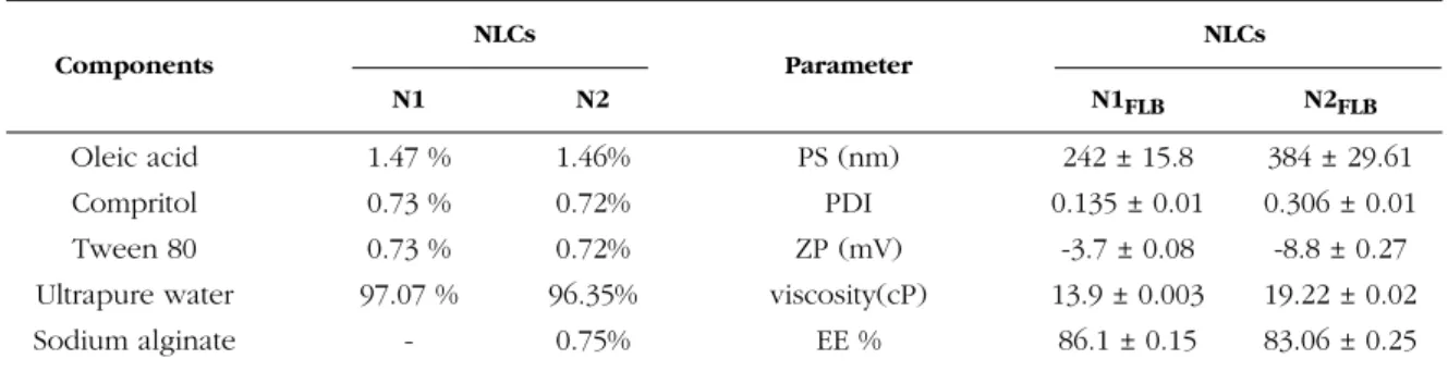
parti-cle size and PDI values which points out the homogeneity of particles. The characterization of FLB loaded NLCs (N1FLBand N2FLB) are given in Table 1. When N1 was modified with sodium alginate, particle size was increased 59 % and PDI was changed from 0.135 to 0.306. This might be due to increment of viscosity of the nanoparticles. When 0.75 % (w/v) sodium algi-nate which is a gel forming agent was added to the N1 formulation, the viscosity of the formula-tion showed an increment as 5.3 cP. Zeta potential values of nanoparticles found 3.7 mV and -8.8 mV. Similarly Jitendra et al. reported that chloramphenicol loaded nanoparticles
formulat-NLCs NLCs Components Parameter N1 N2 N1FLB N2FLB Oleic acid 1.47 % 1.46% PS (nm) 242 ± 15.8 384 ± 29.61 Compritol 0.73 % 0.72% PDI 0.135 ± 0.01 0.306 ± 0.01 Tween 80 0.73 % 0.72% ZP (mV) -3.7 ± 0.08 -8.8 ± 0.27
Ultrapure water 97.07 % 96.35% viscosity(cP) 13.9 ± 0.003 19.22 ± 0.02
Sodium alginate - 0.75% EE % 86.1 ± 0.15 83.06 ± 0.25
ed using sodium alginate showed the negative surface charge of -2 mV 30.
Encapsulation efficiencies of FLB loaded NLCs were determined as 83-86 %. For particle homogeneity of NLCs PDI values were checked and these values were determined smaller than 0.3 with narrow size distribution (Table 1).
There has been a growing interest in using bioadhesive inserts in the ocular cul de sac for enhanced or prolonged localized drug delivery
29. In our previous study NLC based ocular
in-serts were prepared successfully by solvent cast-ing technique 31, therefore this production
method was used in this study. To obtain in-serts, sodium alginate (0.75%) was added into this N1 formulation and glycerin (Ins1) or PEG 400 (Ins2) was added at the concentrations of 5% (w/v) as plasticizers under stirring condi-tions. Then the mixture was poured on a glass petri dish (Fig, 1A) and allowed to evaporate at 40 °C for 36 h. The mixture was dried up to the point where the insert weight was stable. FLB dosage was fixed at 0.3% (w/v).
Sodium alginate has been reported as mu-coadhesive, biodegradable, biocompatible poly-mer and has potential for nupoly-merous pharmaceu-tical and biomedical applications 32. This
poly-mer is used in a variety of ocular pharmaceuti-cal formulations 32-34. A novel ophthalmic gel
Figure 1. Photographs of inserts before drying in a Petri dish (A) and after casting, shaped with a PVC mold (B).
Inserts Parameter
Ins1 Ins2 Ins1FLB Ins2FLB
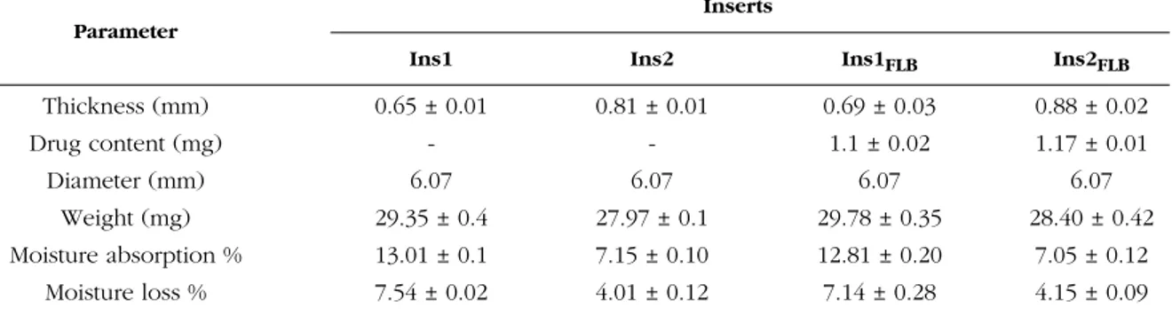
Thickness (mm) 0.65 ± 0.01 0.81 ± 0.01 0.69 ± 0.03 0.88 ± 0.02 Drug content (mg) - - 1.1 ± 0.02 1.17 ± 0.01 Diameter (mm) 6.07 6.07 6.07 6.07 Weight (mg) 29.35 ± 0.4 27.97 ± 0.1 29.78 ± 0.35 28.40 ± 0.42 Moisture absorption % 13.01 ± 0.1 7.15 ± 0.10 12.81 ± 0.20 7.05 ± 0.12 Moisture loss % 7.54 ± 0.02 4.01 ± 0.12 7.14 ± 0.28 4.15 ± 0.09
Table 2. Characterization parameters of developed inserts.
has been developed which is converted to gel in the presence of divalent cations (i.e. calcium ion) which are present in the lachrymal fluid 35.
Moreover alginate which has mucoadhesive property is not easily eroded by tear fluid as it transforms into stable gel upon exposure to di-valent cations 36. Sodium alginate has
advan-tages for ocular drug delivery; however algi-nates are not sufficient enough to increase the retention time of the drug on the precorneal area. Thus a lot of researches used other poly-mers to combine with alginates to overcome this problem. Bhalerao et al. 37formulated an in situ
gelling ophthalmic drug delivery system for the treatment of glaucoma. Sodium alginate in com-bination with hydroxypropyl cellulose (HPC) was used as gelling agent, which also acted as viscosity-enhancer. Other researchers developed a novel in situ gum-based ophthalmic drug de-livery system of linezolid. Hydroxypropyl guar and xanthan were used as gum with the combi-nation of hydroxyethyl cellulose, Carbopol, and sodium alginate as viscosity-enhancing agents 38.
Zhu et al. 39 prepared sodium alginate and
chi-tosan coated nanoparticles for ocular drug deliv-ery. Instead of using another polymer we have decided to make inserts including NLCs inside.
After inserts were prepared and dried at 40 °C for 36 h, they had uniform image and it had simply removable property from the petri dish-es. The inserts are shown in Figure 1b and after being shaped the diameters of the inserts was found as 6.07 mm (Table 2). Thickness of the inserts was found varing from 0.65 ± 0.01 to 0.88 ± 0.02 mm. Ocusert® is commercially drug system by ALZA, has 0.3 mm thickness value and its dimensions are 5.7 × 13.4 mm on its ax-es. The other commercial formulation is Ocu-fit®, its length is 25-30 mm and diameter is 1.9 mm 40. In this study, prepared ocular inserts
have appropriate thicknesses and diameters when compared to the commercial formulations
on the drug market. The developed inserts showed appropriate thickness values with low standard deviation.
To evaluate the suitability of preparation technique of inserts, the drug content of inserts is a very important parameter. It has been found that the insert preparation technique is appro-priate for developing uniform inserts containing FLB (Ins1FLB and Ins2FLB), since FLB content in each insert was found almost the same without any loss (Table 2).
Percentage moisture tests were carried out to check the physical stability of the insert. To evaluate hygroscopicity of developed inserts, they were weighed and retained in desiccators for 3 days. The moisture of the insert was found between 4-7.5 % at the end of the 3rd day. The
detected moisture is necessary for inserts be-cause very dry property will affect flexibility and elasticity of the insert. Ins1FLB which contains glycerin, preserved more moisture than Ins2FLB which contains PEG 400. To find out moisture absorption of the ocular inserts, they were weighed precisely and retained in a cabinet with relative humidity of 75% for 3 days. At the end of the 3 days, Ins1FLB absorbed more moisture than Ins2FLB. These results were thought to be due to the moisturizing properties of glycerin 41.
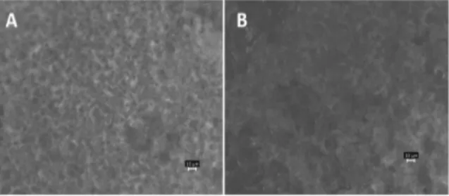
Stereomicroscope was used in order to bet-ter recognize the surface property of inserts. Pic-tures showed the homogeneous appearance of Ins1FLB and Ins2FLB (Fig. 2). Surface homoge-nization is an important factor and it affects the release of drug as a result of erosion and diffu-sion. Intended controlled release might not be feasible if developed inserts have gaps or cracks.
Figure 2. Suface images of Ins1FLB (A) and Ins2FLB (B) after taken with stereomicroscope (1 × 16).
Figure 3. Percentage cumulative release profile of Ins1FLB and Ins2FLB in PBS. Values are means of six experiments ± SD.
the insert surface thus drug is released for longer time 42. FLB release from Ins1
FLB and Ins2FLB is shown at Fig. 3. Ins2FLB showed slightly faster release at the first 1.5 h which was not different than Ins1FLBsignificantly (p > 0.05). At the end of the 4.5 h, in vitro FLB release from Ins1FLB and Ins2FLB was found as 81% and 100%, respectively (p < 0.05). At the end of the 6.5 h, in vitro FLB release from Ins1FLB was found as 100 %.
Aburahma et al. 33 prepared brimonidine
loaded inserts, which consisted of 1.5% sodium alginate, 7% polyvinylpyrrolidone K-90 and 5% propylene glycol and the drug release was de-termined as 80% at 2 h. However for Ins1FLB 80% release was observed at 4th h. The
combi-nation of NLC and alginate inserts sustained the drug release almost 2 times than Ins1 insert. This remarkable result can be an advantage for the controlled release of FLB for ophthalmic ap-plications. Ins1FLB is ideal formulation for in-flammation of the eye if both first fast- acting and then slow-release was desired.
Evaluation of sterility
The final ophthalmic products must be pro-duced under validated conditions in terms of providing sterility. It is important to sterilize the formulations for ophthalmic applications 31,43.
UV radiation was used for sterilizing the devel-oped ocular inserts. The test for sterility is in-tended for detecting the presence of viable forms of bacteria, fungi and yeast in sterilized preparations. Eosin metilen blue agar medium and blood agar medium were used to control the suitability of sterilization procedure. There was no appearance of turbidity and hence no evidence of microbial growth; when all
formula-In-vitrorelease studies
There are two stage of drug release for degradable inserts: 1) Fast release: tear fluid en-ters into the formulation thus drug is released; and 2) Slow release: a gel system is occurred on
tions were incubated for 48 h at 37 °C. The preparations examined; therefore, passed the test for sterility. It can be concluded that the prepared ocular inserts were detected sterile be-cause there is no microorganism growth detect-ed while the microbial growth was observdetect-ed in positive control.
Stability
The stability studies of FLB loaded inserts were performed at 4 ± 1 °C, 25 ± 2 °C at 60% relative humidity and 40 ± 2 °C at 75% relative humidity for 3 months. The accelerated stability studies were carried out in accordance with the ICH guidelines. FLB content, sterility and physi-cal characteristics of inserts did not show any significant change for 3 months. These results concluded that developed ocular inserts were physiochemically and microbiologically stable. Compared to solutions, higher stability obtained with inserts is advantageous for drug industry 43.
Nayak et al. 44 developed ophthalmic insert of
moxifloxacin and evaluated stability of drug content. They found that the inserts were stable for 12 weeks. Since inserts are solid or semisolid dosage forms they do not show the disadvan-tages of traditional ophthalmic liquid dosage forms.
CONCLUSION
FLB (anti-inflammatory agent) was success-fully formulated as NLC and NLC based alginate insert. This study offers a new preparation tech-nique to the ocular inserts by using a combina-tion of NLC and natural polymer sodium algi-nate. The formulation of FLB as NLC or insert for ophthalmic delivery is achievable. To de-crease systemic side effects and inde-crease effec-tive drug concentration in the eye, NLC based ophthalmic inserts could be an alternative ap-proach for ocular drug delivery. The developed NLC based alginate inserts may be useful in clin-ical practice to maintain mydriasis during cataract or other eye surgical treatments.
Conflict of interest. The authors declare no conflict of interest.
Acknowledgements. This study was supported by Ege University Scientific Research Projects No: 13/ECZ/029. The authors would like to thank to Ege University, Faculty of Pharmacy, Department of Mi-crobiology and Pharmaceutical Sciences Research Center (FABAL) for U-HPLC studies. The authors would like to thank to İsmail Öztürk from Ege Uni-versity, Faculty of Pharmacy, Department of Microbi-ology for the assistance at sterility experiments.
REFERENCES
1. Gaudana, R., H.K. Ananthula, A. Parenky &
A.K. Mitra (2010) AAPS Journal 12: 348-60.
2. Kuno, N. & S. Fujii (2011) Polymers 3:
193-221.
3. Maurice, D.M. & S. Mishima (1984) Ocular
pharmacokinetics’, in: Handbook of experimen-tal pharmacology, (M.L. Sears ed.), 69,
Springer Verlag, Berlin-Heidelberg, pp. 16-119.
4. Urtti, A (2006) Adv. Drug Deliv. Revi. 58:
1131–5.
5. Patel, A., K. Cholkar, V. Agrahari & A.K. Mitra
(2013) World J. Pharmacol. 9: 47-64.
6. Quinteros, D.A., L.I. Tártara, S.D. Palma, R.H. Manzo & D.A. Allemandi (2014) J. Pharm. Sci.
103: 3859-68.
7. Pignatello, R., C. Bucolo, G. Spedalieri, A.
Maltese & G. Puglisi (2002) Biomaterials 23:
3247-55.
8. Le Bourlais, C., L. Acar , H. Zia, P.A. Sado, T. Needham & R. Leverge (1998) Progr. Retinal.
Eye Res. 17: 33-58.
9. Gökçe, E.H., G. Sandri, M.C. Bonferoni, S. Rossi, F. Ferrari, T. Guneri, et al. (2008) Int. J.
Pharm. 364: 76-86.
10. Üstündağ Okur, N., E.H. Gökçe, D. İnci Bozbıyık, S. Eğrilmez, Ö. Özer & G. Ertan
(2014) Eur. J. Pharm. Sci. 63: 204-15.
11. Bu, H.Z., H.J. Gukasyan, L. Goulet, X.J. Lou, C. Xiang & T. Koudriakova (2007) Curr. Drug
Metab. 8: 91-107.
12. Rao, M.P.C., M. Nappinnai, S. Raju, V.U. Ma-heshwara Rao & B. Venkateshwara Reddy
(2010) J. Pharm. Sci. Res. 11: 693-9.
13. Karthikeyan, D., M. Bhowmick, V.P. Pandey, J. Nandhakumar, S. Sengottuvelu, S. Sonkar, et al.
(2008) Asian J. Pharm. 2: 192-200.
14. Cronau, H., R.R. Kankanala & T. Mauger
(2010) Am. Fam. Physician 81: 137-44.
15. González-Mira, E., M.A. Egea, M.L. García &
E.B. Souto (2010) Colloids Surf. B. 81: 412-21.
16. Srinivasan, B.D. & P.S. Kulkarni (1989) Prog.
Clin. Biol. Res. 312: 229-49.
17. Araújo, J., E.González, M.A. Egea, M.L. García
& E.B. Souto (2009) Nanomed. Nanotechnol. 5:
394-401.
18. Bhattacherjee, P (1989) Prog. Clin. Biol. Res.
312: 211-27.
19. Shaikh, M.Y., J.S. Mars & C.J. Heaven (2003)
979
20. Kumbhat, S (1994) J. Pharmaceut. Biomed. 12:
131-3.
21. Upadhyaya, N., A. Patidar, S. Agrawal & D.
Gupta (2011) Res. J. Pharm. Biol. Chem. Sci. 2:
411-20.
22. Gassan, J. & A.K. Bledzki (1997) Polym.
Com-posite 18: 179-84.
23. Dandagi, P.M., F.V. Manvi, M.B. Patil, V.S. Mastiholimath & R. Rathod (2004) Indian J.
Pharm. Sci. 66: 309-12.
24. Luo, Q., J. Zhaoa, X. Zhang & W. Pan (2011)
Int. J. Pharm. 403: 185-91.
25. González-Mira, E., S. Nikolic, A.C. Calpena, M.A. Egea, E.B. Souto & M.L. García (2012) J.
Pharm. Sci. 101: 707-25.
26. Üstündağ Okur, N., E.H. Gökçe, S. Eğrilmez, Ö. Özer & G. Ertan (2014) J. Ocul. Pharmacol.
Ther. 30: 319-32.
27. Cohen, S., E. Lobel, A. Trevgoda & Y. Peled
(1997) J. Control. Release 44: 201-8.
28. Séchoy, O., G. Tissié, C. Sébastian, F. Maurin, J.-Y. Driot & C. Trinquand (2000) Int. J.
Pharm. 207: 109-16.
29. Gilhotra, R.M., K. Nagpal & D.N. Mishra
(2011) Int. J. Pharm. Investig. 1: 22-8.
30. Gupta, J., L. Prabakaran, R. Gupta & M.
Govind (2011) Int. J. Pharm. Pharm. Sci. 3:
66-70.
31. Üstündağ Okur, N., E. Homan Gökçe, D. İnci Bozbıyık, S. Eğrilmez, G. Ertan & Ö. Özer
(2015) Expert Opin. Drug Deliv.12: 1-17.
32. Shafie, M.A.A. & H.H.M. Fayek (2013) J. Clin.
Exp. Ophthalmol. 4: 1-11.
33. Aburahma, M.H. & A.A. Mahmoud (2011)
AAPS Pharm. Sci. Tech. 12: 1335-47.
34. Kumar, J.R. & S. Muralidharan (2012) J.
Pharm. Sci. Res. 4: 1973-7.
35. Mandal, S., M.K.M.J. Thimmasetty, G.L. Prab-hushankar & M.S. Geetha (2012) Int. J. Pharm.
Investig. 2: 78-82.
36. Séchoy, O., G. Tissié, C. Sébastian, F. Maurin, J.Y. Driot & C. Trinquand (2000) Int. J. Pharm.
207: 109-16.
37. Bhalerao, A.V. & S.S. Singh (2011) Int. J.
Pharma. Biosci. 2: 7-14.
38. Hiremath, S.S., F.S. Dasankoppa, A. Nadaf, V.G. Jamakandi, J.S. Mulla, S.A. Sreenivas, et
al. (2008) Sci. Pharm. 76: 515-32.
39. Zhu, X., M. Su, S. Tang, L. Wang, X. Liang, F.
Meng, et al. (2012) Mol. Vision. 18: 1973-82.
40. Gavaskar, B (2010) Int. J. Pharm. Pharma. Sci.
2: 152-65.
41. Shinde, A.J., K.C. Garala & H.N. More (2008)
Asian J. Pharm. 2: 265-9.
42. Aldrich, D.S., C.M. Bach, W. Brown, W. Chambers, J. Fleitman, D. Hunt, et al. (2013)
USP Expert Panel Stimuli to the Revision Pro-cess 39(5): 1-21. Available at <http://www.usp. org/sites/default/files/usp_pdf/EN/meetings/wor kshops/ophthalmicpreparations.pdf>.
43. Missel, P.J., J.C. Lang, D.P. Rodeheaver, R. Jani, M.A. Chowhan & T. Dagnon (2009)
De-sign and evaluation of ophthalmic pharmaceuti-cal products, in: Modern pharmaceutics appli-cations and advances (A.T. Florence & J.
Siep-mann, eds.), New York: Informa Healthcare, p. 142.
44. Nayak, B.S., S.C. Patnaik, S. Sethy, P. Ellaiah