Biocompatible Electroactive Tetra(aniline)-Conjugated Peptide
Nano
fibers for Neural Differentiation
Idil Arioz,
†,#Ozlem Erol,
‡,§,#Gokhan Bakan,
‡,∥F. Begum Dikecoglu,
‡Ahmet E. Topal,
‡Mustafa Urel,
‡Aykutlu Dana,
‡Ayse B. Tekinay,
*
,†,‡and Mustafa O. Guler
*
,⊥†Neuroscience Program and‡Institute of Materials Science and Nanotechnology, National Nanotechnology Research Center
(UNAM), Bilkent University, Ankara 06800, Turkey
§School of Chemistry, University of Bristol, Bristol BS8 1TS, U.K.
∥Department of Electrical and Electronics Engineering, Atilim University, Ankara 06836, Turkey ⊥Institute for Molecular Engineering, University of Chicago, Chicago, Illinois 60637, United States
*
S Supporting InformationABSTRACT: Peripheral nerve injuries cause devastating problems for the quality of patients’ lives, and regeneration following damage to the peripheral nervous system is limited depending on the degree of the damage. Use of nanobiomaterials can provide therapeutic approaches for the treatment of peripheral nerve injuries. Electro-active biomaterials, in particular, can provide a promising cure for the regeneration of nerve defects. Here, a supramolecular electroactive nanosystem with tetra(aniline) (TA)-containing peptide nanofibers was developed and utilized for nerve regeneration. Self-assembled TA-conjugated peptide nanofibers demonstrated electroactive behavior. The electroactive self-assembled peptide nanofibers formed a well-defined three-dimensional nanofiber network mimicking the extrac-ellular matrix of the neuronal cells. Neurite outgrowth was improved on the electroactive TA nanofiber gels. The neural differentiation of
PC-12 cells was more advanced on electroactive peptide nanofiber gels, and these biomaterials are promising for further use in therapeutic neural regeneration applications.
KEYWORDS: oligo(aniline), tetra(aniline), peptide amphiphiles, self-assembly, electroactive nanofibers, neural regeneration
■
INTRODUCTIONNervous system injuries and damage cause detrimental consequences for the quality of patients’ lives due to the fact that the nervous system has a much lower regenerative potential compared to that of other tissues. In the peripheral nerves, nerve damage causes loss of connection between the axon bundle and the target tissues. Immediately after damage to the nerves occurs, certain degenerative pathways are induced, which can be overcome by regenerative therapies designed to support neuronal differentiation.1Nerve tissue engineering is a promising strategy to promote regeneration, where biomaterials can be used to accelerate healing of the tissue.2 In particular, electroactive biomaterials have attracted attention for neuronal engineering and regeneration applications,3and electroactive π-conjugated polymers such as polythiophene, polypyrrole, and polyaniline have been used to promote the growth, survival, and differentiation of neuronal cells.4The major drawbacks of π-conjugated polymers in biological applications are the issues of poor biodegradability, low water solubility, and poor processability and flexibility.4,5 Strategies such as preparing their composites or copolymers with biocompatible polymers or immobilizing cell adhesive peptide sequences onto the
polymer backbone can make them more convenient for tissue engineering applications.6 Alternatively, oligomers provide the
opportunity to design and produce defined and
well-characterized materials with defined functionality and proper-ties due to their excellent solubility in common solvents and easier control of their chemical structures.
Supramolecular self-assembly of molecular building blocks allows the “bottom-up” fabrication of nanomaterials with various functionality and morphology. Compared to traditional covalent polymers, supramolecular self-assembled materials show dynamic and reversible transitions.7 The process of self-assembly is based on spontaneous diffusion and specific interactions among molecules governed by noncovalent bonds, including electrostatic, hydrophobic, van der Waals, and metal−ligand interactions, hydrogen bonds, and π−π stacking.8Although the noncovalent interactions, which are the tools for the self-assembly process, are weaker than the covalent
Received: October 30, 2017 Accepted: December 12, 2017 Published: December 12, 2017
Research Article www.acsami.org Cite This:ACS Appl. Mater. Interfaces 2018, 10, 308−317
Downloaded via BILKENT UNIV on February 27, 2019 at 05:41:24 (UTC).
bonds, highly organized and robust structures can be formed from cooperative noncovalent interactions.9
Peptide amphiphile (PA) molecules can be used for development of novel materials due to their molecular self-assembly ability. The PA molecules consist of hydrophilic and β-sheet forming amino acids, which are covalently conjugated to a hydrophobic alkyl tail, triggering self-assembly of the molecules into one-dimensional (1D) nanostructures through hydrogen bonding under physiological conditions.10−12 Nano-fibers of PA molecules can also form three-dimensional (3D) networks through encapsulation of water molecules. Because of their intrinsic biocompatibility, biodegradability, biofunction-ality, rational design, and rich functional groups, short-sequence PAs have been comprehensively utilized in tissue engineering, regenerative medicine, nanofabrication, biomineralization,10 and electronic devices.13For electronics applications, coupling peptides to electroactive units may enable the bottom-up fabrication of self-assembled conductive materials.14
Recently, the design, synthesis, and use of electroactive oligomeric molecules with precise control over the type and number of electroactive units have been an important target.15,16It is interesting to combine electroactive oligomers with nonelectroactive species such as simple alkyl blocks, oligopeptides, and other biological building blocks, inorganic materials, and other structure-directing blocks or functional units.17 This approach can provide striking features such as mechanical and processing properties, functionality, and supramolecular structure formation. Aniline oligomers have been utilized due to their excellent solubility in common solvents and easier control over their chemical structures and oxidation states compared to those of polyaniline.18,19 In addition, their electrical, chemical, and optical properties are comparable to those of polyaniline, making them and their derivatives an attractive class of materials for further applications in anticorrosion coatings,20,21 sensors,22,23 drug delivery,24,25 and tissue engineering.26 On the other hand, fabricating water-soluble electroactive materials present
consid-erable benefits and improved processability compared to that using organic solvent-soluble materials, and intermolecularπ−π interactions facilitate the self-assembly of these molecules in water. To date, a number of oligoaniline-based materials have been investigated in muscle27,28 and bone29 regeneration studies as well as in nerve tissue engineering, including using aniline pentamers30,31 and aniline trimers32 for neural regeneration.
In this work, conductive and electroactive supramolecular nanosystems have been developed by incorporation of the tetra(aniline) (TA) unit as an electroactive unit in self-assembling peptide systems. These materials allow facile processibility of electroactive supramolecular nanofibers for neural cell regeneration by providing electrical, mechanical, and nanotopographical cues for the cells, and the TA-conjugated peptide nanofibers show promising results for neural differ-entiation.
■
EXPERIMENTAL SECTIONMaterials. N-Phenyl-1,4-phenylenediamine, ammonium persulfate (APS), succinic anhydride, 4-(2′,4′-dimethoxyphenyl-Fmoc-amino-methyl)-phenoxyacetamido-norleucyl-MBHA resin (Rink amide MBHA resin), all protected amino acids, lauric acid, N,N,N′,N′-tetramethyl-O-(1H-benzotriazol-1-yl)uronium hexafluorophosphate (HBTU), N,N-diisopropylethylamine (DIEA), and trifluoroacetic acid (TFA) were purchased from Nova-Biochem, Merck, or Sigma-Aldrich. All other chemicals and materials used in this study were purchased from Sigma-Aldrich and used as received.
Synthesis of Tetra(aniline). TA was synthesized according to the literature.33 N-Phenyl-1,4-phenylenediamine (2.5 × 10⁻³ mol) was dissolved in a mixed solution of acetone (50 mL), deionized water (50 mL), and concentrated hydrochloric acid (HCl) (12.5 mL) and stirred in an ice bath to cool down the temperature to 0°C for 30 min. A solution of APS (2.5× 10⁻³mol in 12.5 mL of deionized water) was added dropwise to the reaction system over 30 min under vigorous stirring. After the addition of APS, the resulting solution was stirred for another 3 h at 0°C. The dark green crude product was centrifuged and washed with 0.6 M HCl solution and acetone at least three times, Figure 1. Chemical structures of nonelectroactive (a) lauryl-VVAGEE-Am (E2) and (b) lauryl-VVAGKK-Am (K2) and electroactive (c) TA-VVAGEE-Am (E2-TA) and (d) TA-VVAGKK-Am (K2-TA) peptide amphiphiles. Scanning electron microscopy (SEM) images of (e) E2/K2and (f) E2-TA/K2-TA gels reveal a mesh-like structure. Transmission electron microscopy (TEM) images of (g) E2/K2and (h) E2-TA/K2-TA gels show a nanofibrous structure.
and TA in emeraldine salt state (TA-ES) was obtained. Then, it was dedoped with 0.1 M NH4OH solution for 1.5 h and washed with deionized water until pH reached 7 to get TA in emeraldine base state (TA-EB). After drying at 40°C under vacuum for 24 h, blue-purple TA-EB was obtained. The molecular weight of TA-EB was measured by mass spectrometry to be 365.2 (MH+/e) (Figure S1).
Synthesis of Peptide Amphiphile Molecules. A solid-phase peptide synthesis method was used for the synthesis of non-electroactive oppositely charged lauryl-VVAGEE-Am (E2,Figure 1a) and lauryl-VVAGKK-Am (K2,Figure 1b) and electroactive oppositely charged carboxyl-capped tetra(aniline)-VVAGEE-Am (E2-TA, Figure
1c) and carboxyl-capped tetra(aniline)-VVAGKK-Am (K2-TA,Figure
1d). Peptides were constructed on Fmoc-Rink Amide Resin. Amino acid couplings were performed with 2 equiv of fluorenylmethylox-ycarbonyl (Fmoc)-protected amino acid, 1.95 equiv of HBTU, and 3 equiv of DIEA for at least 4 h. Fmoc removals were performed with 20% piperidine/dimethylformamide (DMF) solution for 20 min. For the synthesis of E2and K2, lauric acid was used as thefinal sequence. For E2-TA and K2-TA, before the TA coupling, the free amino group of the valine was conjugated with the linker by reaction with succinic anhydride (2 equiv) overnight, and free carboxylic acid groups were formed. Then, 2 equiv of TA, 1.95 equiv HBTU, and 3 equiv of DIEA were added as the last coupling. Cleavage of the peptides from the resin was carried out with a mixture of TFA/triisopropylsilane/water in a volume ratio of 95:2.5:2.5 for 2 h. Then, the remaining solution was collected, and the resin was washed with dichloromethane (DCM). Excess TFA and DCM were removed by rotary evaporation. The remaining mixture was triturated with cold ether and precipitate was separated from the ether by centrifugation; the precipitate was dissolved in water, and finally, freeze dried. The PAs were further purified by preparative high-performance liquid chromatography (prep-HPLC). After prep-HPLC treatment and freeze-drying, white-colored E2 and K2, violet-colored E2-TA, and green-colored K2-TA were obtained.
The identity and purity of the PAs were assessed by liquid chromatography−mass spectrometry (Agilent Technologies 6530 Accurate-Mass QTOF system equipped with a Zorbax Extend-C18 column) according to the optical density at 220 nm. A gradient of water (0.1% NH4OH or 0.1% formic acid) and acetonitrile (0.1% NH4OH or 0.1% formic acid) was used as the mobile phase. To purify the peptides, an Agilent preparative reverse-phase HPLC system equipped with a Zorbax Extend-C18 21.2 mm× 150 mm column for basic conditions and a Zorbax SB-C8 21.2 mm× 150 mm column for acidic conditions was used and a gradient of water (0.1% NH4OH or 0.1% TFA) and acetonitrile (0.1% NH4OH or 0.1% TFA) was used as the mobile phase. Positively-charged PAs were treated with 1 mM HCl solution and lyophilized to remove residual TFA. The sample preparation method is explained in detail for each characterization technique.
Fourier Transform Infrared (FTIR) Measurements. Dried samples were mixed with KBr having a KBr/sample ratio of 100:1 (w/w) to obtain a homogeneous mixture and pressed to form transparent pellets. The absorbance of the samples was collected with a Bruker VERTEX 70 FTIR Spectrometer in the 4000−400 cm−1 wavenumber range.
Transmission Electron Microscopy (TEM) Imaging. TEM images of the E2/K2and E2-TA/K2-TA nanofibers were obtained using an FEI Tecnai G2 F30 TEM at 300 kV, and negative staining was performed using 2% (w/v) uranyl acetate.
Scanning Electron Microscopy (SEM) Imaging. For SEM imaging, samples were prepared on a Si-wafer surface by mixing oppositely charged PA solutions. E2 and K2, and E2-TA and K2-TA solutions (13 mM) were mixed at a 1:1 ratio. Fifteen minutes after gel formation, ethanol exchange was carried out and then gels were dried by using a critical point dryer (Tousimis Autosamdri-815B). After the dried samples were coated with 10 nm of Au/Pd, SEM images were taken by using an FEI Quanta 200 FEG scanning electron microscope. Secondary Structure Analysis. The circular dichroism (CD) spectra of E2/K2 and E2-TA/K2-TA solutions and their oppositely charged mixtures at a 1:1 molar ratio, having a concentration value of
0.25 mM, were measured in wavelengths between 190 and 500 nm with a data interval and pitch of 1 nm, digital integration time of 4 s, bandwidth of 1 nm, sensitivity as standard, and scanning rate of 100 nm/min by a Jasco J-815 CD spectrophotometer, and all measure-ments represent three accumulations.
Rheology. Rheology measurements of the E2/K2and E2-TA/K2 -TA samples were carried out with an Anton Paar MCR-301 rheometer equipped with a PP25-SN17979 measuring device with a diameter of 25 mm and the measuring distance was adjusted to 0.5 mm. The oppositely charged peptide solutions (13 mM) were mixed on the bottom stage of the rheometer, and the time sweep test was performed at a constant angular frequency (ω = 10 rad/s) and deformation (γ = 0.1%) for 30 min. Frequency and strain sweep tests were performed after an equilibrium time of gel formation. For the frequency sweep test, the angular frequency was logarithmically ramped fromω = 0.1− 100 rad/s in the linear viscoelastic range (γ = 0.1%). The strain sweep test was carried out at a constant angular frequency of 10 rad/s. Measurements were reported as the average of three repeats for each sample.
UV−Vis−NIR Spectroscopy. UV−vis−NIR absorption spectra of the solutions of dedoped TA and doped TA (with HCl) in DMF, E2 -TA, and K2-TA solutions in water and E2-TA/K2-TA mixtures in water (0.125 mM) were measured from 200 to 1200 nm using a Cary 5000 UV−vis−NIR spectrophotometer.
Conductivity Measurements. For conductivity measurements, 20μL of E2-TA (13 mM) and K2-TA (13 mM) solutions or E2-TA (13 mM) and K2-TA (13 mM) solutions were mixed and cast on the surface of cleaned glass slides. For the TA-doped samples, 13 mM dedoped TA in methanol was doped with HCl and cast on the cleaned glass surface. After waiting for 30 min at room temperature, the sample-coated glass slides were dried in a vacuum oven at 37°C for 24 h. Then, gold electrodes (30 nm) with a channel length of 20μm and channel width of 3750μm were deposited via an Ossila OFET shadow mask using a thermal evaporator. The current−voltage (I−V) characteristics of the samples were measured in an ambient air atmosphere at room temperature using a two-probe configuration by using a semiconductor parameter analyzer (Keithley 4200-SCS). The conductivity of the samples was determined by the followingeq 1
σ = R l dW 1 (1) whereσ is the conductivity of the sample, R is the resistance of the sample, W is channel width, and l is the channel length of the gold electrodes, d is the thickness of the sample, which was determined from atomic force microscopy (AFM) measurements. AFM imaging was performed by an Asylum Research MFP-3D, height profile measurements and roughness analyses were performed with Gwyddion software, and representative height profiles, tilted back-ground subtraction, and 3D visualizations were performed with the AFM software Igor Pro 6.37 (Wavemetrics).
Peptide Coating and Cell Culture. PA solutions were UV sterilized prior to gel formation. For the Live/Dead assay, 50μL of each component was mixed to form 2 mM E2/K2and E2-TA/K2-TA gels on 13 mm glass coverslips. Coverslips were left overnight for drying and UV sterilized for 1 h prior to cell seeding. Poly-L-lysine (PLL, 0.01% (w/v) in phosphate-buffered saline (PBS, R&D Systems)) solution was left in the well for 10 min and washed with 1× PBS at pH 7.4. For differentiation studies, 2 × 10⁴PC-12 cells were seeded in expansion medium onto the coverslips in 24-well plates. On the next day, the medium was replaced with PC-12 induction medium. Minimum Eagle’s essential medium medium (Gibco) containing 2 mM glutamine, 2% horse serum, 1% fetal bovine serum, and 1% P/S medium supplemented with 20 ng/mL rat nerve growth factor (NGF) was used to induce the cells. Three days after induction, half of the medium was refreshed by supplementing with 40 ng/mL NGF (Sino Biological, Cat no. 50385MNAC250).
Biocompatibility Analyses of Materials. Differentiation of PC-12 cells was induced with NGF 24 h after cell seeding. Biocompatibility was assessed before and after the NGF induction: 24 h after cell seeding and 24 h after induction by NGF. 3× 10⁴
12 cells were seeded on E2/K2 and E2-TA/K2-TA coated 13 mm coverslips in expansion medium. Twenty four hours after seeding, 500 ng/mL Calcein-AM (Thermo Fisher Cat no. C3100MP) and 4μg/mL ethidium homodimer-1 (Thermo Fisher Cat no. E1169) in 1× PBS at pH 7.4 were added onto the cells, which were then incubated for 30 min at room temperature. Images were taken with an inverted fluorescent microscope at 100× magnification.
For Alamar Blue Assay (Invitrogen), 40μL of each component was mixed to form 2 mM E2/K2and E2-TA/K2-TA gels in a 96-well plate. Gels were dried overnight and UV sterilized after drying. Coatings were washed with medium, and 3× 10⁴PC-12 cells were seeded on them. For PLL coatings, PLL was left in the well for 10 min and washed with 1× PBS at pH 7.4. 24 h after seeding, 20 μL of Alamar Blue reagent was added to 200μL of medium, and the cells were incubated at 37°C for 4 h. The fluorescent signal was measured by a microplate reader at 570 nm excitation and 585 emission wavelengths. Cell-free controls were used as blanks for individual groups. The PLL group was taken as the control group and statistical significance was determined by the one-way ANOVA test.
Quantification of Neurite Lengths. Seven images per well were taken by an inverted microscope at 200× magnification from three replicates 6 days after induction. Neurite length was quantified by ImageJ software. Neurites that have lengths of at least 10μm were included in the calculations. Average neurite length was determined by dividing the total length by total cell number. Percentages of neurite-bearing cells were determined by calculating the ratio of cells with neurites to the total cell number. Results were analyzed by one-way ANOVA.
Immunocytochemistry. Peptide coating and cell culture were performed as described previously. Cells on day 6 werefixed with 4% paraformaldehyde/PBS for 15 min and permeabilized in 0.3% Triton
X-100 for 15 min at room temperature. Samples were blocked with 10% (w/v) bovine serum albumin/PBS and 10% (w/v) goat serum/ PBS for 30 min at room temperature, followed by incubation inβIII tubulin primary antibody (ab78078, 1:1000) in 0.3% Triton X-100 overnight at 4°C. Goat anti-mouse IgG (H + L) Alexa Fluor 488 was used as the secondary antibody (Millipore AP124JA4, 1:500). The nuclei were stained with 1μM TO-PRO-3 (Invitrogen) in PBS for 15 min at room temperature and samples were mounted with Prolong Gold Anti-fade Reagent (Invitrogen). Negative staining lacking primary antibody was also performed. Sample imaging was performed by using confocal microscopy (Zeiss LSM510).
Western Blotting. Peptide and PLL coating was performed as explained previously. 1.05 × 10⁴ cells/cm2 were seeded on the coatings. Cells were lysed in RIPA buffer with 1× protease inhibitor cocktail (Thermo Scientific) 24 h after induction with NGF. Protein concentrations were determined with BCA protein assay. Equal amounts of protein were loaded in three replicates for each group and separated by 12% sodium dodecyl sulfate polyacrylamide gel electrophoresis gels. Proteins were transferred to a poly(vinylidene difluoride) membrane, blocked with 5% nonfat-milk in TBS-T at room temperature for 2 h and incubated overnight with ERK1/2, (Abcam, ab130004, 1:2000) and pERK1/2 (Abcam, ab50011, 1:1000) primary antibodies at 4 °C, and incubated with horseradish peroxidase-conjugated secondary antibody (Millipore 12-349 Goat Anti-Mouse IgG, 1:1000) at room temperature for 1 h. A chemiluminescent signal enhancement system (Invitrogen, Novex ECL) was used to visualize the expected bands. Mild stripping was used to detect GAPDH (Millipore, MAB374 1990892, 1:600) bands as internal controls. Each band was normalized to its corresponding GAPDH band.
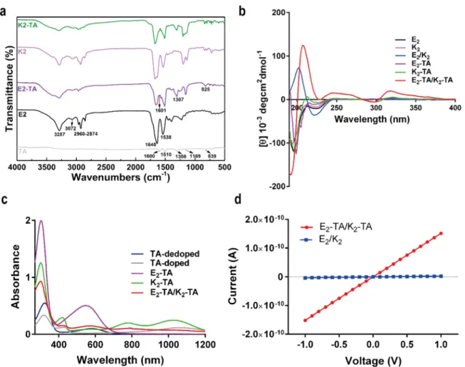
Figure 2.(a) FTIR spectra of TA, and the nonelectroactive and electroactive PAs, (b) CD spectra of the nonelectroactive and electroactive PAs and their corresponding mixtures. (c) UV−vis−NIR spectra of TA-dedoped, TA-doped, E2-TA, K2-TA, and E2-TA/K2-TA. (d) I−V curves of E2-TA/K2 -TA and E2/K2.
■
RESULTS AND DISCUSSIONIn this study, the TA moiety was used as an electroactive core for the fabrication of self-assembling negatively (E2-TA) and
positively (K2-TA) charged electroactive PA molecules. The TA
coupling to the peptide sequence was achieved by using a succinic anhydride linker. The TA unit promotes hydrophobic and π−π stacking interactions; β-sheet forming amino acids (VVA) provide hydrogen bonding, and two glutamic acid or lysine residues facilitate solubility in water and further induce self-assembly into fibrillar nanostructures upon mixing oppositely charged molecules under neutral conditions via charge neutralization. To clarify the effect of the electroactive unit on neural regeneration, the results were compared with those of nonelectroactive PA molecules containing a lauryl group instead of the TA moiety (E2 and K2). The chemical
structures of the electroactive and nonelectroactive PAs are shown inFigure 1.
All PAs were synthesized by using the Fmoc solid-phase peptide synthesis method. Liquid chromatograms and mass spectra of nonelectroactive PAs (E2and K2) and electroactive
ones (E2-TA and K2-TA) are shown in Figure S2a−d,
respectively. The observed molecular mass values were in good agreement with the theoretical molecular mass and confirmed the chemical structures of the desired molecules.
Chemical structures of the TA and PAs were also characterized by FTIR (Figure 2a). The 1600 and 1510 cm−1 peaks were assigned to the CC stretching vibrations of the quinoid and benzenoid rings of TA, respectively. The 1308, 1169, and 839 cm−1 bands correspond to the secondary aromatic C−N stretching vibrations, aromatic C−H in-plane bending vibrations, and the C−H out-of-plane bending deformation in the 1,4-disubstituted benzene rings, respec-tively.34Bands around 3287, 3072, and 2960−2874 cm−1in the FTIR spectrum of E2 were attributed to N−H and C−H
stretching vibrations, indicating the presence of amide and aliphatic C−H groups in the PA structure, respectively. The peaks around 1640 and 1538 cm−1 can be attributed to the amide-I bond and amide-II bond, which are associated with
vibrational stretching of the CO and N−H bonds,
respectively. Similar bands were also observed for K2. In
addition to the peaks observed for E2, the following peaks were
observed for E2-TA: 1601, 1307, and 825 cm−1, which can be
ascribed to CC stretching vibrations of the quinoid ring, aromatic C−N stretching vibrations, and the C−H out-of-plane bending vibrations of two adjacent hydrogen atoms on the 1,4-disubstituted benzene rings, respectively, indicating the successful conjugation of the TA unit to the peptide structure. Distinctive absorption bands demonstrating the presence of TA were also observed for K2-TA.
A positively-charged molecule, K2, was used to induce
nanofiber formation together with the negatively-charged E2
through electrostatic interactions. E2and E2-TA molecules form
nanofibers through self-assembly when mixed at a 1:1 ratio with K2 and K2-TA, respectively. SEM and TEM analyses were
performed to understand the morphological features of the self-assembled nanostructures upon mixing oppositely charged PA molecules. The SEM images of E2/K2(Figure 1e) and E2-TA/
K2-TA (Figure 1f) revealed their self-assembled, well-defined,
and three-dimensional porous nanofiber network structure. TEM images showed that the self-assembled nanofibers have a diameter of ca. 10 nm (Figure 1g,h).
Secondary structures of the electroactive and nonelectroac-tive PAs and their self-assembled corresponding mixtures were studied with circular dichroism spectrometry. In Figure 2b, individual PAs show random coil structures (negative signals at around 197 and 225 nm).35A chiral absorbance maximum at 202 nm and minimum at 222 nm were observed for E2/K2, predominantly corresponding to the β-sheet secondary structure of the nanostructures. On the other hand, E2-TA/ K2-TA showed a negative signal at 295 nm and positive signals
at 207 and 245 nm, where the shape of the CD curves was similar to that ofβ-turn peptides, which are characterized by a negative peak at 180−190 nm and two positive peaks at 200− 205 and 220−230 nm.36 In addition to these peaks, in the region of the characteristic absorption of theπ−π* transition of the benzenoid ring, a new positive signal appeared at 318 nm as a result of the supramolecular chirality of the self-assembled TA motifs, which was not observed for individual electroactive peptides. Increased hydrophobic and H-bonding interactions between self-assembled TA units were responsible for the supramolecular chirality of E2-TA/K2-TA.37,38
The morphological and spectroscopic analyses demonstrated the self-assembly of the PAs under neutral conditions. At neutral pH, individual PA molecules are soluble through electrostatic repulsion of either the positively- or negatively-charged two lysine or glutamic acid segments, respectively, despite the presence of the hydrophobic alkyl or TA moiety. The PA molecules self-assemble into cylindrical nanofibers as the electrostatic repulsion between molecules is neutralized and converted to attractive interactions by adding the oppositely charged counterparts. In the molecular organization of the PA nanofibers, the collapsed hydrophobic alkyl or TA tail are located in the core of the cylindrical micelles whereas the hydrophilic residues of the PA molecules are in contact with the aqueous medium. The β-sheet hydrogen bonding among the peptide sequences adjacent to the hydrophobic core (β-sheet region) oriented parallel to the long axis of the nanofiber results in 1D cylindricalfibers. These 1D nanofibers further entangle into 3D networks at certain concentrations.
Oligoaniline-based materials reversibly switch their emer-aldine base (EB) state to the emeremer-aldine salt (ES) state by acid doping. With the addition of acid, quinoid N atoms of the EB state are protonated, and this ES structure shows remarkable conductivity. This transition was monitored via UV−vis−NIR absorption spectroscopy (Figure 2c). Two peaks around 323 and 578 nm were attributed to the π−π* transition of the benzene ring and an excitonic transition from the benzenoid to quinoid ring (πB−πQ) for TA-dedoped, respectively. After
doping with acid, the peak at 578 nm disappeared because TA was fully doped by HCl. On the other hand, new peaks at 433 and 1053 nm appeared for the TA-doped system due to the formation of polarons and delocalized polaron transitions, respectively.39 After the prep-HPLC purification step, it was obvious that the TA moiety was in its EB state for E2-TA under
basic conditions whereas it was in its ES state for K2-TA under
acidic conditions, according to UV−vis−NIR spectra. E2-TA exhibited two absorption peaks similar to those of TA-dedoped at 304 and 549 nm corresponding to the characteristic absorptions of theπ−π* transition of the benzenoid ring and the excitonic πB−πQ transition, respectively, whereas K2-TA
showed absorption peaks at 420, 781, and 1030 nm, which indicated that TA was in its conductive form and that the TA motif retained its electroactivity after conjugation to the peptide sequence. However, the broad and weak absorption peak at 568
nm (the excitonicπB−πQ transition) indicated that K2-TA in aqueous solution was partially doped. When the oppositely charged electroactive PAs were mixed, E2-TA/K2-TA exhibited absorption peaks at 419 and 781 nm, associated with the existence of polaron species containing cation radicals. The weak adsorption peak at 578 nm indicated partially-doped TA in the structure. Furthermore, characteristic absorption bands of TA showed a hypsochromic shift for electroactive PAs due to the formation of an amide group, which is more electron withdrawing compared to the amino group of TA and leads to a decrease in the electron density of the quinoid ring. In addition, nonelectroactive PAs did not show any absorption peaks in the studied wavelength range.
For conductivity measurements, the current−voltage (I−V) curves of TA-doped, E2/K2, and E2-TA/K2-TA were
determined with the aid of patterned contact gold electrodes in a two-point probe configuration with an electrode separation of 20μm on the sample. The I−V curves for E2-TA/K2-TA and
E2/K2are shown inFigure 2d, thefilm thickness of the samples
was determined via AFM analysis to calculate conductivity values of the samples, and representative AFM images of the samples are shown inFigure S3. In addition, the roughness of the material was also assessed to investigate whether different experimental groups have similar height deviation patterns. Root mean square roughness values, which are described as the average height deviations measured from the mean line, were observed not to be significantly different from each other
(Table S1). This proves that the samples were reliable for
extrapolation of the height of the material. The I−V curves were linear for E2-TA/K2-TA, indicating Ohmic contact.40The
conductivity values of the samples were determined as follows: σE2/K2= 1.10× 10⁻¹⁰S/cm <σE2‑TA/K2‑TA= 6.97× 10
−6S/cm
< σTA‑doped = 2.55 × 10⁻³ S/cm. Nonelectroactive species
including simple alkyl blocks, oligopeptides, and other bio-based blocks, inorganic materials, and other structure-directing
domains or functional units may lead TA to show decreased conductivity depending on the length of the conjugated group.17Nevertheless, it was clearly observed that conjugation of TA to an insulating peptide segment led to obtaining a higher conductivity compared to that of the nonelectroactive E2/K2. Guo et al. reported an electroactive porous tubular
scaffold for neural tissue engineering by blending hyper-branched degradable conducting copolymer and linear polycaprolactone. The conductivity of the blend films doped with (±)-10-camphorsulfonic acid was observed to be between
3.4 × 10⁻⁶ and 3.1 × 10⁻⁷ S/cm depending on the
hyperbranched conducting copolymer content. These con-ductivity values are within the range of those reported to be sufficient for many tissue engineering applications.41Although the conductivity of the E2-TA/K2-TA nanofibers is relatively low due to conjugation of the insulating peptide moiety and the lack of any additional doping agent, the conductivity value of this novel supramolecular electroactive nanofiber is never-theless sufficient to transfer bioelectrical signals in vivo as living activity in the body involves low microcurrents.42
Gel formation upon mixing oppositely charged PAs is shown
in Figure 3a. All individual PA molecules were completely
dissolved, resulting in clear solutions; however, mixing oppositely charged PAs resulted in the immediate formation of a self-supporting gel. Stiffness and elasticity characteristics of the gel systems were studied by oscillatory rheology. The gelation kinetics were first monitored by a time sweep test
(Figure 3b) within the linear viscoelastic region (LVR), and
both gels reached a plateau, indicating complete gelation. The storage moduli (G′) of both PA gels were determined to be significantly higher than the loss moduli (G″) within the LVR, confirming the formation of gels with viscoelastic character. After 30 min, the G′ of the E2/K2gel was observed to be 9 times higher than the G′ of the E2-TA/K2-TA gel, indicating
that it had a more rigid network structure and higher
Figure 3.(a) Photo of the solution of nonelectroactive and electroactive PAs and self-supporting gel behaviors upon their corresponding mixture. (b) Time, (c) strain, and (d) frequency sweep tests to measure the rheological properties of E2/K2and E2-TA/K2-TA gels.
Figure 4.Biocompatibility of PC-12 cells on (a, d) E2-TA/K2-TA and (b, e) E2/K2gels, and (c, f) PLL-coated surfaces before NGF induction (24 h after cell seeding, a−c) and 24 h after NGF induction (d−f). (g) Alamar Blue viability analysis of PC-12 cells on E2-TA/K2-TA and E2/K2gels, and PLL-coated surfaces 24 h after cell seeding. Scale bars are 100μm.
Figure 5.Bright-field images of neurite outgrowth of PC-12 cells on (a) E2-TA/K2-TA and (b) E2/K2gels and (c) PLL-coated surfaces, scale bars are 100μm. Confocal images of βIII tubulin-stained PC-12 cells grown on (d) E2-TA/K2-TA and (e) E2/K2gels and (f) PLL-coated surfaces, scale bars are 20μm. (g) Relative percentage frequency distribution of neurite lengths. (h) Average neurite length and (i) percentage of neurite-bearing PC-12 cells on E2-TA/K2-TA and E2/K2gels and PLL-coated surfaces. Data presented as mean± SEM (n = 3), ***p < 0.0005.
entanglement density of the nanofibers. The difference in rheological properties can be attributed to the internal molecular interactions of the peptide building blocks within the nanofibers. Pashuck et al. found that by manipulating the number and position of β-sheet forming amino acids in the peptide sequence, the highest mechanical stiffness was observed for the PA self-assembled intoβ-sheets with the least amount of twisting and disorder and least red-shift of theβ-sheet signal in CD, even when all of the molecules studied in that study formed 1D nanofibers with similar length and the same amino acid termini.43In our molecular design, all PAs have similar β-sheet forming amino acid sequences and amino acid termini, but they have different hydrophobic tails. Although both PA gels possessed similar supramolecular nanostructures according to the SEM and TEM analyses, the replacement of the lauryl group by a TA moiety possibly affected the molecular packing of β-sheets into the nanofibers, which also had an impact on their CD signal and caused a decrease in mechanical properties. A strain sweep test was performed to investigate the viscoelastic properties and to determine the critical strain value of both gels. InFigure 3c, a change in modulus value was observed with increasing strain amplitude, and the gels maintained their G′ in the LVR. As the strain was increased, the gel network began to break down and collapsed, showing lower G′ than G″, i.e., displaying liquid-like behavior. E2/K2
showed a slightly lower critical strain value (γcwas determined from the intersection of the tangents in the linear regime and the strain-dependent regime)44 than that of E2-TA/K2-TA.
Whereas the value of the moduli is mainly determined by the entanglement density, the stability of the gel is controlled by the strength of the entanglement points. The relatively higher γc of E2-TA/K2-TA can be attributed to the slightly stronger
entanglement points of the nanofibers in the network. Furthermore, the gels maintained their moduli values within the studied frequency range, as shown in Figure 3d, due to having more elastic-dominant gel characteristics compared to those of the viscous one. It can be also concluded that the mechanical properties of the electroactive PA nanofiber were in the range of those of neural tissue as the natural stiffness values of individual neurons and glial cells range from 0.5 to 1.6 kPa, and the overall peripheral nerve tissue stiffness values range from 150−300 kPa.45
For biomedical applications, the biocompatibility of a biomaterial is crucial. Ideally, the biomaterial should support adhesion and survival of the cells. To evaluate the biocompatibility and further investigate the bioactivity of the E2-TA/K2-TA gels, PC-12 cells, derived from rat
pheochromo-cytoma, were used as a model cell line for neural differentiation. Live−dead and Alamar Blue assays were utilized to determine cell viability. We performed live−dead analyses both before and after NGF induction to observe the behavior of the cells before and after neural differentiation.Figure 4a−c shows the live and dead cells stained with green and red, respectively, at 24 h after cell seeding before NGF induction, whereasFigure 4d−f shows the live and dead cells at 24 h after NGF induction. Peptide nanofibers were observed to provide a biocompatible environ-ment for both undifferentiated and differentiated PC-12 cells. A PLL-coated surface is conventionally used to attach the cells to the surface, and thus it was used as a positive control. The cell viability and metabolic activity on E2-TA/K2-TA and E2/K2
coated surfaces were observed to be similar to those of the PLL-coated surfaces, determined by Alamar Blue assay.
Neurites are the projections that arise from the cell body of a neuron or a neuron-like cell before the neuron matures completely. In a mature neuron, projections, later on, become dendrites or axons. PC-12 cells can be induced with NGF to trigger neurite outgrowth. Neurite lengths after NGF induction can give quantitative information about the degree of neural differentiation.46To determine the effect of the E2-TA/K2-TA
gels on neural differentiation, the neurite processes of PC-12 cells were measured 6 days after NGF induction (Figure 5a−c); average neurite length and the percentage of cells with neurites were determined for each group, in addition to representing the relative percentage frequency of neurite length values as a histogram plot. The morphology of the cells and neurite projections in different groups can be visualized by βIII tubulin staining, a neural marker. The neurites on both E2-TA/K2-TA and PLL groups were observed to be slightly thicker and straighter, whereas neurites on E2/K2appeared to be weaker
and curved (Figure 5d−f). The histogram data showed that the frequency of low-length neurites on E2/K2 peaked at around
30−40 μm neurite length, whereas the percentage frequency of longer neurites was higher on E2-TA/K2-TA and PLL. The
histogram statistics proved that neurite lengths on E2-TA/K2 -TA, E2/K2, and PLL have 25% percentile values of 34.5, 23.9,
and 32.8, median values of 57.3, 39.4, and 53.4, and 75% percentile values of 93.5, 68.1, and 90.2, respectively, where the E2-TA/K2-TA samples showed higher values compared to
those of E2/K2 (Table S2). Measurements of the average neurite length showed that there were significantly longer neurites on the E2-TA/K2-TA gels compared to those on the E2/K2gels, as shown inFigure 5h. PLL was used as a positive
control in neurite outgrowth analyses (Figure 5c,f). The rate of neurite-bearing cells did not significantly change on the E2-TA/
K2-TA gels compared to that on the E2/K2gels (Figure 5i).
This is probably due to short neurites on E2/K2gels increasing the number of neurite-bearing cells, but not the average neurite length, which can also be observed by the high frequency of low-length neurites on E2/K2gels in the histogram data (Figure
5g). Neural differentiation of PC-12 cells on E2-TA/K2-TA
scaffolds was also observed to be NGF dependent, as the neurite outgrowth of the cells that were cultured in NGF-free medium was not obvious (Figure S4a−c).
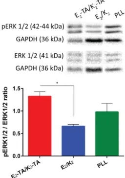
We then investigated the upstream pathways of NGF-induced neural differentiation. Upon binding of NGF to the TrkA surface receptor, a set of signaling pathways, including mitogen-activated protein kinase/extracellular signal-regulated kinase 1/2 (MAPK/ERK) signaling as the major one in addition to AKT and protein kinase C (PKC) pathways, were initiated on the PC-12 cells, resulting in activation of genes that play a role in neuronal differentiation. The ERK1/2 pathway was previously observed to be phosphorylated in the case of electrical stimulation,47 whereas it remains to be determined exactly which pathways take place in electrical stimulation or conductive scaffold-mediated neurite outgrowth. Here, we observed an upregulation in the phosphorylation level of the ERK1/2 pathway as well (Figure 6). Although the cells were primed with the same amount of NGF, upregulation of this pathway on E2-TA/K2-TA might suggest that intrinsic microcurrents of the cells have been enhanced by the conductive scaffold.
Conductive biomaterials have been extensively explored for many biomedical applications. The electrical excitability and the intrinsic electrical connectivity of nerve tissue make conductive biomaterials promising candidates for nerve injury therapies.
These materials can be used as scaffolds, films, or coating materials; however, tuning the mechanical, topographical, and biological properties of the biomaterial expedites its integration of regenerating host tissue. Ideally, the scaffold should mimic the host tissue. Here, we showed an electroactive, nanofibrous peptide hydrogel system for improved neural differentiation of model cell line PC-12. The electroactive gel system shows outstanding properties as it does not require any chemical crosslinker, which could potentially introduce toxicity, or dopants. The porous self-supporting nanofiber gel is also of great significance in soft tissue engineering to allow cell engraftment, and nutrient and waste exchange. The PA nanofibers can be functionalized with diverse bioactive peptide epitopes, which could interact with the cell surface receptors for synthetic extracellular matrix applications.
■
CONCLUSIONSIn this study, we showed that TA-containing peptide amphiphiles can self-assemble intofibrillar network structures having electroactivity by mixing oppositely charged counter-parts at neutral conditions. E2-TA/K2-TA scaffolds showed good biocompatibility toward PC-12 cells and did not alter their metabolic activity. Our results suggest that neural differentiation of PC-12 cells was enhanced when cultured on E2-TA/K2-TA gels compared to that on nonconductive
counterparts. Also, we observed that phosphorylation levels of ERK1/2 were increased on PC-12 cells on the conductive scaffolds compared to that on the nonconductive nanofiber gels. The E2-TA/K2-TA gels could be used as a promising
material in peripheral nerve regeneration. Further studies are necessary using in vivo peripheral nerve injury models and investigating the mechanism of improved neural differentiation.
■
ASSOCIATED CONTENT*
S Supporting InformationThe Supporting Information is available free of charge on the
ACS Publications websiteat DOI:10.1021/acsami.7b16509.
Mass spectra of TA-EB, liquid chromatograms and mass spectra of E2, K2, E2-TA, and K2-TA, AFM 3D images
and height profiles of E2/K2, E2-TA/K2-TA, and TA-doped samples, bright-field images of PC-12 cells on E2/
K2 and E2-TA/K2-TA gels and PLL-coated surfaces,
relative percentage frequency distribution of neurite length values on E2-TA/K2-TA and E2/K2gels and
PLL-coated surfaces (PDF)
■
AUTHOR INFORMATIONCorresponding Authors
*E-mail:[email protected](A.B.T.) *E-mail:[email protected](M.O.G.). ORCID Ozlem Erol:0000-0003-2156-537X Ahmet E. Topal:0000-0001-9951-0171 Ayse B. Tekinay:0000-0002-4453-814X Mustafa O. Guler:0000-0003-1168-202X Author Contributions
#I.A. and O.E. contributed equally.
Notes
The authors declare no competingfinancial interest.
■
ACKNOWLEDGMENTSThe authors thank Mr. M. Guler for TEM imaging. I.A. acknowledges support from the TUBITAK-BIDEB 2210-C fellowship.
■
REFERENCES(1) Huebner, E. A.; Strittmatter, S. M. Axon Regeneration in the Peripheral and Central Nervous Systems. In Cell Biology of the Axon; Springer, 2009; pp 305−360.
(2) Subramanian, A.; Krishnan, U. M.; Sethuraman, S. Development of Biomaterial Scaffold for Nerve Tissue Engineering: Biomaterial Mediated Neural Regeneration. J. Biomed. Sci. 2009, 16, 108.
(3) Ghasemi-Mobarakeh, L.; Prabhakaran, M. P.; Morshed, M.; Nasr-Esfahani, M. H.; Baharvand, H.; Kiani, S.; Al-Deyab, S. S.; Ramakrishna, S. Application of Conductive Polymers, Scaffolds and Electrical Stimulation for Nerve Tissue Engineering. J. Tissue Eng. Regener. Med. 2011, 5, e17−e35.
(4) Balint, R.; Cassidy, N. J.; Cartmell, S. H. Conductive Polymers: Towards a Smart Biomaterial for Tissue Engineering. Acta Biomater. 2014, 10, 2341−2353.
(5) Lyu, W.; Feng, J.; Yan, W.; Faul, C. F. Self-Assembly of Tetra (Aniline) Nanowires in Acidic Aqueous Media with Ultrasonic Irradiation. J. Mater. Chem. C 2015, 3, 11945−11952.
(6) Qazi, T. H.; Rai, R.; Boccaccini, A. R. Tissue Engineering of Electrically Responsive Tissues Using Polyaniline Based Polymers: A Review. Biomaterials 2014, 35, 9068−9086.
(7) Fleming, S.; Ulijn, R. V. Design of Nanostructures Based on Aromatic Peptide Amphiphiles. Chem. Soc. Rev. 2014, 43, 8150−8177. (8) Cavalli, S.; Albericio, F.; Kros, A. Amphiphilic Peptides and Their Cross-Disciplinary Role as Building Blocks for Nanoscience. Chem. Soc. Rev. 2010, 39, 241−263.
(9) Aida, T.; Meijer, E.; Stupp, S. Functional Supramolecular Polymers. Science 2012, 335, 813−817.
(10) Zhao, X.; Pan, F.; Xu, H.; Yaseen, M.; Shan, H.; Hauser, C. A.; Zhang, S.; Lu, J. R. Molecular Self-Assembly and Applications of Designer Peptide Amphiphiles. Chem. Soc. Rev. 2010, 39, 3480−3498. Figure 6.Protein expression levels of ERK1/2 phosphorylation of
PC-12 cells cultured on E2-TA/K2-TA and E2/K2 gels and PLL-coated surfaces. Data presented as mean± SEM (n = 3), *p < 0.05.
(11) Han, S.; Cao, S.; Wang, Y.; Wang, J.; Xia, D.; Xu, H.; Zhao, X.; Lu, J. R. Self-Assembly of Short Peptide Amphiphiles: The Cooperative Effect of Hydrophobic Interaction and Hydrogen Bonding. Chem.− Eur. J. 2011, 17, 13095−13102.
(12) Toksoz, S.; Acar, H.; Guler, M. O. Self-Assembled One-Dimensional Soft Nanostructures. Soft Matter 2010, 6, 5839−5849.
(13) Cipriano, T.; Knotts, G.; Laudari, A.; Bianchi, R. C.; Alves, W. A.; Guha, S. Bioinspired Peptide Nanostructures for Organic Field-Effect Transistors. ACS Appl. Mater. Interfaces 2014, 6, 21408−21415. (14) Eakins, G. L.; Pandey, R.; Wojciechowski, J. P.; Zheng, H. Y.; Webb, J. E.; Valéry, C.; Thordarson, P.; Plank, N. O.; Gerrard, J. A.; Hodgkiss, J. M. Functional Organic Semiconductors Assembled Via Natural Aggregating Peptides. Adv. Funct. Mater. 2015, 25, 5640− 5649.
(15) Bell, O. A.; Wu, G.; Haataja, J. S.; Brömmel, F.; Fey, N.; Seddon, A. M.; Harniman, R. L.; Richardson, R. M.; Ikkala, O.; Zhang, X.; Faul, C. F. J. Self-Assembly of a Functional Oligo (Aniline)-Based Amphiphile into Helical Conductive Nanowires. J. Am. Chem. Soc. 2015, 137, 14288−14294.
(16) Faul, C. F. Ionic Self-Assembly for Functional Hierarchical Nanostructured Materials. Acc. Chem. Res. 2014, 47, 3428−3438.
(17) Udeh, C. U.; Fey, N.; Faul, C. F. Functional Block-Like Structures from Electroactive Tetra (Aniline) Oligomers. J. Mater. Chem. 2011, 21, 18137−18153.
(18) Wei, Z.; Faul, C. F. Aniline Oligomers−Architecture, Function and New Opportunities for Nanostructured Materials. Macromol. Rapid Commun. 2008, 29, 280−292.
(19) Shao, Z.; Rannou, P.; Sadki, S.; Fey, N.; Lindsay, D. M.; Faul, C. F. Delineating Poly (Aniline) Redox Chemistry by Using Tailored Oligo (Aryleneamine) S: Towards Oligo (Aniline)-Based Organic Semiconductors with Tunable Optoelectronic Properties. Chem. − Eur. J. 2011, 17, 12512−12521.
(20) Qu, G.; Li, F.; Berda, E. B.; Chi, M.; Liu, X.; Wang, C.; Chao, D. Electroactive Polyurea Bearing Oligoaniline Pendants: Electrochromic and Anticorrosive Properties. Polymer 2015, 58, 60−66.
(21) Zhang, W.; Yu, Y.; Chen, L.; Mao, H.; Wang, C.; Wei, Y. Synthesis and Study of Phenyl-Capped Tetraaniline as an Anti-corrosion Additive. In Electroactive Polymers for Corrosion Control; American Chemical Society, 2003; Chapter 9, pp 156−165.
(22) Yeh, L.-C.; Huang, T.-C.; Huang, Y.-P.; Huang, Y.; Chen, H.-H.; Yang, T.-I.; Yeh, J.-M. Synthesis Electroactive Polyurea with Aniline-Pentamer-Based in the Main Chain and Its Application in Electrochemical Sensor. Electrochim. Acta 2013, 94, 300−306.
(23) Wang, S.; Chao, D.; Berda, E. B.; Jia, X.; Yang, R.; Wang, X.; Jiang, T.; Wang, C. Fabrication of Electroactive Oligoaniline Functionalized Poly (Amic Acid) Nanofibers for Application as an Ammonia Sensor. RSC Adv. 2013, 3, 4059−4065.
(24) Wu, Y.; Liu, S.; Tao, Y.; Ma, C.; Zhang, Y.; Xu, J.; Wei, Y. New Strategy for Controlled Release of Drugs. Potential Pinpoint Targeting with Multiresponsive Tetraaniline Diblock Polymer Vesicles: Site-Directed Burst Release with Voltage. ACS Appl. Mater. Interfaces 2014, 6, 1470−1480.
(25) Hardy, J. G.; Mouser, D. J.; Arroyo-Currás, N.; Geissler, S.; Chow, J. K.; Nguy, L.; Kim, J. M.; Schmidt, C. E. Biodegradable Electroactive Polymers for Electrochemically-Triggered Drug Delivery. J. Mater. Chem. B 2014, 2, 6809−6822.
(26) Cui, H.; Liu, Y.; Cheng, Y.; Zhang, Z.; Zhang, P.; Chen, X.; Wei, Y. In Vitro Study of Electroactive Tetraaniline-Containing Thermo-sensitive Hydrogels for Cardiac Tissue Engineering. Biomacromolecules 2014, 15, 1115−1123.
(27) Li, L.; Ge, J.; Wang, L.; Guo, B.; Ma, P. X. Electroactive Nanofibrous Biomimetic Scaffolds by Thermally Induced Phase Separation. J. Mater. Chem. B 2014, 2, 6119−6130.
(28) Ma, X.; Ge, J.; Li, Y.; Guo, B.; Ma, P. X. Nanofibrous Electroactive Scaffolds from a Chitosan-Grafted-Aniline Tetramer by Electrospinning for Tissue Engineering. RSC Adv. 2014, 4, 13652− 13661.
(29) Dong, R.; Zhao, X.; Guo, B.; Ma, P. X. Self-Healing Conductive Injectable Hydrogels with Antibacterial Activity as Cell Delivery
Carrier for Cardiac Cell Therapy. ACS Appl. Mater. Interfaces 2016, 8, 17138−17150.
(30) Hu, J.; Huang, L.; Zhuang, X.; Zhang, P.; Lang, L.; Chen, X.; Wei, Y.; Jing, X. Electroactive Aniline Pentamer Cross-Linking Chitosan for Stimulation Growth of Electrically Sensitive Cells. Biomacromolecules 2008, 9, 2637−2644.
(31) Wu, Y.; Wang, L.; Guo, B.; Shao, Y.; Ma, P. X. Electroactive Biodegradable Polyurethane Significantly Enhanced Schwann Cells Myelin Gene Expression and Neurotrophin Secretion for Peripheral Nerve Tissue Engineering. Biomaterials 2016, 87, 18−31.
(32) Guo, Y.; Li, M.; Mylonakis, A.; Han, J.; MacDiarmid, A. G.; Chen, X.; Lelkes, P. I.; Wei, Y. Electroactive Oligoaniline-Containing Self-Assembled Monolayers for Tissue Engineering Applications. Biomacromolecules 2007, 8, 3025−3034.
(33) Sun, Z.; Kuang, L.; Jing, X.; Wang, X.; Li, J.; Wang, F. Synthesis of Phenyl/Amino-Capped Tetraaniline by Chemical and Electro-chemical Methods. Chem. J. Chin. Univ. 2002, 23, 496−499.
(34) Lv, W.; Feng, J.; Yan, W.; Faul, C. F. Self-Assembly and Ph Response of Electroactive Liquid Core−Tetra (Aniline) Shell Microcapsules. J. Mater. Chem. B 2014, 2, 4720−4725.
(35) Greenfield, N. J.; Fasman, G. D. Computed Circular Dichroism Spectra for the Evaluation of Protein Conformation. Biochemistry 1969, 8, 4108−4116.
(36) Huang, R.; Su, R.; Qi, W.; Zhao, J.; He, Z. Hierarchical, Interface-Induced Self-Assembly of Diphenylalanine: Formation of Peptide Nanofibers and Microvesicles. Nanotechnology 2011, 22, No. 245609.
(37) Huang, X.; Li, C.; Jiang, S.; Wang, X.; Zhang, B.; Liu, M. Self-Assembled Spiral Nanoarchitecture and Supramolecular Chirality in Langmuir-Blodgett Films of an Achiral Amphiphilic Barbituric Acid. J. Am. Chem. Soc. 2004, 126, 1322−1323.
(38) Garifullin, R.; Guler, M. O. Supramolecular Chirality in Self-Assembled Peptide Amphiphile Nanostructures. Chem. Commun. 2015, 51, 12470−12473.
(39) Wang, Q.; He, W.; Huang, J.; Liu, S.; Wu, G.; Teng, W.; Wang, Q.; Dong, Y. Synthesis of Water Soluble, Biodegradable, and Electroactive Polysaccharide Crosslinker with Aldehyde and Carbox-ylic Groups for Biomedical Applications. Macromol. Biosci. 2011, 11, 362−372.
(40) Nalluri, S. K. M.; Shivarova, N.; Kanibolotsky, A. L.; Zelzer, M.; Gupta, S.; Frederix, P. W.; Skabara, P. J.; Gleskova, H.; Ulijn, R. V. Conducting Nanofibers and Organogels Derived from the Self-Assembly of Tetrathiafulvalene-Appended Dipeptides. Langmuir 2014, 30, 12429−12437.
(41) Guo, B.; Sun, Y.; Finne-Wistrand, A.; Mustafa, K.; Albertsson, A.-C. Electroactive Porous Tubular Scaffolds with Degradability and Non-Cytotoxicity for Neural Tissue Regeneration. Acta Biomater. 2012, 8, 144−153.
(42) Huang, H.; Li, W.; Wang, H.; Zeng, X.; Wang, Q.; Yang, Y. Conducting Hydrogels of Tetraaniline-G-Poly (Vinyl Alcohol) in Situ Reinforced by Supramolecular Nanofibers. ACS Appl. Mater. Interfaces 2014, 6, 1595−1600.
(43) Pashuck, E. T.; Cui, H.; Stupp, S. I. Tuning Supramolecular Rigidity of Peptide Fibers through Molecular Structure. J. Am. Chem. Soc. 2010, 132, 6041−6046.
(44) Niu, L.; Song, J.; Li, J.; Tao, N.; Lu, M.; Fan, K. Solvent Effects on the Gelation Performance of Melamine and 2-Ethylhexylphos-phoric Acid Mono-2-Ethylhexyl Ester in Water−Organic Mixtures. Soft Matter 2013, 9, 7780−7786.
(45) Chang, Y.-J.; Hsu, C.-M.; Lin, C.-H.; Lu, M. S.-C.; Chen, L. Electrical Stimulation Promotes Nerve Growth Factor-Induced Neurite Outgrowth and Signaling. Biochim. Biophys. Acta, Gen. Subj. 2013, 1830, 4130−4136.
(46) Harrill, J. A.; Mundy, W. R. Quantitative Assessment of Neurite Outgrowth in Pc12 Cells. In In Vitro Neurotoxicology; Methods and Protocols; Humana Press: Totowa, NJ, 2011; pp 331−348.
(47) Wang, L.; Li, X.; Tsou, Y.; Xu, X. Applications of Conductive Materials for Tissue Engineering. In Smart Materials for Tissue Engineering; Royal Society of Chemistry, 2017; pp 110−143.