Endoscopic Endonasal Management of Cerebrospinal
Fluid Rhinorrhea
Ozmen Ozturk, MD,* Senol Polat, MD,Þ and Cuneyd Uneri, MDþ
Abstract: The authors review their 5 years’ experience withendo-nasal endoscopic repair of the anterior skull base fistulas presenting with cerebrospinal fluid (CSF) rhinorrhea. A total of 12 patients were managed endoscopically between 2004 and 2008. Seven patients (58.3%) had nonsurgical posttraumatic CSF rhinorrhea, 2 patients (16.7%) had CSF rhinorrhea due to surgical/iatrogenic trauma, and 3 patients (25%) had spontaneous onset of CSF rhinorrhea. Radio-surgical correlation for CSF fistula identification was positive in all patients. The most common site of leak was the fovea ethmoidalis. The repair method consisted of an extradural underlay closure of a defect with fascia lata. The largest diameter of a defect to be closed was 15 mm. Immediate results were good in all patients, but later in the follow-up, CSF rhinorrhea recurred in 2 patients, and each patient had a revision 2 times. In the first revisions, transcranial approach was used, whereas in the second revisions endonasal endoscopic route was resorted. The primary closure rate was 83.3%, and the over-all closure rate was 100%. The average follow-up period thus far is 21 months. Endonasal endoscopic technique well known to otolaryn-gologists should be considered as the first choice of surgery in the repair of CSF rhinorrhea because of low morbidity and a higher closure rate. The possibility of revision with the same technique makes this approach ideal for the repair of cranionasal osteodural defects.
Key Words: Cerebrospinal fluid rhinorrhea, skull base, paranasal sinuses, endoscopic surgical procedure, complications
(J Craniofac Surg 2012;23: 1087Y1092)
C
erebrospinal fluid (CSF) rhinorrhea management has been a challenging problem for the surgeons dealing with one of the most complex anatomic regions of the human body, the anterior skull base and nasal cavity. The surgical techniques used for the closure of a cranionasal CSF fistula are highly demanding, depending on eti-ology, location, and size of the defect.Novelties in paranasal sinus surgery in conjunction with the technological improvements in endoscopes and the trend of refining minimally invasive approaches have pioneered significant advances in the management of CSF rhinorrhea.1,2The current use of endo-scopes in endonasal CSF rhinorrhea surgery facilitates excellent visualization of the fistula site providing ease of graft placement and shortened operating time.2With a consequent decrease in periopera-tive morbidity and surgical expense, this method has been preferred to more invasive intracranial and extracranial approaches.3Y12
In this retrospective study, a review of 12 patients who under-went endonasal endoscopic repair of anterior skull base CSF fistula during a period of 5 years is presented.
MATERIALS AND METHODS
Between January 2004 and December 2008, 12 patients un-derwent endonasal endoscopic repair for the cranionasal defects of the anterior skull base presenting with CSF rhinorrhea in the Oto-rhinolaryngology Department, Acibadem Kozyatagi Hospital, Aci-badem Healthcare Group. There were 8 men (66.7%) and 4 women (33.3%), ranging in age from 8 to 47 years (mean, 27 years). During the review of the medical records, several parameters were consid-ered including history, etiology, radiographic studies, localization of the defect, surgical procedure used, postoperative complications, and success rate.
After thorough history taking, CSF rhinorrhea was investi-gated in all patients by preoperative endoscopic examination. At nasal endoscopy, a CSF fistula presence was confirmed when an osseous skull base or a paranasal sinus defect was identified with clear fluid drainage, either pulsatile or elicited with a Valsalva maneuver.
With high-resolution computed tomography (CT) imaging of the paranasal sinuses and skull base, apparent signs of a skull base fracture and unspecific signs arousing the suspicion of a defect with CSF leak (eg, any opacity filling a sinus cavity or covering a poten-tial osteodural defect) were sought. When the strength of precision had to be increased, magnetic resonance imaging (MRI) was used. In cases of suspect,A2 transferrin assay was performed. A small sample (È0.3 mL) of draining fluid was collected after bending the head of the patient forward and sent in a sterile container to the laboratory.
Repair of a CSF fistula was performed with endonasal en-doscopic approach under general anesthesia. Intravenous ceftriaxone (15 mg/kg) was injected during the induction phase of anesthesia. After nasal mucosa decongestion and middle turbinate manipulation, endoscopic investigation by using rigid telescopes (0 and 30 degrees) showed CSF collection in the suspected approximate location of the bone defect. If the site of the leak was unclear, a Valsalva maneuver with positive pressure ventilation identified pulsating and glistening areas of CSF leaking through a defect. When needed, lumbar puncture was performed preoperatively in the operating room by a neurosur-geon. Intraoperatively, the lumbar drain was kept closed until verifi-cation of the release of CSF through the cranionasal defect.
The selection of a surgical technique for exposure was de-pendent on the size and location of the skull base defect. For a bone defect in the medial portion of the cribriform plate, a routine eth-moidectomy was performed in an anterior-to-posterior fashion, and
From the *Otorhinolaryngology Department, School of Medicine, Istanbul Medipol University; and †Otorhinolaryngology Department, Faculty of Medicine, Acibadem University, and ‡Otorhinolaryngology Department, Acibadem Kozyatagi Hospital, Acibadem Health Group, Istanbul, Turkey. Received October 20, 2011.
Accepted for publication January 2, 2012.
Address correspondence and reprint requests to Ozmen Ozturk, MD, Istanbul Medipol Universitesi, Istanbul Medipol Hastanesi, Kulak Burun Bogaz Klinigi, Haydarpasa Harem Yolu, Kosuyolu Kadikoy 34718, Istanbul, Turkey; E-mail: [email protected]
This article was presented at the First Congress of the Confederation of the European ORL-HNS, July 2Y6, 2011, Barcelona, Spain. The authors report no conflicts of interest.
Copyright* 2012 by Mutaz B. Habal, MD ISSN: 1049-2275
the middle turbinate was rotated laterally. For CSF fistulas located in the lateral part of the cribriform plate and fovea ethmoidalis, the ethmoidectomy cavity was enlarged after middle turbinate manipu-lation. Sphenoid sinus defects were visualized after enlargement of sphenoid ostium to the extent providing an adequate exposure.
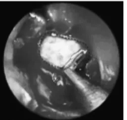
After identifying the exact localization of the osteodural de-fect, mucosa remnants, granulation tissue, and bone lamellae were removed from the edges for at least 5 mm circumferentially with a forceps to define the site and provide a raw base for graft contact (Fig. 1). The defect was closed with an appropriately trimmed au-togenous fascia lata graft laid into the groove between the bone and the dura in an ‘‘extradural underlay method’’ (Fig. 2) with supportive layers of fat tissue placed inferiorly. When dealing with a sphenoid sinus fistula, after stripping the mucosa from the sinus walls, fascia lata was placed properly, and the cavity was obliterated entirely with autogenous fat. During surgical manipulations, care was given for protecting the patency of the frontal sinus ostium.
In case of an obvious graft detachment risk, fibrin glue was applied directly to ensure stabilization of the graft and fat tissue. Nasal packing with Nasopore (Polyganics BV, Groningen, the Netherlands) and Merosel (Pope Epistaxis Packing; Medtronic Xomed, Jacksonville, FL) supported the graft and fat tissue. With its fine biocompati-bility and elasticity, Nasopore was placed in the superior part of the nasal cavity as the first line of support in contact with fat tissue. Merosel was positioned low in the nasal cavity as the second line of support firmly pushing the graft, fat tissue, and Nasopore. The preserved and manipulated middle turbinate assisted in reinforce-ment of the whole area.
Postoperatively, the patients were confined to bed rest with head elevation, and they were advised to avoid nose blowing and coughing and to sneeze with their mouths open. Laxatives were pre-scribed to reduce straining. Merosel packs and Nasopore remnants were gradually removed from the nasal cavity between the second and the fifth days, depending on the size and the location of the reconstructed skull base defect. Prophylactic antibiotics were used for a period of 5 days. Lumbar drainage was terminated as early as pos-sible. Six months later, an endonasal endoscopic examination was performed to assess success by checking the graft and its appearance. This report was approved by the ethics committee of Acibadem Health Group, Acibadem University, Faculty of Medicine, with the file number B.30.2.ACU.0.00.00.900/454 and carried out in accor-dance with the Declaration of Helsinki. Informed consent and full consent have been received from the patients or their parents for sharing and publishing the data of this study.
RESULTS
Patients with CSF fistula were classified according to etiol-ogy. In our series, 9 patients (75%) had posttraumatic CSF rhinorrhea (nonsurgical/craniofacial trauma [n = 7, 58.3%] and surgical trauma as a complication of endoscopic nasal polypectomy [n = 2, 16.7%]), and 3 patients (25%) had spontaneous onset of CSF rhinorrhea with no history of antecedent surgery or trauma (Table 1).
The duration of CSF rhinorrhea from diagnosis to man-agement varied from immediate (the iatrogenic fistula of the fovea ethmoidalis as a complication of endoscopic nasal polypectomy was managed immediately in the same operation, in patient 8) to 13 months after trauma (patient 9 with a posttraumatic CSF fistula of the sphenoid sinus superior wall) (Table 1). Eight patients (66.7%) reported permanent CSF leak, whereas rhinorrhea was recurrent in the remaining 4 patients (33.3%). Two patients with spontaneous CSF rhinorrhea (patients 2 and 3) had meningitis before diagnosis of a CSF fistula. Although patient 2 had recurrent meningitis with 3 attacks during the preceding 4 years, the leak was diagnosed after the referral of the patient to neurosurgery clinic and was managed 2 months after diagnosis.
Radiosurgical correlation for CSF fistula identification was positive in all patients. Computed tomography scan identified the precise site of the bony defect in 8 patients (66.7%) (Fig. 3). In the remaining 4 patients (33.3%) (patients 1Y3 and 9), the bone defect itself was not precisely confirmed; however, the presence of any opacity in the definite sinus or a suspicious skull base site and local tissue inflammation indirectly suggested the fistula’s location. To increase precision in evaluation, MRI provided useful information about the defect site in these patients (patients 1Y3 and 9) (Fig. 4). Biochemical testing with A2 transferrin performed in 1 patient (patient 3) was proved to be positive.
After manipulating the middle turbinate and performing eth-moidectomy, intraoperative nasal endoscopy located a fistula site in all patients with posttraumatic CSF rhinorrhea (n = 9, 75%). In the patients with spontaneous CSF rhinorrhea (n = 3, 25%) (patients 2, 3, and 5), nasal endoscopy allowed direct visualization of the exact fistula site in 1 patient (patient 5, with a fistula of the posterior fovea ethmoidalis), but in the remaining 2 patients (patients 2 and 3), an initial examination revealed a mass of granulation tissue and a shinier mucosa with pulsatile clear fluid drainage from the suspected area and thus indirectly confirmed the diagnosis.
Ten patients (83.3%) had 1 anatomic site involved (Table 1). In 5 patients (41.7%) of this subset, the location of the skull base defect was fovea ethmoidalis (anterior ethmoid region [n = 3, 25%] and posterior ethmoid region [n = 2, 16.7%]). In 3 patients (25%), cribriform lamina was the fistula site. The superior wall of sphenoid sinus was detected to be defective in 2 patients (16.7%).
Two posttrauma patients (16.7%) had more than 1 site in-volved (Table 1). One of these patients (patient 10) had fovea eth-moidalis and cribriform lamina involved, and the remaining patient (patient 4) had simultaneous involvement of cribriform lamina, posterior fovea ethmoidalis, and superior wall of sphenoid sinus.
The defects were classified according to size. In the subset of patients with 1 fistula site, the size of the skull base osteodural defect was smaller than 5 mm in 5 patients (41.7%), between 6 and 10 mm in 3 patients (25%), and between 11 and 15 mm in 2 patients (16.7%) (Table 1).
FIGURE 1. The margin of the defect (dashed lines) is defined circumferentially and made raw by removing bone chips and granulation tissue.
FIGURE 2. Placement of fascia lata graft between the elevated dural margins and bony skull base (extradural underlay technique) with a ball probe after middle turbinate medialization and ethmoidectomy (dashed lines define the defect borders).
Two remaining patients (16.7%) (patients 4 and 10) in the subset with more than 1 site involvement had defect sizes between 3 and 15 mm. Patient 10 had simultaneous fistulas of fovea ethmoidalis and cribriform lamina, 5 and 3 mm in diameter, respectively. The second patient (patient 4) had a large fistula involving posterior fovea ethmoidalis and cribriform lamina with a fistula diameter of 15 mm and an additional defect in the superior wall of the sphenoid sinus with a diameter of 3 mm.
Fascia lata was the graft of choice for closure of the defects, and fat tissue was used as a supportive layer to increase the stability in all patients. In patient 4 with more than 1 site involvement, 2 sepa-rate fascia lata grafts were used. Fibrin glue was applied in 6 patients (50%). Lumbar drainage was used in 6 patients (50%), with 3.25 days (range, 2Y7 days) of average application time.
The average hospital stay varied from 2 to 57 days. Four poly-trauma patients (33.3%) remained hospitalized for a period longer than 1 week for medical care unrelated to the repair of a CSF fistula or any complication related to skull base.
In 1 patient (patient 2), periorbital ecchymosis due to lamina papyracea violation developed as an early complication. The ecchy-mosis improved without any sequela in a period of 10 days. As a late complication, patient 3 had frontal sinusitis due to the obstruction of the left frontal sinus ostium developing 4 months after the CSF fistula repair. The patient was operated on to create a patent frontal sinus outflow tract.
Immediate results were good in all patients, but later in the follow-up, CSF rhinorrhea recurred in 2 patients (16.7%) (patients 2 and 9). In the first patient (patient 2), CSF rhinorrhea recurred at
10 months after endonasal endoscopic fistula management. In the revision operation performed by the neurosurgery team, the patient underwent cranioplasty and duraplasty with bifrontal craniotomy approach to manage the fovea ethmoidalis fistula. But 4 months after the secondary operation, CSF rhinorrhea recurred a second time. In the second and final revision, otolaryngology team resorted to the procedure of positioning the fascia lata graft by endonasal endo-scopic approach. After a follow-up period of 24 months, no CSF rhinorrhea is detected.
The second patient (patient 9) with a spontaneous-type sphenoid sinus CSF rhinorrhea had a recurrence 9 months after an endonasal endoscopic CSF rhinorrhea management. For the revision operation, the patient was referred to the neurosurgery team, and fascia lata was positioned to the skull base by using the intracranial approach. But 2 months after the revision operation, the patient had meningitis. After an appropriate medical therapy, CSF rhinorrhea recurrence was detected at the same defect site (superior wall of sphenoid sinus), and a second revision operation was attempted si-multaneously by 2 approaches: galeal graft was placed, and dur-aplasty was performed intracranially by the neurosurgery team, while a fascia lata graft was positioned endonasally by the otolaryngology team. After a follow-up period of 18 months, no CSF rhinorrhea was detected. Although a final recovery has been achieved after 2 revi-sion surgeries for each patient, these patients are considered un-successful for the primary operations.
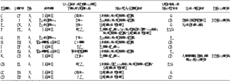
TABLE 1. Synopsis of Patients Presenting With CSF Rhinorrhea Patient Age, y Sex Etiology
Duration of Rhinorrhea
Before Diagnosis Fistula Localization
Diameter of
Fistula, mm Complication Recurrence 1 14 F Trauma 1.5 mo Anterior fovea ethmoidalis 5
2 8 F Spontaneous 2 mo Posterior fovea ethmoidalis 10 Periorbital edema 2 Recurrences 3 19 M Spontaneous 6 mo Cribriform lamina 5 Frontal sinusitis
4 43 M Trauma G1 d Sphenoid sinus (superolateral wall), posterior fovea ethmoidalis, cribriform lamina
3, 15 5 47 M Spontaneous 4 mo Posterior fovea ethmoidalis 3 6 36 F Iatrogenic trauma 1 week Anterior fovea ethmoidalis 10 7 24 M Trauma 2 d Sphenoid sinus 10 8 35 M Iatrogenic trauma Immediately Anterior fovea ethmoidalis 3
9 12 M Trauma 13 mo Sphenoid sinus 12 Meningitis after the first recurrence
2 Recurrences 10 38 M Trauma G1 d Anterior and posterior fovea ethmoidalis,
cribriform lamina
3, 5 11 25 F Trauma 1.5 mo Cribriform lamina 5 12 22 M Trauma 8 d Cribriform lamina 12
F indicates female; M, male.
FIGURE 3. Preoperative coronal CT scan showing bone defect in the superolateral wall roof of sphenoid sinus (arrow).
FIGURE 4. T2-weighted coronal MRI through the anterior ethmoid shows the communication (arrow) between the subarachnoid space and the ethmoid cavity on the right side, with an opaque nasal cavity (arrowhead) and maxillary sinus (star).
With confirmation of the correct position of the grafts with endoscopic and CT scan examinations at the postoperative sixth month, overall success rate was accepted to be achieved in 10 (83.3%) of 12 patients after the primary operations. Absence of CSF rhinorrhea was evaluated routinely with endoscopic examinations of the nasal cavity. The average follow-up period thus far is 21 months (range, 12Y52 months).
DISCUSSION
Nonsurgical (blunt or penetrating craniofacial injuries) and surgical (eg, due to endoscopic sinus or skull base surgeries) traumas are the main causes of a CSF rhinorrhea. A leak may also be clas-sified as spontaneous when it is idiopathic in origin or emerges secondary to an intracranial pathology presenting with either a high-pressure or a normal-high-pressure CSF drainage.5,12Y15 In our series, although most of the cases (n = 9, 75%) were caused by trauma (nonsurgical/craniofacial [n = 7, 58.3%] and surgical/iatrogenic [n = 2, 16.7%]), 3 cases (25%) were classified in the spontaneous CSF fistula subset.
Diagnostic workup of a CSF rhinorrhea usually involves a number of investigations.3,12,16Y20 Preoperative endoscopic nasal examination rarely identifies the site of the leak, but with an addi-tional information from the biochemical testing of the draining fluid, it helps in the differential diagnosis.3,12 Determining the glucose concentration is rapid and simple but obsolete because this assay frequently gives false-positive results among various sinonasal pa-thologies (ie, rhinitis and rhinosinusitis).17A2 transferrin testing is much more reliable with high sensitivity and specificity, but it is not widely available.18In our study,A2 transferrin test was used in only 1 case to increase reliability. In the remaining patients, the imaging techniques were satisfactory for the diagnosis of a fistula. In our study, the diagnosis of a CSF fistula was based on clinical history, preoperative and intraoperative endoscopic exam-inations, and imaging techniques. Our patients mostly consisted of trauma cases (n = 9, 75%), and with high-resolution CT, we have been able to define the osteodural defects of the skull base and paranasal sinuses, evaluate surgical landmarks, and plan surgery independent of an activity of CSF drainage. In 8 patients (66.7%), the site of the bony defect was accurately identified with CT, and in the remaining 4 patients, the indirect imaging findings (ie, any opacity filling the definite sinus or covering a suspected fistula site) were suggestive of a fistula presence. However, these radiologic findings cannot be regarded solely as a reliable indicator of the site of the fistula.19T
2-weighted MRI has been used as a complementary technique in the evaluation of these patients because of its superior demonstration of CSF and its continuity through a fistula (Fig. 4). Conventional and fast spin-echo T2-weighted MRI sequences have been reported to have an accuracy of 100% in depicting CSF fis-tulas.21Magnetic resonance imaging is also useful for differentiating between mucosal edema or soft tissue herniation such as meningo-cele and meningoencephalomeningo-cele. In suspected cases of a defect with CSF leak, cisternographic techniques may be complementary by virtue of their ability to enhance CSF signal without the need to use any contrast media.19In our study, we have used MRI and/or MRI cis-ternography in 4 patients (33.3%) for providing a detailed evaluation. Investigation with any intrathecal dye (ie, fluorescein) injection was not used in our series because this method is reported to be associated with serious neurologic complications (eg, transverse myelitis, seizure, and allergic reactions) unless a careful protocol is conducted.20
In most series, fovea ethmoidalis and the cribriform lamina were reported to be the usual sites for an anterior skull base fis-tula.4,12,22In our study, fovea ethmoidalis was the most common site, being involved in 5 cases (41.7%) (Table 1).
Posttraumatic nonsurgical CSF leaks commonly originate from the cribriform plate and fovea ethmoidalis.5,12In our series,
cribriform lamina was the most frequently involved site in 4 patients (33.3) of a total of 7 patients (58.3%) in the posttraumatic nonsur-gical CSF fistula subset (Table 1).
Surgical CSF rhinorrhea is due to an iatrogenic violation of the lateral lamella, cribriform plate, and fovea ethmoidalis usu-ally encountered during endoscopic sinus surgery in the presence of an inflammatory polypoid erosion of the skull base.5,12 The surgical trauma risk is closely related to the variability of skull base thickness increasing from anterior to posterior.23 In our series, 2 patients (16.7%) had iatrogenic trauma of the anterior fovea eth-moidalis during endoscopic nasal polypectomy, and one of these fistulas was repaired immediately during the operation (patient 9) (Table 1).
The spontaneous CSF fistulas may occur subsequent to rup-ture of a preexisting defect associated with increased intracranial pressure.6,15The area of the cribriform plate where dura around the olfactory nerves appears to extend, fovea ethmoidalis, and an over-pneumatized sphenoid sinus are the typical sites for a spontaneous CSF leak.6In our study, 3 patients (25%) had spontaneous-type CSF fistulas, and the defects were detected to be in posterior fovea eth-moidalis (n = 2, 16.7%) and cribriform lamina (n = 1, 8.3%). Two patients (patients 2 and 3) in this classification subset had meningitis before diagnosis, a condition that is thought to be due to intermittent reopening of an antecedent fistulous tract because of nose blowing, a Valsalva maneuver, or a possible minor head trauma.
The spontaneous healing of a CSF defect with fibrous scar tissue is a possible source of an ascending infection with a perma-nence of fistulous pathway.7,12,16,24 Surgery overcomes this defi-ciency by providing a watertight repair of the fistula. Intracranial and extracranial approaches have been used to reach this goal, but the high perioperative morbidity rates directed the search for alter-native methods.2,5,14Endonasal endoscopic CSF rhinorrhea closure technique has been developed to provide a secure and efficient ap-proach with low morbidity.2,5A widened endoscopic field of vision in the intricate anatomy of the nose allows the surgeon to localize the defect, remove fibrous tissue, and scarify the defect borders to provide optimum graft adherence.1,2
Various grafts including muscle, fascias (temporalis, lata, and rectus abdominis), bone, mucoperiosteum, mucochondrium, dural flaps, fat, and various synthetic dural substitutes may be positioned with different techniques (eg, onlay and/or underlay, dumbbell, bath plug, and fat obliteration) to provide a stable scaffold at the site of the defect, with almost identical postoperative success rates.7Y9,25 Free grafts (eg, fascia lata) are advantageous by providing an easily manageable mass allowing satisfactory field of intraoperative vision with decreased tissue tension.7,26Fascia lata harvesting from thigh through a small incision provides a thick and a large free graft with plentiful fat tissue, and its application was adopted as a standard tech-nique to all our patients.
A critical point in reconstruction is the dissection of mucosa and bone lamellae around the defect for providing a suitable graft adherence. In experimental studies, evidence has been found that a stable graft becomes incorporated with the dura after 1 week.24,27
Various materials are used for supporting the secure posi-tion of a graft. Fat is a useful adjunct and can act like a glue to hold fascia in place and usually makes fibrin glue unnecessary.7Also, free fat is the preferred material to obliterate the sphenoidal sinus and mount the graft to the sinus wall.5,7Fibrin glue can be used as an adjunct to enhance the adhesion of the graft and secure the graft into the correct position.5In our series, we have used free fat tissue to all patients, and fibrin glue reinforcement was necessary in 6 patients (50%). Bone or cartilage taken from the nasal septum can be har-vested for rigid support if there is a risk of graft slippage.5Nasopore, a degradable and biocompatible packing material, is used to support the graft and fat tissue and, by providing an intermediate media,
avoids displacement of the graft during the postoperative removal of the solid nasal packing.
The surgical method used to close a leak is largely determined by the site of the leak. Although it has been postulated that the size of the defect is not the most critical factor of success for CSF rhi-norrhea repair, the upper limit for the size of the osteodural defect managed by the endoscopic approach remains controversial.4,7,10,24 In our series, the largest diameter of a fistula successfully closed in the primary surgery was 15 mm (patient 4, Table 1). A fistula of the sphenoid sinus, 3 mm in diameter, accompanied this large fistula of the ethmoid roof (Fig. 3).
The necessity of perioperative lumbar drainage is controver-sial according to most series.7,28The decision to use lumbar drainage may be given according to the size and multiplicity of the skull base defects or profuseness of the CSF leak. It has been stated that endo-scopic repair of smaller defects (G2 cm2
) can be safely performed without lumbar drainage in the absence of an intracranial trauma.7
Although many major complications have been listed (eg, meningitis, pneumoencephalus, intracranial hematomas, and anos-mia) as a sequela of endonasal endoscopic CSF closure, the risk is low.25,29Periorbital ecchymosis in the early postoperative period and frontal sinus obstruction 4 months after the fistula repair are the 2 complications reported in our series. In one of our patients (patient 3), CSF fistula recurred 2 times, and meningitis was an im-portant sign of second fistula recurrence.
The success rate of endonasal endoscopic approach in closing CSF rhinorrhea is satisfactory, reaching 90% after the first attempt and rising to 96% after a secondary procedure independently of the etiology, site, and size of defect; choice of material; and surgical technique used.3,8,11In our series, overall success rate was accepted to be achieved in 10 (83.3%) of 12 patients after the primary operations, which is still a high rate of success.
Failures after surgery are probably due to improper posi-tioning of the graft due to under tension or displacement. The presence of a large defect, an extensive fracture, and an undetected neighboring defect are the other probable causes of insufficient graft adherence.24In every patient, an evaluation of the skull base for excluding a secondary defect is strongly recommended.24 Tech-nically, the defects located at the lateral extensions of the sphenoid sinus and the presence of a high-pressure CSF drainage are related to poor surgical outcomes.3,5
Endonasal approach is also a distinguished approach in dealing with failures. In our series, this approach failed in 2 patients, and each fistula was revised 2 times. These 2 patients were referred to otolaryngology clinic from pediatric neurosurgery clinic, and the choice of the surgical technique for the revisions was decided by 2 surgical teams. In first recurrences, these 2 patients were op-erated on by the neurosurgery team, and the intracranial approach was the choice of surgery in both patients. The first revisions were not successful. The second revisions were performed with endonasal approach for the first patient and with a combination of endonasal and intracranial approaches for the second patient. The second re-vision operations were successful, proving the efficacy of endo-scopic endonasal CSF repair alone or in combination with another approach for recurrences. Endoscopic endonasal approach has an important advantage that, in case of a failure, a second endoscopic repair can be attempted safely and easily.
CONCLUSIONS
Endoscopic endonasal surgery for the closure of an anterior skull base defect is an elegant technique. Precise localization of the defect site with endoscopic examinations and imaging is a prereq-uisite for a successful surgical repair. Endoscopic technique, well known to otolaryngologists, should be considered as the first choice
of surgery in the repair of CSF rhinorrhea because of low patient morbidity and a higher closure rate. Although the recurrences prove that our technique needs further perfection, the possibility of revision with the same technique makes this approach ideal for an anterior skull base osteodural defect and yields paramount importance over the transcranial approaches.
ACKNOWLEDGMENT
The authors thank the Neurosurgery Clinic, Acibadem Kozyatagi Hospital, Acibadem Health Group, for its close cooperation.
REFERENCES
1. Wigand WE. Transnasal ethmoidectomy under endoscopic control. Rhinology 1981;19:7Y15
2. Stankiewics JA. Cerebrospinal fluid fistula and endoscopic sinus surgery. Laryngoscope 1991;101:250Y256
3. Lund VJ. Endoscopic management of cerebrospinal fluid leaks. Am J Rhinol 2002;16:17Y23
4. Dodson EE, Gross CW, Swerdloff JL, et al. Transnasal endoscopic repair of cerebrospinal fluid rhinorrhea and skull base defects: a review of twenty-nine cases. Otolaryngol Head Neck Surg 1994;111:600Y605
5. Gendeh BS, Mazita A, Selladurai BM, et al. Endonasal endoscopic repair of anterior skull-base fistulas: the Kuala Lumpur experience. J Laryngol Otol 2005;119:866Y874
6. Lopatin AS, Kapitanov DN, Potapov AA. Endonasal endoscopic repair of spontaneous cerebrospinal fluid leaks. Arch Otolaryngol Head Neck Surg 2003;129:859Y863
7. Schmerber S, Righini C, Lavielle JP, et al. Endonasal endoscopic closure of cerebrospinal fluid rhinorrhea. Skull Base 2001;11:47Y58 8. Hegazy HM, Carrau RL, Synderman CH, et al. Transnasal
endoscopic repair of cerebrospinal fluid rhinorrhea: a meta-analysis. Laryngoscope 2000;110:1166Y1172
9. Zweig JL, Carrau RL, Celin SE, et al. Endoscopic repair of cerebrospinal fluid leaks to the sinonasal tract: predictors of success. Otolaryngol Head Neck Surg 2000;123:195Y201
10. Gjuric M, Goede U, Keimer H, et al. Endonasal endoscopic closure of cerebrospinal fluid fistulas at the anterior cranial base. Ann Otol Rhinol Laryngol 1996;105:620Y623
11. Schick B, Ibing R, Brors D, et al. Long term study of endonasal duraplasty and review of the literature. Ann Otol Rhinol Laryngol 2001;110:142Y147
12. Simmen D, Jones N. Skull base surgery: management of skull base lesions with a CSF leak. In: Simmen D, Jones D, eds. Manual of Endoscopic Sinus Surgery. Stuttgart, Germany: Thieme, 2005:240Y251
13. Ommaya AK, Di Chiro G, Baldwin M, et al. Non- traumatic cerebrospinal fluid rhinorrhea. J Neorol Neurosurg Psychiatry 1968;31:214Y225
14. Schlosser RJ, Bolger WE. Nasal cerebrospinal fluid leaks: critical review and surgical considerations. Laryngoscope 2004:114:255Y265 15. Har-El G. What is spontaneous cerebrospinal fluid rhinorrhea?
Classification of cerebrospinal fluid leaks. Ann Otol Rhinol Laryngol 1999;108:323Y326
16. Lund VJ, Savy L, Lloyd G, et al. Optimum imaging and diagnosis of cerebrospinal fluid rhinorrhea. J Laryngol Otol 2000;114:988Y992 17. Kosoy J, Trieff NM, Winkelmann P, et al. Glucose in nasal secretions.
Diagnostic significance. Arch Otolaryngol 1972;95:225Y229 18. Zapalac JS, Marple BF, Schwade ND. Skull base cerebrospinal fluid
fistulas: a comprehensive diagnostic algorithm. Otolaryngol Head Neck Surg 2002;126:669Y676
19. El Gammal T, Sobol W, Wadlington VR, et al. Cerebrospinal fluid fistula: detection with MR cisternography. AJNR Am J Neuroradiol 1998;19:627Y631
20. Moseley JI, Carton CA, Stern WE. Spectrum of complications in the use of intrathecal fluorescein. J Neurosurg 1978;48:765Y767
21. Johnson DB, Brennan P, Toland J, et al. Magnetic resonance imaging in the evaluation of cerebrospinal fluid fistulae. Clin Radiol 1996;51:837Y841 22. Wax MK, Ramadan HH, Ortiz O, et al. Contemporary management
of cerebrospinal fluid rhinorrhea. Otolaryngol Head Neck Surg 1997;116:442Y449
23. Charles DA, Snell D. Cerebrospinal fluid rhinorrhea. Laryngoscope 1979;89:822Y826
24. Draf W, Schick B. How I do it: endoscopic-microscopic anterior skull base reconstruction. Skull Base 2007;17:53Y58
25. Wormald PJ, McDonogh M. ‘‘Bath-plug’’ technique for the endoscopic management of cerebrospinal fluid leaks. J Laryngol Otol 1997;111:1042Y1046
26. Zeitouni AG, Frenkiel S, Mohr G. Endoscopic repair of anterior skull base cerebrospinal fluid fistulas: an emphasis on postoperative nasal function maximization. J Otolaryngol 1994;23:225Y227
27. Hosemann W, Goede U, Sauer M. Wound healing of mucosal autografts for frontal cerebrospinal fluid leaksVclinical and experimental investigations. Rhinology 1999;37:108Y112
28. Casiano RR, Jassir D. Endoscopic cerebrospinal fluid rhinorrhea repair: is a lumbar drain necessary? Otolaryngol Head Neck Surg
1999;121:745Y750
29. Senior BA, Jafri K, Benninger M. Safety and efficacy of endoscopic repair of CSF leaks and encephaloceles: a survey of the members of the American Rhinologic Society. Am J Rhinol 2001;15:21Y25