1 Department of Dentomaxillofacial Radiology, Faculty of Dentistry, Pamukkale University, Denizli, Turkey 2 Department of Dentomaxillofacial Radiology, Faculty of Dentistry, Selcuk University, Konya, Turkey 3 Department of Periodontology, Faculty of Dentistry, Pamukkale University, Denizli, Turkey
4 Department of Dentomaxillofacial Surgery, Faculty of Dentistry, Pamukkale University, Denizli, Turkey
209
ACCURACY OF DIGITAL PANORAMIC RADIOGRAPHS ON THE VERTICAL MEASUREMENTS OF DENTAL IMPLANTS
Dijital Panoramik Radyografilerin Dental İmplantların Dikey Ölçümleri Üzerindeki Doğruluğu
Burak Kerem APAYDIN1, Füsun YASAR2,
Alper KIZILDAĞ3, Osman Ufuk TASDEMİR4
Makale Kodu/Article Code : 421453
Makale Gönderilme Tarihi : 17.05.2018
Kabul Tarihi : 17.09.2018
ABSTRACT
Objectives: Panoramic radiography (PR) is the most
commonly used technique to evaluate the dental and associated structures. The aim of this study was to determine the accuracy of panoramic radiographic images (PRIs) in planning the dental implant treatment and the magnification rate of a panoramic device for anterior, premolar and posterior regions.
Materials and Methods: Eighty-eight patients with PRIs
were taken after implant surgery were included to the study. A total of 240 dental implants (53 anterior, 69 premolar, and 118 molar regions) of which actual vertical lengths were known, were re-measured on post-operative radiographic images using the scaling tools of the panoramic system to determine the magnification rate and the accuracy of PRIs. Because the data had normal distribution, the paired t test was used for the statistical analysis (p<0.05). The magnification rates of the three regions were calculated as the ratio of the radiographically measured vertical length of the implants to the actual vertical length of the implants.
Results: A statistically significant difference was found
between the actual and measured vertical length of the implants on the PRI (p<0.05). However, the correlation rate was found close to 1 for all regions. The difference between the actual and measured vertical length of the implants on the PRI was 0.50 mm for the anterior region, 0.97 mm for the premolar region, and 0.83 mm for the molar region. The magnification rate of the panoramic system corrected by CliniviewTM
(Instrumentarium Corp., Tuusula, FINLAND) software was found around 1 for all the regions.
Conclusions: Due to their readily accessible nature and low
radiation dose, PRIs can be used in implant surgery for vertical measurements with 1 mm confidence interval.
Keywords: Dental implant, Panoramic radiography,
Radiographic examination, Radiographic magnification, Vertical measurement
ÖZ
Amaç: Panoramik radyografi (PR), dental ve ilişkili yapıları
değerlendirmek için en sık kullanılan tekniktir. Bu çalışmanın amacı, dental implant tedavisinin planlanmasında panoramik radyografik görüntülerin (PRI) doğruluğunu ve anterior, premolar ve posterior bölgeler için bir panoramik cihazın büyütme oranını belirlemekti.
Gereç ve Yöntem: İmplant cerrahisi sonrası PRI'ları alınmış
olan 83 hasta çalışmaya dahil edildi. Önceden dikey uzunlukları bilinen toplam 240 adet dental implant (53 anterior, 69 premolar ve 118 molar bölge), PRI’nın büyütme oranını ve doğruluğunu belirlemek için, panoramik sistemin ölçüm araçları kullanılarak ameliyat sonrası radyografik görüntüler üzerinde tekrar ölçüldü. Verilerin normal dağılıma sahip olması nedeniyle, istatistiksel analiz için eşleştirilmiş t testi kullanıldı (p <0,05). Üç bölgenin büyütme oranı, implantların ölçülen dikey uzunluğunun, implantların gerçek dikey uzunluğuna oranı bulunarak hesaplandı.
Bulgular: İmplantların gerçek dikey uzunluğu ve PRI'dan
ölçülen dikey uzunluğu arasında istatistiksel olarak anlamlı fark bulundu (p <0,05). Bununla birlikte, korelasyon oranı tüm bölgeler için 1'e yakın bulundu. İmplantların gerçek dikey uzunluğu ve PRI'den ölçülen dikey uzunluğu arasındaki fark, ön bölge için 0.50 mm, premolar bölge için 0,97 mm ve molar bölge için 0,83 mm idi. CliniviewTM (Instrumentarium Şti.,
Tuusula, FİNLANDİYA) yazılımı tarafından düzeltilen panoramik sistemin büyütme oranı, tüm bölgeler için 1 civarında bulundu.
Sonuçlar: PRI'lar, kolay ulaşılabilir olmaları ve düşük
radyasyon dozları sayesinde, implant cerrahisi planlamasındaki dikey ölçümler için 1 mm'lik güven aralığı ile kullanılabilir.
Anahtar Kelimeler: Dental implant, Panoramik radyografi,
INTRODUCTION
Prior to dental implant surgery, it is necessary to determine the vertical height and width of the bucco-lingual bone in which the implant will be placed, location of the nasal and maxillary sinus floor, mandibular canal site, mental foramen, the submandibular gland fossa and the location of the possible lesions in the maxilla and
mandible.1-4 Determining the exact location of
anatomical structures in relation to the size of dental implant increases the success rate of the implant surgery and avoids damage during surgery. This can only be possible with proper
and careful radiological examination.2, 5
To date, radiographic methods such as panoramic, periapical and occlusal radiographs,
conventional tomography (T), computed
tomography (CT) and cone beam computed tomography (CBCT) have been used for the pre-surgical implant planning. Clinicians should determine the optimal imaging method
for every patient.6-8 Naturally, choosing a
radiographic method that provides sufficient diagnostic information for treatment planning with least possible radiation dose (ALARA principle: as low as reasonably achievable)
should be the goal.9
Determination of the bucco-lingual width of edentulous region needs cross-sectional images that can be obtained from T, CT, and
CBCT.9 Recently, the use of CT and CBCT has
been increased.1 Studies showed that Ts and
CTs are more reliable than intraoral and
panoramic radiographs.10,11 However,
expensive imaging devices have several disadvantages including having high radiation dose, formation of CT streak artifacts in the presence of pins or metal restorations, and inability of the patient to move during long exposure time. In addition, CBCT performs highly to visualize anatomical structures, periodontal and periapical bone defects, and
evaluation of the implant sites.1, 5, 9
Non-uniform magnification rate causes dimensional measurements to be restricted in
PRDs.4,5,12 Nevertheless, PR is an easily
accessible and widely used technique. It provides imaging of both maxillary and mandibular dental arches along with their neighboring tissues; residual dental roots, apical or bone lesions and distance between remaining
teeth with a low radiation dose in a short time.5,
13 Studies have reported that PRIs are reliable to
assess the posterior mandibular bone height when the patient is appropriately positioned, and cross- sectional imaging is not necessary for
each patient.1,14-17 Frei et al.18 stated that PR
provided sufficient information for implant
length selection. Sakakura et al.7 reported that
the majority of dentists (82.6%) chose PR in the dental implant planning because of the broad coverage and economical reasons. However, having image distortion, and not giving the width of alveolar bone and the cross-sectional dimension of the bone are shortcomings of PRIs. The width of alveolar bone can be determined by various clinical tests. The width of alveolar ridge, the presence and size of lingual undercuts can be examined manually in an edentulous region. If necessary, an impression can be made for an accurate
evaluation.1 A well-trained surgeon can
evaluate the width of the posterior mandible with his experience and decide whether it is suitable for implant placement. However, muscles and connective tissues can affect the
clinical appearance.18
Vazquez et al.14 have studied the
magnification rate of the Scanora® unit
(Soredex Orion Corp., Helsinki, Finland) and they recommended that the magnification rate of other panoramic devices should be verified. Some authors have reported that the reference objects of known dimensions should be used to
precisely determine the magnification rate.18, 19
The purpose of this study was to determine the reliability of PR in vertical bone measurements prior to dental implant surgery and to determine the magnification rate of the
211
ORTHOPANTOMOGRAPHTM OP200D
(Instrumentarium Corp., Tuusula, FINLAND) (OP200D) instrument for anterior, posterior and molar regions. The null hypothesis of this study was ’there is no statistically significant difference between actual and measured vertical length of the implant on the PRI’.
MATERIALS AND METHODS
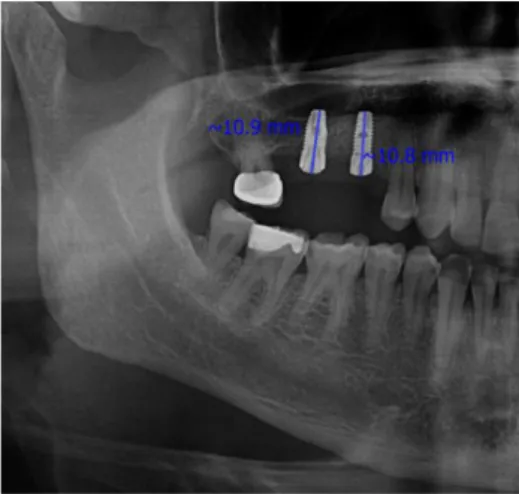
A letter confirming ethical approval for the study was obtained from Pamukkale University, Faculty of Medicine, Ethics Committee (number: 60116787-020/32012). Eighty-eight patients with no pathological lesions or bone disease who had undergone implant surgery at the Pamukkale University, Faculty of Dentistry (Denizli, Turkey) with a clear PRI taken in the correct position after the implant surgery were included in the study. PRIs were randomly chosen from the patient archive of the Dentomaxillofacial Radiology Clinic. None of the PRIs were obtained specifically for this study. All radiographs were taken by using the same panoramic machine (OP 200D) and by the same x-ray technician (S.C.) who used a standard exposure protocol (66kV/10mA/16s). All patients were positioned using a bite block according to the manufacturer’s instructions during the exposure of PRIs. The 240 dental implants (53 anterior, 69 premolar and 118 molar region), which dimensions were previously known, were measured on the postsurgical PRIs by a dentomaxillofacial radiologist (BK. A. who didn’t participate in the surgery) with the scaling tool of the panoramic system (Figure 1).
Using postsurgical PRIs, the magnification rate was determined for each region by the following formula:
Measured length of the implant on PRI /Actual length of the implant
Statistical data analysis was performed using a computer software (SPSS 21.0 version IBM Corp., Chicago, IL, USA). The Kolmogorov-Smirnov test was performed to determine whether the data showed a normal distribution. The paired t test was used to analyze the difference between the measured and actual vertical length of the implants (p<0.05). Pearson correlation analysis was used to determine the correlation between the actual length and the measured length of the implant (p<0.05).
RESULTS
A statistically significant difference was found between the measured and actual size of the implants in all regions (p<0.05). The difference between the actual size and measured size of the implants were 0.50 mm in the anterior region, 0.97 mm in the premolar region and 0.83 mm in the molar region (Table 1).
Table 1. The relationship between the actual size of the implants
and the measured size on the PRI of the implants according to the regions (M ±SD).
M: Mean; SD: Standard Deviation
Region Measured size (M ±SD) Actual size (M ±SD) n Averages of differences t p Anterior 11.89 ±1.55 11.84 ±1.55 53 0.50 ±0.12 -2.97 0.005 Premolar 11.12 ±1.46 11.02 ±1.44 69 0.97 ±0.12 -6.40 <0.001 Molar 10.40 ±1.53 10.32 ±1.50 118 0.83 ±0.12 -7.47 <0.001
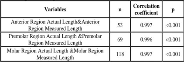
212 The correlation coefficient was close to 1 for all regions (Table 2).
Table 2. Pearson correlation between actual and measured values
according to the regions.
The magnification rates was close to 1 for all regions (Table 3).
Table 3. Magnification rates according to the regions.
In the user manual of OP200D, the magnification rate is specified as 1.3. However
the manual also states that CliniviewTM software
automatically corrects the growth rate. In this
study values corrected with CliniviewTM
software were accounted.
DISCUSSION
The choice of implant length and width is generally determined by the volume and density of the existing alveolar ridge, the location of adjacent teeth and vital anatomical structures, the type of prosthesis, and the treatment protocol. Theoretically, longer and wider implants should be preferred in order to resist the loaded forces and avoid fracture risk after
prosthetic treatment.9 On the other hand, studies
involving new implant designs/surfaces have reported that the failure rate of short and long implants is comparable and there is no relationship between implant diameter and
survival ratio.2, 9, 20 On the contrary, it has also
been argued that cases with fresh sockets require larger and/or longer implants to ensure primary implant stability and to reduce the distance between implant and bone socket
walls.21 However, Vazquez et al. pointed out
that protecting the mandibular canal was always
their priority. They reported that the use of short implants does not jeopardize the long-term implant success rate and may reduce nerve injury risk associated with implant placement in the posterior segment of the mandible, especially when the mandibular canal is
difficult to localize on the radiograph.22
Schropp et al.9 argued that the choice of
implant size was greatly influenced by the radiographic technique used for presurgical treatment planning. They also reported that the lack of cross-sectional information may lead to
the use of shorter and narrower implant sizes.23
However, they reported that the implants planned with cross-sectional imaging could not be considered more successful. In addition, they reported that there was no consensus on the need for cross-sectional imaging when a
panoramic radiograph was already present.9
The information provided by cross-sectional images are valuable in giving detailed and 1:1 images, detecting lingual undercuts in the posterior zone of the mandible. If lingual perforation occurs during implant operation, this can result in life-threatening bleeding and/or airway obstruction due to the impact on
the large arteries of the mandibular base.18 In
order to avoid the complications associated with mandibular canal, it is recommended that the distance between the lower limit of the implant and the mandibular canal should be at least 2mm taking the magnification rate of the PR in
consideration.6, 9, 14 Although CT and CBCT
images are known to give detailed and 1:1 rearranged images, it is recommended that the distance between the implant and mandibular canal should be at least 1.7 mm. This value is
similar to the recommended value for PR.15
CBCT may be preferred when 3D imaging is required, but effective radiation dose of CBCT is much higher and more expensive than
traditional dental radiographs.1, 9 For these
reasons; we preferred to plan this study on PR. PRIs tend to underestimate the distance between the alveolar crest and the upper border
Variables n Correlation
coefficient p
Anterior Region Actual Length&Anterior
Region Measured Length 53 0.997 <0.001 Premolar Region Actual Length &Premolar
Region Measured Length 69 0.996 <0.001 Molar Region Actual Length &Molar Region
Measured Length 118 0.997 <0.001
Region of implant Magnification rate
Anterior region 1.004 (ranging from 0.98 to 1.02)
Premolar region 1.008 (ranging from 0.98 to 1.03)
213
of the mandibular canal.24 Underestimation of
this distance is less harmful than
overestimation. In the case of an
overestimation, a long implant may damage the
nerve.15 Frie et al.18 found a magnification rate
of 1.27 ± 0.01. They also showed that the height of the vertical bone measured by spiral tomogram was 1 mm longer than the height of the bone measured by PR. Because spiral tomograms tend to overestimate the distance, assessment of the vertical bone height can be
risky if measured only by spiral CT.22
There are also studies reporting that PR shows overestimation in posterior mandibular
measurements.4, 5 Rockenbach et al.4 estimated
that PR overestimated linear measurements on the mandibular field. Nevertheless, they stated that PR and linear tomography of implant site measurements can be used safely with a safety margin of 2 mm.
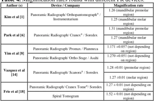
The accuracy of bone measurements on PR
has been questioned because of the
magnification and distortion (Table 4).14, 25
Table 4. Magnification rates found with different PR devices.
Knowing the correct magnification rate will allow more precise selection of implant size. A previous study on imaging procedures of implant treatment has suggested that the correct magnification rate should be determined for the calibration of the region to which the implant is
to be applied. 2, 9 For this reason, we aimed to
determine the correct magnification rate for the OP200D in this study.
It has also been reported that the use of bite blocks reduces positional errors while PRIs are
taken.5, 6, 14 Therefore, in our study the patients
used bite blocks and their positioning was done according to the manufacturer's instructions.
No single implant trademark was used in this study. In addition, grouping of maxilla and mandible was not performed in implant measurements. These are the shortcomings of this study.
CONCLUSIONS
Within the limitations of this study, following conclusions can be drawn; the magnification rate of OP200D is close to 1 and it gives errors less than 1 mm in vertical measurements made
with CliniviewTM software. Therefore, except
the cases where the location of the mandibular canal and lingual undercuts in the posterior region of the mandible cannot be clearly detected and cross sectional images are required; PRs with known magnification rates can be used with easy access, low radiation dose and low cost, in the vertical measurements with a 1mm confidence interval.
REFERENCES
1. Kim YK, Park JY, Kim SG, Kim JS, Kim
JD. Magnification rate of digital panoramic radiographs and its effectiveness for pre-operative assessment of dental implants. Dentomaxillofac Radiol 2011;40:76-83.
2. Schropp L, Stavropoulos A, Gotfredsen E,
Wenzel A. Calibration of radiographs by a reference metal ball affects preoperative selection of implant size. Clin Oral Investig 2009;13:375-381.
3. Mehra A, Pai KM. Evaluation of
dimensional accuracy of panoramic cross-sectional tomography, its ability to identify the inferior alveolar canal, and its impact on estimation of appropriate implant dimensions in the mandibular posterior region. Clin Implant Dent Relat Res 2012;14:100-111.
4. Rockenbach MI, Sampaio MC, Costa LJ,
Costa NP. Evaluation of mandibular implant sites: correlation between panoramic and linear tomography. Braz Dent J 2003;14:209-213.
Author (s) Device / Company Magnification rate
Kim et al [1] Panoramic Radiograph/ Orthopantomograph ® / Instrumentarium 1.26 (mandibular premolar region) 1.25 (mandibular molar region) Park et al [6] Panoramic Radiograph/ Cranex® / Soredex
1.31 (mandibular premolar region) 1.27 (mandibular molar
region) Yim et al [8]
Panoramic Radiograph/ Promax / Planmeca 1.171 ±0.057 (not depending on region) Panoramic Radiograph/ Ortho Stage / Asahi 1.270 ±0.051 (not depending on region) Vazquez et al
[14] Panoramic Radiograph/ Scanora ® / Soredex
1.28 ±0.01 (premolar region) 1.27 ±0.01 (molar region) Frie et al [18]
Panoramic Radiograph/ Cranex Tome®/ Soredex 1.27 ± 0.01 (not depending on region) Spiral Tomogram 1.52 ± 0.01 (not depending on region)
214
5. Haghnegahdar A, Bronoosh P. Accuracy of
linear vertical measurements in posterior mandible on panoramic view. Dent Res J (Isfahan) 2013;10:220-224.
6. Park JB. The evaluation of digital panoramic
radiographs taken for implant dentistry in the daily practice. Med Oral Patol Oral Cir Bucal 2010;15:663-666.
7. Sakakura CE, Morais JA, Loffredo LC, Scaf
G. A survey of radiographic prescription in dental implant assessment. Dentomaxillofac Radiol 2003;32:397-400.
8. Yim JH, Ryu DM, Lee BS, Kwon YD.
Analysis of digitalized panorama and cone beam computed tomographic image distortion for the diagnosis of dental implant surgery. J Craniofac Surg 2011;22:669-673.
9. Schropp L, Stavropoulos A, Gotfredsen E,
Wenzel A. Comparison of panoramic and conventional cross‐sectional tomography for preoperative selection of implant size. Clin Oral Implants Res 2011;22:424-429.
10. Lindh C, Petersson A, Klinge B.
Visualisation of the mandibular canal by different radiographic techniques. Clin Oral Implants Res 1992;3:90-97.
11. Sonick M. A comparison of the accuracy of
periapical, panoramic, and computerized
tomographic radiographs in locating the mandibular canal. Int J Oral Maxillofac Implants 1994;9:455-460.
12. Kitai N, Mukai Y, Murabayashi M,
Kawabata A, Washino K, Matsuoka M, Shimizu I, Katsumata A. Measurement accuracy with a new dental panoramic radiographic technique based on tomosynthesis. Angle Orthod 2013;83:117-126.
13. Nikneshan S, Sharafi M, Emadi N.
Evaluation of the accuracy of linear and angular measurements on panoramic radiographs taken at different positions. Imaging Sci Dent 2013;43:191-196.
14. Vazquez L, Nizam Al Din Y, Christoph
Belser U, Combescure C, Bernard JP. Reliability of the vertical magnification factor on panoramic radiographs: clinical implications
for posterior mandibular implants. Clin Oral Implants Res 2011;22:1420-1425.
15. Vazquez L, Nizamaldin Y, Combescure C,
Nedir R, Bischof M, Dohan Ehrenfest DM, Carrel JP, Belser UC. Accuracy of vertical
height measurements on direct digital
panoramic radiographs using posterior
mandibular implants and metal balls as reference objects. Dentomaxillofac Radiol 2013;42:20110429.
16. Tal H, Moses O. A comparison of panoramic
radiography with computed tomography in the planning of implant surgery. Dentomaxillofac Radiol 1991;20:40-42.
17. Volohansky A, Cleaton-Jones P, Drummond
S, Bönecker M. Technique for linear measurement on panoramic and periapical radiographs: a pilot study. Quintessence Int 2006;37:191-197.
18. Frei C, Buser D, Dula K. Study on the
necessity for cross-section imaging of the posterior mandible for treatment planning of standard cases in implant dentistry. Clin Oral Implants Res 2004;15:490-497.
19. Stramotas S, Geenty JP, Petocz P,
Darendeliler MA. Accuracy of linear and
angular measurements on panoramic
radiographs taken at various positions in vitro. Eur J Orthod 2002;24:43-52.
20. Renouard F, Nisand D. Impact of implant
length and diameter on survival rates. Clin Oral Implants Res 2006;17:35-51.
21. Degidi M, Piattelli A, Iezzi G, Carinci F. Do
longer implants improve clinical outcome in immediate loading? Int J Oral and Maxillofac Surg 2007;36:1172-1176.
22. Vazquez L, Saulacic N, Belser U, Bernard
JP. Efficacy of panoramic radiographs in the preoperative planning of posterior mandibular implants: a prospective clinical study of 1527 consecutively treated patients. Clin Oral Implants Res 2008;19:81-85.
23. Schropp L, Wenzel A, Kostopoulos L.
Impact of conventional tomography on prediction of the appropriate implant size. Oral Surg Oral Med Oral Pathol Oral Radiol Endod 2001;92:458-463.
215
24. Lindh C, Petersson A, Klinge B.
Measurements of distances related to the mandibular canal in radiographs. Clin Oral Implants Res 1995;6:96-103.
25. Yassaei S, Ezoddini-Ardakani F, Ostovar N.
Predicting the actual length of premolar teeth on the basis of panoramic radiology. Indian J Dent Res 2010;21:468-473.
Corresponding Author
Burak Kerem Apaydın Pamukkale University Faculty of Dentistry,
Department of Dentomaxillofacial Radiology, Denizli, Turkey
Tel : +90 258 2964225
Fax : +90 258 2961763
E-mail : [email protected]
Conflict of interest: The authors declare that