INTRODUCTION
Chronic urticaria (CU) is defined as the persistence of an urti-carial episode for at least 6 weeks.1 The prevalence of CU in children is one-tenth of that in adults (0.1% vs 1%).2,3 Autoim-mune disorders, physical stimuli, infections, vasculitis, and al-lergies are the primary causes of CU. However, an underlying cause can only be identified in 20% to 55% of pediatric cases in spite of thorough clinical and laboratory investigations.4,5 CU is classified according to whether it is inducible or not into chron-ic spontaneous urtchron-icaria (CSU) or chronchron-ic inducible urtchron-icaria (CIU). CSU may be attributed to known or unknown causes.6,7
Matrix-metalloproteinases (MMPs) are a large family of zinc-dependent endopeptidases that can degrade many compo-nents of the extracellular matrix and other extracellular pro-teins.8 MMP-9 belongs to the gelatinase subfamily of MMPs and cleaves gelatin and type 4 collagen, which is the main com-ponent of the basement membrane. This cleavage helps in-flammatory cells reach the site of inflammation.9 Additionally,
MMPs can cleave proinflammatory chemokines, thereby regu-lating their functions and moduregu-lating inflammatory process-es.10,11 MMP-9 can be synthesized by many types of cells, in-cluding macrophages, neutrophils, T cells, and mast cells, and play a substantial role in autoimmune diseases, cancer, cardio-vascular diseases, and allergic diseases.9,12,13
A few studies (with conflicting results) exist on the role of MMP-9 in the pathogenesis of CSU in adults; however, to our knowledge no studies have been performed in children. This study aims to clarify this topic.
Plasma Levels of Matrix Metalloproteinase-9 in Children With
Chronic Spontaneous Urticaria
Fatih Dilek,
1* Deniz Ozceker,
2Emin Ozkaya,
1Zeynep Tamay,
2Mebrure Yazici,
1Siddika Kesgin,
3Abdurrahim Kocyigit,
3Nermin Guler
41Department of Pediatric Allergy and Immunology, Bezmialem Vakif University Medical Faculty, Istanbul, Turkey 2Department of Pediatric Allergy, Istanbul University Istanbul Medical Faculty, Istanbul, Turkey
3Department of Clinical Biochemistry, Bezmialem Vakif University Medical Faculty, Istanbul, Turkey 4Department of Pediatrics, Istanbul Bilim University, Istanbul, Turkey
This is an Open Access article distributed under the terms of the Creative Commons Attribution Non-Commercial License (http://creativecommons.org/licenses/by-nc/3.0/) which permits unrestricted non-commercial use, distribution, and reproduction in any medium, provided the original work is properly cited.
Purpose: Chronic spontaneous urticaria (CSU) is a disease that is primarily seen in adults and is comparatively rare in children. Consequently, only a few studies have focused on the pathogenesis of the disease in children. This study investigated the possible role of metalloproteinase-9 (MMP-9) in the pathogenesis of CSU in children. Methods: The study group was composed of 54 children with CSU; 34 healthy children comprised the con-trol group. The demographic and clinical features of the study group were extensively evaluated, and laboratory assessments were also performed. An enzyme-linked immunosorbent assay was used to evaluate levels of plasma MMP-9. Disease activity was quantified using the urticaria activity score (UAS). Results: The median value of plasma MMP-9 was 108.9 ng/mL (interquartile range, 93.3-124.1) in the CSU group and 87.8 ng/mL (69.4-103.0) in the control group. The difference between the 2 groups was statistically significant (P<0.001). Also, MMP-9 levels showed a signifi-cant positive correlation with UAS (rho=0.57, P<0.001). Twenty-six percent of patients had positive autologous serum skin test (ASST) results. Nei-ther UAS nor plasma MMP-9 levels were significantly different between ASST-positive and -negative patients (P>0.05). Conclusions: Plasma MMP-9 levels were elevated in children with CSU and were positively correlated with disease activity. MMP-9 may be both a good biomarker of disease activity and a potential therapeutic target in CSU.
Key Words: Chronic spontaneous urticaria; children; disease severity; MMP-9
Correspondence to: Fatih Dilek, MD, Department of Pediatric Allergy and Immunology, Bezmialem Vakif University Medical Faculty, Adnan Menderes Bulvari Vatan Caddesi 34093 Fatih, Istanbul, Turkey.
Tel: +902125232288; Fax: +902124531870; E-mail: [email protected] Received: December 8, 2015; Revised: March 19, 2016;
Accepted: March 23, 2016
•This work was supported by a grant from the Bezmialem Vakif University.
•There are no financial or other issues that might lead to conflict of interest.
Allergy Asthma Immunol Res. 2016 November;8(6):522-526. http://dx.doi.org/10.4168/aair.2016.8.6.522 pISSN 2092-7355 • eISSN 2092-7363
MATERIALS AND METHODS Study population
Seventy-two consecutive patients who were referred between January and June of 2015 to the pediatric allergy outpatient clinics of 2 university hospitals (Istanbul University/Istanbul Medical Faculty and Bezmialem Vakif University) with the di-agnosis of CSU were assessed for study eligibility. Patients who were diagnosed with CIU and whose potential causes of CU had been defined (except autoimmunity) were excluded. Diag-nosis was based on the European Academy of Allergy and Clin-ical Immunology guidelines.6 None of the patients were taking corticosteroids, omalizumab, or immunosuppressive drugs. Detailed medical histories were obtained, and physical exami-nations were performed. Children with diseases that influence plasma MMP-9 levels, including concomitantly diagnosed asthma, allergic rhinitis, atopic dermatitis, or other identified autoimmune disorders, were excluded from the study.9,13,14 Af-ter exclusion, 54 patients with CSU were finally enrolled.
The control group included 34 healthy children who were pe-riodically attending pediatric welfare clinics in the same hospi-tals for regular checkups. Children in the control group did not have a history of either acute or chronic urticaria, had never been diagnosed with any allergic disease, and were not taking any medications. Additionally, no signs of infectious disease were present in any of the subjects in the control group. The study was performed in accordance with the tenets of the Dec-laration of Helsinki and good clinical practice, and was ap-proved by the Bezmialem Vakif University Ethical Committee. All study patients and their parents were given information about the study, and signed consent was obtained from the parents.
Assessment of disease activity
Disease activity was evaluated by the same pediatric allergist using the urticaria activity score (UAS) according to the EAACI/ GA2LEN/EDF/WAO Guidelines.15 UASs were calculated in in-terviews with patients and families as well as during physical examination. UAS consisted of the sum of the wheal number score and the pruritus score, which ranged from 0 to 6. The wheal numbers are graded as follows: 0, none; 1, Mild (<20 wheals/24 hours); 2, Moderate (20-50 wheals/24 hours); 3, In-tense (>50 wheals/24 hours or large confluent areas of wheals). Moreover, the severity of itching is graded as follows: 0, none; 1, Mild (present but not annoying or troublesome); 2, Moderate (troublesome but does not interfere with normal daily activity or sleep); 3, Intense (severe pruritus, which is sufficiently trou-blesome to interfere with normal daily activity or sleep).15 Blood sample collection
Antihistamines and montelukast were discontinued at least 24 hours before blood sampling. After overnight fasting,
pe-ripheral blood samples (total, 4 mL) were collected from an an-tecubital vein into heparinized tubes; thereafter, blood was cen-trifuged at 1,500×g for 10 minutes to obtain the plasma. The separated plasma was stored at -80°C until further analysis of MMP-9 levels.
Laboratory investigation
For standard laboratory procedures, the following tests were obtained: thyroid-stimulating hormone, free thyroxin, anti-thy-roid peroxidase and anti-thyroglobulin antibodies, liver func-tion tests, complete blood count, erythrocyte sedimentafunc-tion rate, serum total immunoglobulin E (IgE), urinalysis with test strips and urine cultures, microscopic investigation of stool for parasites, and stool enzyme immunoassay for Helicobacter py-lori antigen.
Skin prick tests and autologous serum skin tests (ASSTs) were also performed in the CSU group, but not for the control group. Commercial allergen solutions manufactured by Stallergenes (Paris, France) were used for skin prick tests. Among the test al-lergens were 10 different aeroalal-lergens consisting of grass pol-len mixture, Dermatophagoides pteronyssinus, Dermatophagoi-des farinae, Alternaria, Aspergillus mixture, weed pollen mix-ture, birch, cypress, cat epithelia, dog epithelia, and 6 food aller-gens consisting of milk, egg white, egg yolk, peanut, wheat, and cocoa. Skin prick tests were considered positive with the pres-ence of a wheal with at least 3-mm maximum diameter after subtraction of the negative value. ASST was performed using the method described by Sabroe et al.16 We were not able to perform skin prick tests and ASST on 8 patients because their antihistamine-containing medications could not be discontin-ued due to the intensity and severity of their symptoms. Measurement of plasma MMP-9 levels
Plasma levels of 9 were assessed using a human MMP-9 enzyme linked immunosorbent assay (ELISA) kit (Bender Med Systems, Vienna, Austria). Samples were thawed at room temperature and were then centrifuged at 3,000 rpm for 10 minutes. Samples were diluted to obtain the appropriate con-centrations, and an ELISA was performed according to the manufacturer’s instructions. Supplied standards were used to generate standard curves. The samples and standards were added to the wells. Unbound protein was removed by washing, and conjugate was added. After a color reaction with a sub-strate, the optical density was recorded using an automated ELISA reader at a wavelength of 450 nm. The absorbance at 450 nm was converted to ng/mL for MMP-9. The minimal detec-tion limit was 0.05 ng/mL for MMP-9.
Statistical analysis
Statistical analyses were performed using IBM SPSS 19 (IBM, Armonk, NY, USA). The Shapiro-Wilk test was used to test dis-tributions for normality. Parametric data are expressed as the
mean±standard deviation (SD), and non-parametric data are expressed as the median, interquartile range (IQR). The Mann-Whitney U test was used to calculate the differences in vari-ables between groups. The correlation between 2 varivari-ables was assessed using the Spearman rank correlation coefficient. Cate-gorical data were evaluated using the chi square test; P<0.05 was accepted as statistically significant.
RESULTS
General characteristics
The CSU group consisted of 33 boys and 21 girls, and the con-trol group consisted of 18 boys and 16 girls. The mean ages of the CSU and control group participants were 10.8±4.2 and 10.3±3.9 years, respectively. No significant differences in age or gender were present between the 2 groups (P>0.05). The me-dian duration of disease was 13 months (7-23.3 months), and the median value of UAS was 4 (2-5). Some demographic and clinical features of the patients and the control subjects are shown in Table.
Plasma MMP-9 levels
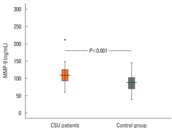
The median values of plasma MMP-9 were 108.9 ng/mL (93.3-124.1) in the CSU group and 87.8 ng/mL (69.4-103.0) in the control group. The difference between the 2 groups was sta-tistically significant (P<0.001; Fig. 1). Additionally, plasma MMP-9 levels showed a significant positive correlation with UAS (rho=0.57, P<0.001; Fig. 2). We detected ASST positivity in
26% of CSU group patients; plasma MMP-9 levels were not dif-ferent between patients with ASSTs positivity and those without (P>0.05).
DISCUSSION
Our results clearly showed that children with CSU had elevat-ed MMP-9 levels comparelevat-ed to healthy control subjects and that plasma MMP-9 levels were positively correlated with disease activity. Although some studies exist that evaluate the plasma MMP-9 levels in adult patients with CSU, this is the first study that focuses on the pediatric age group. This focus is especially important because CSU is rare in children, and recommenda-tions for its management and also because treatment in chil-dren are based on data obtained from studies conducted on
Table. Some demographic and clinical features of the study and control groups CSU patients
(n=54) Control group (n=34) P value Age, year (mean±SD) 10.8±4.2 10.3±3.9 >0.05
Gender (male/female) 33/21 18/16 >0.05
Disease duration mon
(median, [IQR]) 13 (7-23.3)
-Aeroallergen sensitization (n [%]) 8/46 (17%) ND
ASST positivity (n [%]) ND
ASST-positive 12/46 (26%)
ASST-negative 32/46 (74%)
UAS (median, [IQR]) 4 (2-5)
-Treatment (n [%]) -Second-generation antihistamines at licensed doses 29 (54%) Second-generation antihistamines at high doses 9 (17%) Combination of second-generation antihistamines+montelukast 16 (29%)
CSU, chronic spontaneous urticaria; SD, standard deviation; IQR, interquartile range; ASST, autologus serum skin test; UAS, urticaria activity score; ND, not
done. Fig. 2.levels and urticaria activity scores (UASs) in patients with CSU. Correlation graphs between plasma matrix metalloprotease-9 (MMP-9)
MMP-9 (ng/mL) 1 2 3 4 5 6 UAS 250 200 150 100 50 0
Fig. 1. Plasma matrix metalloprotease-9 (MMP-9) levels in the study and con-trol groups. MMP-9 (ng/mL) CSU patients * Control group 300 250 200 150 100 50 0 P<0.001
adults.17 Even comprehensive guidelines written on this topic include little information about children.1,6,15 Undoubtedly, it is not the proper approach to interpret children as “small adults.”17 Therefore, further studies are needed that focus on children, so the necessity of adapting the experiences of adults with the dis-ease (and with treatment) is lessened or eliminated.
The activity of CSU can be assessed both by the physician during the examination (UAS) or by patients measuring and documenting urticaria through 24-hour self-evaluation scores once daily for 7 days (UAS7).6 UAS7 includes full family-derived information and scores, and provides an overview of the overall activity of the disease. We preferred to use UAS in this study be-cause we attempted to compare momentary activity scores and cytokine levels. In addition, we aimed to reduce the influence on scoring of subjective assessments provided by families as much as possible. Although UAS has some limitations, it is sim-ple, widely accepted, and only validated for assessing the sever-ity of CU.6,15
The exact pathogenesis of CSU is not well delineated. The presence of persistent activation of dermal mast cells is a hall-mark of CSU pathogenesis, but the underlying mechanisms of mast cell activation are an enigma.1,18 Urticarial wheals are characterized by dermal edema, vasodilatation, and perivascu-lar infiltrates comprising lymphocytes, monocytes, neutrophils, eosinophils, and basophils.18,19 Altered numbers and imbal-anced cytokine production of T cells have also been reported in patients with CSU.20,21
Kessel et al.22 first reported that adult patients with CU have el-evated plasma MMP-9 levels compared to healthy subjects, be-sides the presence of a positive correlation between MMP-9 levels and UAS. They also demonstrated that the CD4+ T cells of these patients were highly activated, concluding that activat-ed CD4+ T cells residing in urticarial lesions may cause dermal mast cell activation and MMP-9 secretion.22 Tedeschi et al.23 and Altricher et al.24 also reported elevated plasma MMP-9 lev-els in patients with CU compared to healthy subjects in their studies conducted in adults. Conversely, in study by Altricher et al. the authors stated that disease activity is not correlated with MMP-9 levels, and that MMP-9 should not to be used to moni-tor disease activity or the efficacy of treatment.24
Wheals, flares, and angioedema, which are characteristics of CSU, develop as a result of increased vascular permeability.25 The literature provides some evidence that the presence of MMPs leads to increased vascular permeability, primarily through the disruption of tight junction proteins.26 Further-more, MMPs strongly induce leukocyte migration.27 These fea-tures of MMP-9 could explain some of the key points behind the pathogenesis of CSU. The therapeutic efficacy of MMP-9 activity inhibition is a highly active area of research in the treat-ment of cancer and ischemic strokes.28,29 Several methods and drugs have been proposed for this purpose in the literature, in-cluding lentiviral-mediated MMP-9 gene silencing, MMP-9
neutralizing antibodies, recombinant tissue inhibitor of matrix metalloproteinases-1, gold nanoparticles, minocycline, and synthetic MMP inhibitors.28,29 Therefore, MMP-9 seems not only to be a good biomarker of disease activity, but also to be a promising therapeutic target.
CONCLUSIONS
Plasma MMP-9 levels were elevated in pediatric patients with CSU and were positively correlated with the UAS. Therefore, MMP-9 seems to be both a good biomarker for disease activity and a promising therapeutic target. Several drugs and methods that can inhibit MMP-9 activity are described in the literature. Additionally, we emphasize that this is the first and only study conducted on children. We need further studies to say the last word on this subject.
REFERENCES
1. Powell RJ, Leech SC, Till S, Huber PA, Nasser SM, Clark AT; British Society for Allergy and Clinical Immunology. BSACI guideline for the management of chronic urticaria and angioedema. Clin Exp Allergy 2015;45:547-65.
2. Maurer M, Weller K, Bindslev-Jensen C, Giménez-Arnau A, Bous-quet PJ, BousBous-quet J, et al. Unmet clinical needs in chronic spontane-ous urticaria. A GA²LEN task force report. Allergy 2011;66:317-30. 3. Khakoo G, Sofianou-Katsoulis A, Perkin MR, Lack G. Clinical
fea-tures and natural history of physical urticaria in children. Pediatr Allergy Immunol 2008;19:363-6.
4. Volonakis M, Katsarou-Katsari A, Stratigos J. Etiologic factors in childhood chronic urticaria. Ann Allergy 1992;69:61-5.
5. Ghosh S, Kanwar AJ, Kaur S. Urticaria in children. Pediatr Derma-tol 1993;10:107-10.
6. Zuberbier T, Aberer W, Asero R, Bindslev-Jensen C, Brzoza Z, Ca-nonica GW, et al. The EAACI/GA(2) LEN/EDF/WAO guideline for the definition, classification, diagnosis, and management of urti-caria: the 2013 revision and update. Allergy 2014;69:868-87. 7. Vestergaard C, Deleuran M. Chronic spontaneous urticaria: latest
developments in aetiology, diagnosis and therapy. Ther Adv Chron-ic Dis 2015;6:304-13.
8. Wells JM, Gaggar A, Blalock JE. MMP generated matrikines. Matrix Biol 2015;44-46:122-9.
9. Ram M, Sherer Y, Shoenfeld Y. Matrix metalloproteinase-9 and au-toimmune diseases. J Clin Immunol 2006;26:299-307.
10. Van den Steen PE, Proost P, Wuyts A, Van Damme J, Opdenakker G. Neutrophil gelatinase B potentiates interleukin-8 tenfold by ami-noterminal processing, whereas it degrades CTAP-III, PF-4, and GRO-alpha and leaves RANTES and MCP-2 intact. Blood 2000; 96:2673-81.
11. Song J, Wu C, Zhang X, Sorokin LM. In vivo processing of CXCL5 (LIX) by matrix metalloproteinase (MMP)-2 and MMP-9 promotes early neutrophil recruitment in IL-1β-induced peritonitis. J Immu-nol 2013;190:401-10.
12. Yabluchanskiy A, Ma Y, Chiao YA, Lopez EF, Voorhees AP, Toba H, et al. Cardiac aging is initiated by matrix metalloproteinase-9-me-diated endothelial dysfunction. Am J Physiol Heart Circ Physiol
2014;306:H1398-407.
13. Belleguic C, Corbel M, Germain N, Léna H, Boichot E, Delaval PH, et al. Increased release of matrix metalloproteinase-9 in the plas-ma of acute severe asthplas-matic patients. Clin Exp Allergy 2002;32: 217-23.
14. Harper JI, Godwin H, Green A, Wilkes LE, Holden NJ, Moffatt M, et al. A study of matrix metalloproteinase expression and activity in atopic dermatitis using a novel skin wash sampling assay for func-tional biomarker analysis. Br J Dermatol 2010;162:397-403. 15. Zuberbier T, Asero R, Bindslev-Jensen C, Walter Canonica G,
Church MK, Giménez-Arnau A, et al. EAACI/GA(2)LEN/EDF/WAO guideline: definition, classification and diagnosis of urticaria. Aller-gy 2009;64:1417-26.
16. Sabroe RA, Grattan CE, Francis DM, Barr RM, Kobza Black A, Greaves MW. The autologous serum skin test: a screening test for autoantibodies in chronic idiopathic urticaria. Br J Dermatol 1999; 140:446-52.
17. Church MK, Weller K, Stock P, Maurer M. Chronic spontaneous ur-ticaria in children: itching for insight. Pediatr Allergy Immunol 2011;22:1-8.
18. Jain S. Pathogenesis of chronic urticaria: an overview. Dermatol Res Pract 2014;2014:674709.
19. Caproni M, Giomi B, Volpi W, Melani L, Schincaglia E, Macchia D, et al. Chronic idiopathic urticaria: infiltrating cells and related cy-tokines in autologous serum-induced wheals. Clin Immunol 2005; 114:284-92.
20. Sun RS, Sui JF, Chen XH, Ran XZ, Yang ZF, Guan WD, et al. Detec-tion of CD4+ CD25+ FOXP3+ regulatory T cells in peripheral blood of patients with chronic autoimmune urticaria. Australas J Derma-tol 2011;52:e15-8.
21. Dos Santos JC, Azor MH, Nojima VY, Lourenço FD, Prearo E,
Maru-ta CW, et al. Increased circulating pro-inflammatory cytokines and imbalanced regulatory T-cell cytokines production in chronic idio-pathic urticaria. Int Immunopharmacol 2008;8:1433-40.
22. Kessel A, Bishara R, Amital A, Bamberger E, Sabo E, Grushko G, et al. Increased plasma levels of matrix metalloproteinase-9 are asso-ciated with the severity of chronic urticaria. Clin Exp Allergy 2005; 35:221-5.
23. Tedeschi A, Asero R, Lorini M, Marzano AV, Cugno M. Plasma lev-els of matrix metalloproteinase-9 in chronic urticaria patients cor-relate with disease severity and C-reactive protein but not with cir-culating histamine-releasing factors. Clin Exp Allergy 2010;40:875-81.
24. Altrichter S, Boodstein N, Maurer M. Matrix metalloproteinase-9: a novel biomarker for monitoring disease activity in patients with chronic urticaria patients? Allergy 2009;64:652-6.
25. Carr TF, Saltoun CA. Chapter 21: urticaria and angioedema. Aller-gy Asthma Proc 2012;33 Suppl 1:S70-2.
26. Bauer AT, Bürgers HF, Rabie T, Marti HH. Matrix metalloprotein-ase-9 mediates hypoxia-induced vascular leakage in the brain via tight junction rearrangement. J Cereb Blood Flow Metab 2010;30: 837-48.
27. Song J, Wu C, Korpos E, Zhang X, Agrawal SM, Wang Y, et al. Focal MMP-2 and MMP-9 activity at the blood-brain barrier promotes chemokine-induced leukocyte migration. Cell Rep 2015;10:1040-54.
28. Herszényi L, Hritz I, Lakatos G, Varga MZ, Tulassay Z. The behavior of matrix metalloproteinases and their inhibitors in colorectal can-cer. Int J Mol Sci 2012;13:13240-63.
29. Chaturvedi M, Kaczmarek L. Mmp-9 inhibition: a therapeutic strategy in ischemic stroke. Mol Neurobiol 2014;49:563-73.