Dermoscopic Findings in Patients with Pigmented
Purpuric Dermatoses
Seray Külcü Çakmak
1, Arzu Kılıç
2, Ahu Yorulmaz
1, Duru Onan
1,
Derya Yayla
1, Ferda Artüz
11Ankara Numune Education and Research Hospital, Dermatology Clinic, Ankara,
Turkey, 2Balıkesir University Faculty of Medicine, Dermatology Clinic, Ankara, Turkey Corresponding author:
Seray Külcü Çakmak, MD
Aykon Park sit. A Blok no:14 742.sokak Yıldızevler Çankaya
Received: May 25, 2016 Accepted: October 5, 2016
ABSTRACT Pigmented purpuric dermatoses (PPD) are a group of chronic and relapsing cutaneous disorders characterized by a distinct purpuric rash. The di-agnosis is made with clinical and histopathological findings. Dermoscopy has rarely been used in the diagnosis of PPD. The aim of our study is to describe the dermoscopic findings in patients with PPD.
Eighteen patients who were clinically and histopathologically diagnosed with PPD were studied prospectively. The type and duration of PPD, associated dis-eases, and medication history of the patients were noted. Dermoscopic exami-nation was performed in all of the patients.
Four of the patients were women and 14 of them were men. 16 (88.8%) of them had Shamberg’s disease, 1 of them had lichen aureus, and 1 had purpura an-nularis telangiectoides. Dermoscopic examination revealed multiple irregular red dots, globules and/or patches and brown-coppery coloration on the back-ground in all of the patients, a network of interconnected brown lines in 8, lin-ear vessels in 9, brown dots in 3, grey dots in 3, twisted red loops in 5, comma-like vessels in 2, and red lacunae in 1 patient.
The dermoscopic examination of PPD might improve the accuracy of clinical diagnosis.
Key woRDS: pigmented purpuric dermatoses, dermoscopy, Schamberg’s dis-ease
INTRoDUCTIoN
Pigmented purpuric dermatoses (PPD) are a rela-tively uncommon group of diseases characterized by petechia, purpura, and brown, red, or orange pigmentation, mainly confined to the lower limbs (1,2). Five types of PPD have been defined, including Shamberg’s disease, lichen aureus, purpura annularis telangiectoides, eczematid-like purpura of Doucas and Kapetanakis and pigmented purpuric lichenoid dermatitis of Gougerot and Blum. Dermoscopy is a non-invasive method which can be used to enhance clinical examination (1). Dermoscopic features of PPD have very rarely been reported (3-8). The aim of our study was to describe the dermoscopic findings in patients with PPD.
MeTHoDS
The study was conducted in Numune Education and Research Hospital, Ankara, Turkey. The study was approved by institutional ethical committee (approv-al number: 733/2014). The contents of the approv(approv-al included dermoscopy of patients with PPD who gave informed consent. The study was performed on 18 consecutive patients who applied to our outpatient clinic and were clinically and histopathologically di-agnosed with PPD and were studied prospectively. The type and duration of PPD, associated diseases, and the medication history of the patients were re-corded. For each patient, a dermoscopic evaluation
was performed with Mole Max I Plus® (Derma Medical Systems GmbH, Vienna, Austria).
Table 1. Clinicoepidemiological characteristics and dermoscopic features of patients with pigmented
pur-puric dermatoses (PPD)
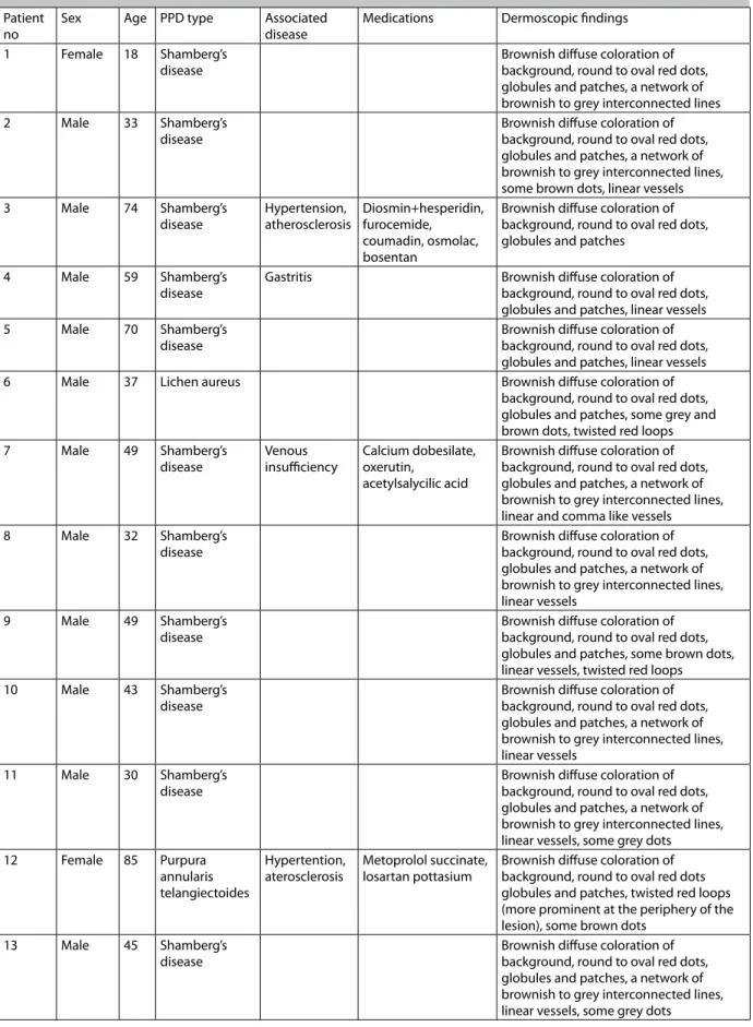
Patient
no Sex Age PPD type Associated disease Medications Dermoscopic findings
1 Female 18 Shamberg’s
disease Brownish diffuse coloration of background, round to oval red dots, globules and patches, a network of brownish to grey interconnected lines
2 Male 33 Shamberg’s
disease Brownish diffuse coloration of background, round to oval red dots, globules and patches, a network of brownish to grey interconnected lines, some brown dots, linear vessels
3 Male 74 Shamberg’s
disease Hypertension, atherosclerosis Diosmin+hesperidin, furocemide, coumadin, osmolac, bosentan
Brownish diffuse coloration of background, round to oval red dots, globules and patches
4 Male 59 Shamberg’s
disease Gastritis Brownish diffuse coloration of background, round to oval red dots, globules and patches, linear vessels
5 Male 70 Shamberg’s
disease Brownish diffuse coloration of background, round to oval red dots, globules and patches, linear vessels
6 Male 37 Lichen aureus Brownish diffuse coloration of
background, round to oval red dots, globules and patches, some grey and brown dots, twisted red loops
7 Male 49 Shamberg’s
disease Venous insufficiency Calcium dobesilate, oxerutin, acetylsalycilic acid
Brownish diffuse coloration of background, round to oval red dots, globules and patches, a network of brownish to grey interconnected lines, linear and comma like vessels
8 Male 32 Shamberg’s
disease Brownish diffuse coloration of background, round to oval red dots, globules and patches, a network of brownish to grey interconnected lines, linear vessels
9 Male 49 Shamberg’s
disease Brownish diffuse coloration of background, round to oval red dots, globules and patches, some brown dots, linear vessels, twisted red loops
10 Male 43 Shamberg’s
disease Brownish diffuse coloration of background, round to oval red dots, globules and patches, a network of brownish to grey interconnected lines, linear vessels
11 Male 30 Shamberg’s
disease Brownish diffuse coloration of background, round to oval red dots, globules and patches, a network of brownish to grey interconnected lines, linear vessels, some grey dots
12 Female 85 Purpura
annularis telangiectoides
Hypertention,
aterosclerosis Metoprolol succinate, losartan pottasium Brownish diffuse coloration of background, round to oval red dots globules and patches, twisted red loops (more prominent at the periphery of the lesion), some brown dots
13 Male 45 Shamberg’s
disease Brownish diffuse coloration of background, round to oval red dots, globules and patches, a network of brownish to grey interconnected lines, linear vessels, some grey dots
The statistical analysis was performed using SPSS software (version 20; SPSS Inc., Chicago IL, USA). Frequencies were calculated for variables related to demographic and clinical patient characteristics. Chi-square test or Fisher’s exact tests were used to deter-mine if there were statistical associations between the type of PPD and sex the type of PPD and age group, and the type of PPD and dermoscopic findings.
ReSULTS
Four (22.2%) of the patients were women, and 14 (77.7%) of them were men. The ages of the patients ranged between 18 and 85 years (mean ± standard deviation (SD) 47.055±18.092 years). When the ages of the patients were grouped as 18-35 years, 36- 50 years, and 50 years and over, there were 6 (33.3%) pa-tients in each group.
Sixteen (88.8%) of them had Shamberg’s disease, 1 (5.5%) had lichen aureus, and 1 (5.5%) had purpura annularis telangiectoides.
Six (33.3%) of the patients had associated diseas-es, which were hypertension (3 patients), hyperlipid-emia (1 patient), coronary artery disease (1 patient), and venous insufficiency (1 patient) (Table 1). The medications of the patients are also listed in Table 1. Notably, none of the patients had a history of onset of sign or symptoms of PPD after the initiation of these medications. Dermoscopic examination revealed multiple irregular red dots, globules, and/or patches and brown-coppery coloration on the background in all of the patients, a network of interconnected brown lines in 8 (44.4%), linear vessels in 9 (50.0%), brown dots in 3 (16.6%), grey dots in 3 (16.6%), twisted red loops in 5 (27.7%), comma-like vessels in 2 (11.1%), and red lacunae in 1 (5.5%) patient (Figure 1, 2 , 3). No significant association was found between the type of PPD and sex, and the type of PPD and age group, and the type of PPD and dermoscopic findings (P> 0.05).
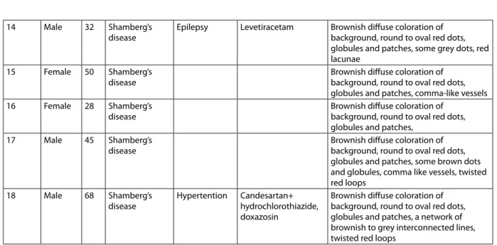
14 Male 32 Shamberg’s
disease Epilepsy Levetiracetam Brownish diffuse coloration of background, round to oval red dots, globules and patches, some grey dots, red lacunae
15 Female 50 Shamberg’s
disease Brownish diffuse coloration of background, round to oval red dots, globules and patches, comma-like vessels
16 Female 28 Shamberg’s
disease Brownish diffuse coloration of background, round to oval red dots, globules and patches,
17 Male 45 Shamberg’s
disease Brownish diffuse coloration of background, round to oval red dots, globules and patches, some brown dots and globules, comma like vessels, twisted red loops
18 Male 68 Shamberg’s
disease Hypertention Candesartan+ hydrochlorothiazide, doxazosin
Brownish diffuse coloration of background, round to oval red dots, globules and patches, a network of brownish to grey interconnected lines, twisted red loops
Figure 1. (patient no 6) Brownish diffuse coloration
of the background, round to oval red dots, globules and patches, some grey and brown dots, and twisted red loops.
Figure 2. (Patient no 12) Brownish diffuse coloration
of the background, round to oval red dots globules and patches, twisted red loops (more prominent in the periphery of the lesion), and some brown dots.
DISCUSSIoN
Pigmented purpuric dermatoses are a group of chronic and recurrent diseases that have overlap-ping clinical and histopathological features (3,9). Symmetrical, petechial, and macular brown, red, and patchy pigmentation is seen clinically (2). The histo-pathological features include superficial lymphocytic infiltration and marked hemosiderin deposition with erythrocyte extravasation (1).
PPD are more commonly observed in men be-tween their third and fifth decades of life (2,9). Simi-larly, 82.4% of our patients were men, and the mean age was 44.82±1.6 years. PPD are generally seen in the lower limbs, and in our patients the lesions were confined to the lower limbs except in one patient with PAT who had generalized lesions all over her body (2). Shamberg’s disease is the most commonly observed type of PPD: 88.2% of our patients had Shamberg’s disease.
The etiology of PPD is unknown, however venous hypertension, exercise, gravitational dependency, capillary fragility, focal infections, alcohol ingestion, and drugs such as acetaminophen, aspirin, adalin, glipizide, glybuzole, hydralazine, and rezerpine have been reported in the etiopathogenesis of PPD (1,2,9). Some disorders have also been found to be associat-ed with PPD, including diabetes mellitus, rheumatoid arthritis, and lupus erythematous (1,2,9). In our study, 3 patients had hypertension, 1 had venous hyper-tension, 1 had gastritis, and 1 had epilepsy; all were taking medications for their diseases. None of the pa-tients experienced onset of lesions after the initiation of the medications.
The diagnosis of PPD is based on with clinical and histopathological findings (1). Dermoscopy is a non-invasive procedure used for evaluating pigmented
lesions of the skin surface (4). It has also been used in the evaluation non-pigmented skin lesions such as psoriasis, lichen planus, eczema, and urticaria (5). There have been few reports describing the dermo-scopic features of PPD, of which the majority are case reports or case series. In these reports, the most com-monly observed features were brownish or coppery-red diffuse coloration of the background, round to oval red dots, globules, and patches, gray dots, and a network of grey interconnected lines. Punctate and linear vessels were also features of PPD that were de-scribed (3-8).
The dermoscopic features we observed in our patients were: multiple irregular red dots, globules and/or patches, and brown-coppery coloration on the background in all of the patients, a network of in-terconnected brown lines, linear vessels, brown dots, grey dots in twisted red loops, comma-like vessels, and red lacunae.
Brownish or coppery-red diffuse coloration of the background is thought to correlate with the typical histopathological finding of PPD, which is the pres-ence of dermal lymphocyte and histiocytes, extrava-sated red blood cells, and hemosiderin-laden mac-rophages. The round to oval red dots, globules, and patches are assumed to correspond to the extravasa-tion of red blood cells and increased number of blood vessels. Grey dots are accepted as the dermoscopic counterpart of hemosiderin-laden macrophages in the dermis and the network of brownish to gray in-terconnected lines are thought to correlate with the presence of hyperpigmentation of the basal cell layer and pigmentary incontinence of the upper dermis. Brown dots and globules may correspond to mela-nocytes and melanophages in the dermoepidermal junction. Linear vessels, twisted red looped comma-like vessels, and red lacunae may correspond to an increased number of dilated and swollen blood ves-sels (3-8).
The differential diagnosis of PPD includes angio-ma serpiginosum, stasis derangio-matitis, leukocytoclastic vasculitis, purpura, and scurvy (8).
In angioma serpiginosum, small, relatively well-demarcated, round to oval red lagoons can be seen, which may be accompanied by comma, hairpin-like vessels and patchy pigmentation dispersed through the background (10).
The presence of glomerular-like vessels, red glob-ules, and a scaly surface has been observed in stasis dermatitis (11).
Linear vessels, a homogeneous erythematous purple-brown background, red-purple dots, or glob-ules and development of a prominent purplish hue
Figure 3. (Patient no 13) Brownish diffuse coloration
of the background, round to oval red dots, globules and patches, a network of brownish to grey intercon-nected lines, linear vessels, and some grey dots.
over time have been observed in vasculitic lesions in vasculitis, which may resemble the dermoscopic fea-tures of PPD (12).
Purpuric globules, wide, homogeneous, structure-less purpuric areas, are observed in senile or steroid purpura and purpura due to bleeding diathesis (13).
The dermoscopic pattern of scurvy consists of purpuric halos centered by hair follicles, “corkscrew hairs”; follicular hyperkeratosis can also be observed (13).
We believe that by using dermoscopy it may be possible to avoid performing cutaneous biopsy in PPD cases, as we can use dermoscopy to differentiate between the major diseases in the differential diag-nosis as mentioned above.
It has also been suggested that pigmented pur-puric eruption could be an early manifestation of mycosis fungoides; alternatively, over the years PPD may progress to cutaneous T-cell lymphoma, and the two conditions may extremely rarely co-exist (14). We do not know if we can differentiate between PPD-like mycosis fungoides and PPD by using dermoscopy, as there are no reports on the dermoscopic findings of PPD-like mycosis fungoides. In further reports, if any differentiating dermoscopic findings between the two diseases are found, dermoscopy might be a use-ful tool in the follow-up of the patients as well.
We believe that it is beneficial to define and recog-nize the dermoscopic features of PPD to differentiate them from other causes of purpura and to improve the accuracy of the diagnosis.
References:
1. Kim DH, Seo SH, Ahn HH, Kye YC, Choi JE. Charac-teristics and clinical manifestations of pigmented purpuric dermatosis. Ann Dermatol 2015;27:404-10.
2. Sharma L, Gupta S. Clinicoepidemiological study of pigmented purpuric dermatoses. Indian Der-matol Online J 2012;3:17-20.
3. Zaballos P, Puig S, Malvehy J. Dermoscopy of pig-mented purpuric dermatoses (lichen aureus): a
useful tool for clinical diagnosis. Arch Dermatol 2004;140:1290-1.
4. Portela PS, Melo DF, Ormiga P, Oliveira FJ, Freitas NC, Bastos Júnior CS. Dermoscopy of lichen au-reus. An Bras Dermatol 2013;88:253-5.
5. Vázquez-López F, Kreusch J, Marghoob AA. Der-moscopic semiology: further insights into vascu-lar features by screening a vascu-large spectrum of non-tumoral skin lesions. Br J Dermatol 2004;150:226-31.
6. Tortelly VD, Silva RS, Mota AN, Piñeiro-Maceira J. Agminate lichen aureus. An Bras Dermatol 2013;88:143-5.
7. Fujimoto N, Nagasawa Y, Tachibana T, Inoue T, Tanaka M, Tanaka T. Dermoscopy of lichen aureus. J Dermatol 2012;39:1050-2.
8. Diego PZ. Pigmented purpuric dermatoses. In: Mi-cali G, Lacarrubba F, editors. Dermoscopy in ge-neral practice 1st ed. Essex: Informa Healthcare; 2010. pp. 112-5.
9. Gönül M, Külcü Çakmak S, Ozcan N, Oğuz ID, Gül U, Bıyıklı Z. Clinical and laboratory findings of pigmented purpuric dermatoses. Ann Dermatol 2014;26:610-4.
10. Ghanadan A, Kamyab-Hesari K, Moslehi H, Abasi A. Dermoscopy of angioma serpiginosum: a case report. Int J Dermatol 2014;53:1505-7.
11. Zaballos P, Salsench E, Puig S, Malvehy J. Dermos-copy of venous stasis dermatitis. Arch Dermatol 2006;142:1526.
12. Suh KS, Kang DY, Lee KH, Han SH, Park JB, Kim ST,
et al. Evolution of urticarial vasculitis: a clinical,
dermoscopic and histopathological study. J Eur Acad Dermatol Venereol 2014;28:674-5.
13. Vazquez-Lopez F, García-García B, Sanchez-Martin J, Argenziano G. Dermoscopic patterns of purpu-ric lesions. Arch Dermatol 2010;146:938.
14. Riyaz N, Sasidharanpillai S, Abdul Latheef EN, Da-vul H, Ashraf F. Pigmented purpuric dermatosis or mycosis fungoides: A diagnostic dilemma. Indian Dermatol Online J. 2016;7:183-5.