This content has been downloaded from IOPscience. Please scroll down to see the full text.
Download details:
IP Address: 193.48.163.136
This content was downloaded on 12/11/2014 at 16:45
Please note that terms and conditions apply.
Plasmon-based photopolymerization: near-field probing, advanced photonic nanostructures
and nanophotochemistry
View the table of contents for this issue, or go to the journal homepage for more 2014 J. Opt. 16 114002
Review
Plasmon-based photopolymerization:
near-field probing, advanced photonic
nanostructures and nanophotochemistry
Xuan Zhou
1, Olivier Soppera
2, Jérôme Plain
1, Sa
fi Jradi
1, Xiao Wei Sun
3,
Hilmi Volkan Demir
3,5, Xuyong Yang
3, Claire Deeb
1,6, Stephen K Gray
4,
Gary P Wiederrecht
4and Renaud Bachelot
11Institut Charles Delaunay-Laboratoire de Nanotechnologie et d’Instrumentation Optique (ICD-LNIO),
CNRS UMR 6281, Université de Technologie de Troyes, Troyes, France
2
Institut de Science des Matériaux de Mulhouse IS2M, CNRS UMR 7361, Université de Haute-Alsace, Mulhouse, France
3
LUMINOUS! Center of Excellence for Semiconductor Lighting and Displays, School of Electrical and Electronic Engineering, Nanyang Technological University, 639798, Singapore
4
Center for Nanoscale Materials, Argonne National Laboratory, Argonne, IL 60439, USA
5
Department of Electrical and Electronics Engineering, Bilkent University, Bilkent, Ankara TR-06800 Turkey E-mail:[email protected]
Received 12 August 2014, revised 9 September 2014 Accepted for publication 12 September 2014 Published 3 November 2014
Abstract
Hybrid nanomaterials are targeted by a rapidly growing group of nanooptics researchers, due to the promise of optical behavior that is difficult or even impossible to create with nanostructures of homogeneous composition. Examples of important areas of interest include coherent coupling, Fano resonances, optical gain, solar energy conversion, photocatalysis, and
nonlinear optical interactions. In addition to the coupling interactions, the strong dependence of optical resonances and damping on the size, shape, and composition of the building blocks provides promise that the coupling interactions of hybrid nanomaterials can be controlled and manipulated for a desired outcome. Great challenges remain in reliably synthesizing and characterizing hybrid nanomaterials for nanooptics. In this review, we describe the synthesis, characterization, and applications of hybrid nanomaterials created through plasmon-induced photopolymerization. The work is placed within the broader context of hybrid nanomaterials involving plasmonic metal nanoparticles and molecular materials placed within the length scale of the evanescentfield from the metal surface. We specifically review three important applications of free radical photopolymerization to create hybrid nanoparticles: local field probing, photoinduced synthesis of advanced hybrid nanoparticles, and
nanophotochemistry.
J. Opt. 16 (2014) 114002 (17pp) doi:10.1088/2040-8978/16/11/114002
6
Current address: Department of Chemistry, Northwestern University 2145 Sheridan Road, Evanston, IL 60208 USA.
Content from this work may be used under the terms of the Creative Commons Attribution 3.0 licence. Any further distribution of this work must maintain attribution to the author(s) and the title of the work, journal citation and DOI.
plasmonics
(Somefigures may appear in colour only in the online journal) 1. Introduction
Localized surface plasmons (LSPs) are quanta of energy asso-ciated with collective oscillation of electrons in metal nano-particles [1]. In the case of interaction with light, the rich angular spectrum of plane waves resulting from diffraction by small objects makes possible the LSP–light coupling. This coupling results in single photonic sources and radiation pre-senting, in particular, local field enhancement and extremely confined evanescent waves. When coupled with an external system (molecules, semiconductor quantum dots (QDs), etc), the LSP energy transfer and related Einstein coefficient (i.e. probabilities of events: rates of absorption, transition, emission, etc) can be either radiative or non-radiative. Weak or strong coupling is then possible [2]. In the case of weak coupling, energy transfer is slower than intrinsic damping or relaxation of the different involved excited states. The Purcell effect, which predicts the way an emitter emits light inside a nanocavity (in this case the metal particle itself) [3], is an example of weak coupling. In the case of strong coupling, damping is weaker and slower than the transfer of energy, allowing the energy to go back and forth between excited states of the LSP and those of the molecular system. This effect corresponds to Rabi oscilla-tions and can result in spectral splitting. These energy transfers have been widely studied and used for many applications including light emission [4], sensing [5] and photovoltaics [6]. Over the past ten years, LSPs have been exploited for triggering and controlling photochemical and photophysical reactions. This coupling is of weak nature in the sense that the photonic LSP energy is absorbed and directly transferred into the chemical system for triggering reactions. These reactions include free radical photopolymerization involving local photon and charge transfer and solidifying the initial liquid organic formulation. This article highlights important achievements and applications of this approach. We will show that photopolymerization can be used for probing the plasmonic near-field or for synthesizing new polymer–metal hybrid nanostructures revealing unique optical properties. In particular, incorporating active species in the polymer allows for the development of a new generation of hybrid nano emitters for potential nanophotonic applications.
The article is organized as follows. In section2wefirst describe the context and framework of this work by citing and commenting on important achievements in the hybrid plas-monics community. We emphasize the possibilities for boosting and controlling photochemical and photophysical reactions via plasmonics routes. We then introduce our approach of nanoscale plasmon-based free radical photo-polymerization (section 3) and present in section 4 three important applications: (i) local field probing, (ii) photo-induced synthesis of advanced hybrid nanoparticles, and (iii) nanophotochemistry. We finally conclude in section 5 and highlight some perspectives of this work.
2. Context
Our efforts to create photopolymerized, composite metal– molecule nanostructures are motivated by the novel optical effects produced by the physical interactions between organic and metal nanoparticle constituents. However, as will be seen throughout the paper, this work also requires a deep under-standing of the chemistry of the free radical polymerization reaction initiated by plasmon excitation. Such a multi-disciplinary knowledge request is currently the challenge of many researchers in the nanoscience community. In this section we place our work in the context of many outstanding efforts underway worldwide to create, understand, and apply molecule–plasmon interactions at the nanoscale.
The study of the interactions between plasmons and molecules in nanostructures is a rapidly growing, multi-disciplinaryfield [7–11]. The goals of researchers in thisfield can be broadly separated along the lines of those striving to achieve plasmon-induced chemical reactions, and those seeking to understand the unique properties derived from coupling interactions between plasmons and molecules. A specific motivation for pursuing either goal with nanos-tructures is the strong correlation between nanoparticle size, shape, and composition with optical properties. This enables an extraordinarily varied, yet precise, degree of control over the optical properties of the nanoparticle building block to target a specific interaction with the molecular building block. This hybridization approach can strongly modify the static and dynamic optical behaviors of hybrid nanostructures as compared to isolated nanoparticles. Hybridization with organic materials thus presents the basis for additional control and a greater range of modified optical behavior, as compared to isolated nanoparticles, through careful fabrication and a deep understanding of the molecule–plasmon interactions.
How does hybridization specifically enable a greater range of optical properties? What additional handles are made available to researchers to change the optical properties of the new materials? Afirst example is the control over the spatial distribution of the organic molecules between the metallic surface and the mole-cules. By means of fabrication methods that allow a precise spatial positioning of an organic material next to a plasmonic nanostructure, one can change not only the resonance of the plasmon, but also induce anisotropy in the absorption and scat-tering cross-section of the nanoparticle. Such an effect does not necessarily require a resonant interaction between the molecules and nanoparticle, but resonance effects can produce additional anisotropy in emission lifetime and decay pathways. A focus of our work, discussed in subsequent sections, is to induce structural anisotropy at the nanoscale to induce near-field and far-field optical anisotropies, both resonantly and nonresonantly.
A second handle is control over the spacing between the metal nanoparticle surface and molecules with resonant interactions. There have been extensive efforts in
understanding the interactions between metal surfaces and molecules, led by seminal works such those of Drexhage, Chance et al and Pockrand et al [12–14]. Many modern studies continue to explore new structures and architectures to optimize molecular and artificial molecules (QDs) responses in the evanescentfields of nanostructures [15–17]. In general, at small molecule–metal spacing, typically below 5 nm or so, there are dipole–dipole interactions that open a strong, non-radiative quenching channel following excitation. At larger spacing, radiative rates can be enhanced because of the pre-sence of an enhanced optical near field that decays expo-nentially from the metal surface, while dipole–dipole interactions decay with a much higher order distance dependence.
A third handle is control over the degree of aggregation of molecular species. For example, the aggregation of mole-cules to form j-aggregates can produce polarizable excitons which couple strongly to surface plasmons. This has produced novel effects such as coherent coupling between plasmons and excitons, and Fano-like transparencies result [18–22].
The metal–molecule interface also plays a key role in the range of possible applications of hybrid nanostructures due to the possibility of optically-induced energy and charge migration across the interface. Additionally, the chemical nature of the interface changes with nanoparticle size and shape, once again providing control over both the degree of chemical reactivity, but also over key physical properties such as altered electronic conductivity or charge transfer at the surface of the nanoparticles [23, 24]. These observables provide clear opportunities for sensing, photocatalysis,
nonlinear optics, solar energy conversion, optical gain/lasing, etc [25–31].
Within the context of these control handles, plasmon-induced photochemistry, and particularly plasmon-plasmon-induced polymerization, provides a possible rational fabrication route to create hybrid nanostructures. We should note that very nice accounts and reviews of photochemistry utilizing plasmonic nanoparticles have recently appeared in the literature [28,32,33].
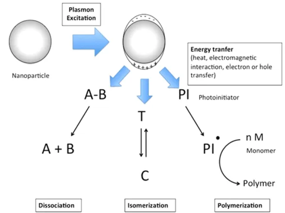
The primary plasmon-assisted chemical reactions can be classified into the three following categories, schematically illustrated in figure1:
- Molecular dissociation.
- Photoinduced switching of photochromes. - Photopolymerization.
The last section will focus on the applications of polymerization. In particular, the case of free-radical photo-polymerization will be emphasized, in order to stress the specificities of the plasmon-induced photopolymerization versus far-field photopolymerization.
2.1. Plasmon-assisted chemical reactions
Thefirst report on plasmon-enhanced photochemical reaction was provided by Chen et al in 1983 [34]. The photoreaction consisted of a dissociation reaction of a cadmium complex (dimethyl cadmium) on cadmium nanoparticles. The wave-length (257 nm) was chosen to overlap with the plasmon resonance of the Cd nanoparticles. Such irradiation of the Cd
nanoparticles in contact with the Cd complexes gave rise to elliptical nanoparticles. Following this example, many other reactions have been driven by plasmons. Other important examples of small molecules dissociation assisted by plas-mons are photodissociation of alkanes on Pd nanoparticles, photodesorption of NO and site conversion of CO on Pd
nanoparticles and, more recently, water splitting and photo-decomposition of organic dyes on metal nanoparticles loaded on TiO2surfaces. Examples can be found in a recent review article from Ueno and Misawa [35].
More recently, the concept was extended to other inter-esting photoinduced reactions like switching of
Isomerization cycles → Translation along molecule axis
B C
D
F G
E
Figure 2.Near-field photochemical imaging of noble metal nanostructures using PMMA-DR1. (A) Principle: the active azobenzene molecule moves under local optical illumination. (B) AFM image of a bow–tie plasmonic antenna covered with a thin layer of PMMA-DR1 (the left image is a scanning electron micrograph of the antenna). (C) Same as (B) after optical exposure with a plane wave linearly polarized along the antenna axis. (D) and (E) Zoom out of (B) and (C). (F) AFM image of an array of silver circular nanoparticles covered with a thin layer of PMMA-DR1. (G) Same as (B) after optical exposure with a plane wave circularly polarized. (Adapted with permission from [36,39]. Copyright 2005 American Chemical Society.)
photochromes. In particular, azobenzene derivatives are known to undergo a photoinduced transition between trans and cis isomers upon visible light irradiation. This transition is reversible and can lead to the initiation of a chemical reaction by thermal and photochemical pathways. Grafting azobenzene moieties on a polymer material such as PMMA allows designing a material that can be patterned at the micro and nanoscale upon spatially controlled irradiation. This physico-chemical system has been used in different irradia-tion configurations and Hubert et al have demonstrated that it can be coupled to a metal nanoparticle surface plasmon to induce nanostructuring [36] (figure2). In this case, the local trans–cis isomerization after photoexcitation gives rise to a mass diffusion process due to thermal reorganization and reconfiguration of the chromophores by the external electro-magneticfield [37]. Such an experimental approach was used on arrays of metal nanoparticles, producing nanostructures that varied depending of the polarization of light and the metal structure. A wide variety of nanostructures geometries were used [37–40] showing that this approach is well adapted to image the near-field distribution around metal nanostructures.
Diarylethene is another class of photochromes that has been applied to optical near-field irradiation in order to gen-erate molecular isomerization at the nanoscale. Such mole-cules can switch between two separate states, namely an open and a closed form. The isomerization is reversible and can be light induced (figure3). Such systems were coupled to plas-monic nanostructures and the near-field isomerization was investigated quantitatively by Tsuboi et al [41]. The effect of the distance between the metal nanoparticle and the dyes was also examined [42].
A third very important example of plasmon-induced photoreaction is photopolymerization. Photopolymerization consists of forming macromolecules from monomers or mixtures of monomers by free radical or ionic reactions
triggered by light. Combining the optical near-field with photopolymerization opens new doors towards nanophoto-lithography using visible light sources, without being limited by the diffraction limit. Many efforts have been dedicated during recent years to propose innovative methods to push the limits and constraints of nanofabrication processes [43]. Among them, some processes take advantage of the possi-bility to perform far-field imaging beyond the diffraction limit [44–47].
Photochemical reactions can be confined at the nanoscale by controlling the diffusion rates of reactive species. Near-field photopolymerization appears in this context as an alternative approach for reducing the size of features and/or fabricating sophisticated hybrid nanostructures.
2.2. Photopolymerization
The two main routes for photopolymerization are cationic and free-radical processes, depending on the nature of the reactive species involved in the polymer chain growth.
Cationic photopolymerization in the context of micro-nanopatterning usually includes an epoxy-based monomer. The epoxy group can be opened in acidic conditions, leading to the polymerization of epoxy monomers. The photoinitiator for such polymerization is a photoacid generator species. SU8, a commercially available cationic negative tone photo-resist [48], was used in the framework of plasmon-induced nanostructuring in several examples. Among thefirst exam-ples, Srituravanich et al produced sub-wavelength dots using aluminum hole arrays as a plasmonic photomask [49]. Structures as small as 90 nm with a period of 170 nm could be generated using 365 nm irradiation. Silver 2D hole array masks were also used to generate 60 nm half-pitch resolution dots using SU8 [50].
2.3. One-photon versus two-photon photopolymerization
Nonlinear processes are frequently desired for nanofabrica-tion approaches since the incident light profile usually does not have a sharp spatial distribution. The response function of the material is, therefore, used to sharpen the object produced by lithographic process. Several chemical or physical prin-ciples can be involved. Two-photon absorption is a way to generate highly nonlinear responses. Using this principle, Sundaramurthy et al achieved the polymerization of SU8 photoresist under femtosecond laser irradiation [51]. The multiphoton absorption regime is reached when a high den-sity of photons (either temporally or spatially) interacts with an organic molecule. These specific conditions are usually obtained using a femtosecond pulsed laser and lenses with high numerical apertures. The enhancement of the electro-magnetic field due to the plasmon resonance is enough in some configurations to obtain multiphoton absorption with incoherent light. Ueno et al demonstrated that photo-polymerization of SU8 can be triggered by a halogen lamp emitting in the W cm−2range (600–900 nm) (figure 4). The authors stressed that in the nanogap between metal nano-particles, the enhancement factor is strong enough to reach
Open-form 0.4 0.2 0.0 Absorbance 400 600 800 Open-form Vis Vis UV F FF FF F F FF FF F UV Closed-form Closed-form Wavelength/ nm S S S S
Figure 3.Molecular structure of diarylethene and spectra of open and closed forms. Reprinted with permission from [41]. Copyright 2009 American Chemical Society.
regimes for which two-photon absorption and subsequent polymerization is possible [52].
3. Our approach: plasmon based free-radical photopolymerization
Over the past decade, we have been developing an approach of plasmon-assisted free-radical photopolymerization. The advantage of this reaction is to offer a wider range of pho-toinitiators, especially in the visible range, able to trigger the photopolymerization reaction. Thus, it is possible to finely
adjust the wavelength sensitivity of the photopolymer with the resonance of metal nanoparticles. Additionally, we will see that it is possible to functionalize the material to make it active.
The principle of our approach is illustrated in figure5 [53]. The photopolymerizable formulation (PPF) is made up of three basic components: a sensitizer dye, a co-synergist amine, and a multifunctional acrylate monomer, pentaery-thritol triacrylate (PETIA). PETIA is used as received from the supplier and forms the backbone of the polymer network. The co-synergist amine is the methyldiethanolamine (MDEA). For example, the Eosin-Y (2
′,4′,5′,7′-Figure 4.(Top) SEM images of photopolymerized regions on gold nanoblock dimers for two polarization states. (Bottom) Theoretical results of the distribution of the electromagneticfield around the nanogap showing the good agreement with polymerization. Reprinted with permission from [52]. Copyright 2008 American Chemical Society.
tetrabromofluorescein disodium salt) can be used as the sen-sitizer dye that makes the system sensitive to the spectral region from 450 nm to 550 nm. As another example, methy-lene blue (MB) molecules make the formulation red-sensitive. This liquid system is veryflexible as it is possible to modify the components independently to adjust the physical and the chemical properties of the formulation, namely, viscosity, spectral sensitivity, and polymerization threshold energy. A typical mixture contains 0.5 wt% of Eosin-Y and 4 wt% of MDEA. After optical absorption from the excited LSP, the Eosin-Y is excited to its single state, followed by non-radia-tive intersystem crossing to create a triple state. The excited molecule interacts through electron and proton transfer with the amine to create radicals that interact with the monomer, and trigger the polymerization. An important parameter that characterizes this formulation is the threshold dose of energy, Dth, generally expressed in mJ cm−2. This parameter is the minimum dose of photonic energy that should be provided to the system to start the polymerization. Below Dth, the energy is lost through a couple of reactions including formation of peroxides from radicals. We will show that our method relies on this parameter.
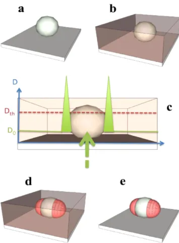
Metal nanoparticles on a substrate (figure 5(a)) are homogeneously covered by a synthesized free radical PPF possessing high-resolution visible-light sensitivity (figure5(b)).
The polymerization is activated by laser irradiation (plane wave of controlled polarization state) with wavelengths overlapping with both the PPF absorption spectrum and the NPs plasmon resonance. The exposure dose (D0) is chosen to be weaker than Dth, the threshold dose necessary for reticu-lation (figure 5(c)). Therefore, photopolymerization is not expected to occur in the absence of NPs: because of thefield enhancement at the plasmon resonance (figure 5(c)), the effective dose near the metallic nanoparticles can get higher than the threshold Dthto initiate the chain reaction leading to local polymerization (figure 5(d)). After irradiation, any monomer that is not reticulated is removed by a rinsing procedure (figure5(e)) and the resulting hybrid nanostructure is characterized by an atomic force microscope (AFM) and optical spectroscopy. The size of the polymer wings attached to NPs is related to the strength and the depth of the plas-monic optical near-field.
4. Achievement and applications
4.1. Studying and quantifying plasmonic near-fields
The method constitutes an efficient way of probing the intensity of the optical near-field. It can be viewed as a near-field photographic approach where the ‘pictures’ are read by AFM, ruling out any diffraction-limited farfield effects. This idea wasfirst introduced in [53].
By analyzing precisely the dimension of the polymerized regions as a function of the relevant parameters (incident energy, local position, direction of polarization, etc), it is possible to get valuable quantitative information about the plasmonic near-field intensity. It would be very difficult to acquire such information by using alternative methods like scanning near-field optical microscopy [54], photo emission electron microscopy [55], or electron energy loss spectro-scopy [56].
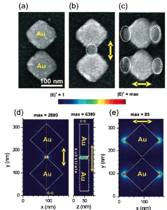
Two illustrating examples are shown infigures 6and 7. Thefirst example concerns gold nanocubes [57]. The optical properties of metal nanocubes have been the focus of recent experimental and theoretical works (e.g. see [58–60]). Most of these works have focused on the unique properties of the cubes, especially enhanced local electromagnetic fields, par-ticularly at the eight vertices and along the twelve edges of the cube, when illuminated with light. To date, only little is known about the local near-field optical properties of metal nanocubes, since most of the plasmonic studies were carried out in the far-field. However, to pave the way for tailoring nanostructured electromagnetic fields using nanocubes, a detailed characterization of their near-field properties is essential. Our method relying on nanoscale photo-polymerization allows one to address this challenge. Figure6(A) shows a scanning electron micrograph (SEM) of a single, 60 nm edge, gold nanocube. It presents a dipolar
Figure 5.Scheme of the approach. (a) Metal NP deposited on a functionalized glass substrate. (b) Deposition of the photopolymer-izable formulation. (c) and (d) Plasmon based near-field photo-polymerization of PPF leading to polymer nanostructures corresponding to the LSP intensity distribution. (e) The resulting hybrid nanoparticle is revealed by rinsing procedure. Reprinted with permission from [53]. Copyright 2010 American Chemical Society.
plasmon resonance in the 500–600 nm spectral range, corre-sponding to the spectral sensitivity of the PPF used. The polarization used for photopolymerization is vertical and the orientation of the cube side edge relative to this polarization is described by the angleθ. The experiment was performed with θ = 45°, i.e. with the cube corner parallel to the polarization. In this situation, the localfield enhancement factor is expected to be significant due to the lighting rod effect.
An AFM image of the hybrid ‘nanocube + polymer’ system is acquired, from which a background AFM image of the nanoparticle preceding the photoexcitation has been subtracted, leading to a differential AFM image that high-lights the integrated polymer nanostructures, regardless of the initial topography, as a signature of the local light. A repre-sentative differential AFM image can be seen infigure6(B). In particular, polymer elongation is seen along the polariza-tion direcpolariza-tion, while no polymerizapolariza-tion is induced where the unit surface normal vector is perpendicular to the laser polarization direction. Figure 6(B) was obtained with an incident dose of 25% of Dth. It is worth comparing thisfigure with the expected calculated near-field intensity (figure6(C))
where the iso-intensity contour line represents the threshold to be exceeded for polymerization. Fromfigures6(B) and (C), it turns out that the polymerization occurred inside the zone defined by the iso-intensity contour, while the energy outside was not high enough to trigger the polymerization. Based on the knowledge of the polymer extending along the corner (yp in figure 6(B)), it has been possible to quantify valuable features related to the nanocube plasmonic field. This quan-tification is made in the following way. Let us consider that the near-field of the excited nanocube is characterized by an exponential decay. Along the y-axis, the local dose D at the cube corner can be expressed as
α
= −
D FmaxD0exp( y), (1)
where Fmax is the maximum (at the surface) intensity enhancement factor related to the LSP resonance, α is the spatial decay rate of thefield intensity (α−1is its characteristic decay length), and y is the distance from the surface of the metal nanoparticle in the y-direction.
We have already established that photopolymerization only occurs when D > Dth. By applying this condition to
Figure 6.Investigation of the near-field plasmonic intensity at the surface of a gold nanocube. (A) SEM image of one single nanocube. θ is the cube orientation. The arrow represents the direction of the incident polarization used for photopolymerization. (B) Differential AFM image of the hybrid nanosystem obtained by plasmonic near-field photopolymerization. (C) Calculated near-field intensity enhancements
E2/E 0
2
in an n = 1.5 medium. Light is incident from below along the z-axis and is polarized along the y-axis. The observation plane is at z = 6 nm below the top of each cube. The contour lines correspond to E2/E =40
0 2
, illustrating the threshold to be exceeded for D0= 0.025 × Dth. (D) Demonstrated intensity enhancement factor as a function of the measured polymer extension along the cube corner. The
equation (1), one can obtain α − ⩾ × y D F D exp( ) th . (2) max 0
By simplifying equation (2), one can get the maximum dis-tance ymaxaway from the Au nanocube at which the effective dose overcomes Dthas ⎛ ⎝ ⎜ ⎞ ⎠ ⎟ α = − × y F D D ln . (3) max 1 max 0 th Equation (3) can be rewritten as
α
= −
(
×)
ymax 1ln Fmax f , (4) where f (in %) represents the incident dose, normalized by the threshold.
ymax is equal to ypthat is experimentally measured. For example, an elongation of yp= 3.5 nm was measured for an incident dose of f = 5%, which implies that an enhancement factor of at least 20 (=1/f) is expected at the corner of the nanocube at the ypdistance. This (3.5 nm; 20) plot is repre-sented infigure6(D). In order to accurately determine Fmax
andα, many exposures of different doses were performed on numerous identical nanocubes, resulting in figure6(D) that shows the values of 1/f, deduced from 1/f = Dth/D0, as a function of the polymer elongation yp= ymax. The data follow a single exponential decay reflecting the near-exponential decay of the plasmon near-field. 1/f can be deduced from equation (4) as = F e−α
f
y
1
max max. Thus, when fitting the experimental data with an exponential function, one can deduce the values of thefitting parameters Fmaxand α−1as 35 and 5 nm, respectively. This simple example illustrated well the high capability of the method for quantitative near-field analysis. The analysis of the influence of θ was performed as well, allowing one to discuss the importance of incidentfield excitation in such structures [57]. This point will be discussed in the text relevant tofigure7.
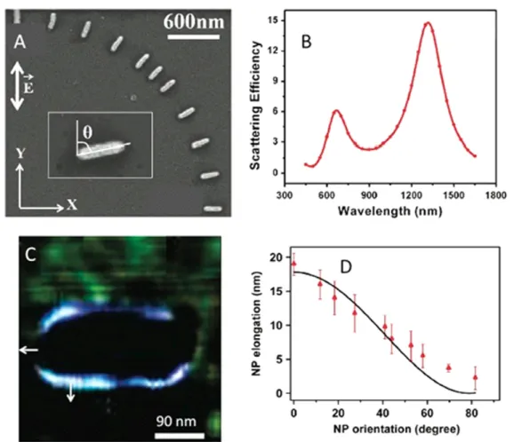
The second example deals with non-resonant metal nanosystems. While resonant surface plasmons have been widely studied over the past 20 years, only few experimental studies on non-resonant effects have been reported (e.g. [61]) because off-resonant excitations generate weak and very localizedfields that are extremely difficult to measure. In that
Figure 7.Mapping non-resonant electromagnetic effects. (A) Representative SEM showing eleven differently oriented GNRs. The incident field polarization direction is represented by the double white arrow. The inset of (A) shows the angle θ between the nanorod major axis and the polarization direction. (B) Calculated far-field scattering spectrum for a single rod, embedded in a medium of refractive index 1.485, corresponding to that of the photopolymerizable solution, illustrating the localized surface plasmon response of both nanorod axes. (C) Differential AFM image of the hybrid structure resulting from a plasmonic phopolymerization withθ = 90°. The white arrows represent the surface normal unit vector at two locations. (D) Polymer elongation at the rod extremity (along the long axis) as a function ofθ. Red dots: experimental data. Black curve: analytical model (see text). Adapted with permission from [62]. Copyright 2011 American Chemical Society.
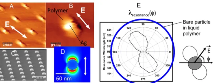
Figure 8.Structuring the surrounding environment by plasmonic photopolymerization (A) and (B) AFM image taken after procedure applied to silver hemispherical nanoparticles. (C) SEM image of the nanoparticles before exposure. (D) Calculated intensity distribution during the photopolymerization (γ = 514 nm). (E) Polar diagram showing the position of the plasmon resonance peak of the hybrid particle as a function of the direction of incidentfield. The blue circle illustrates what would be obtained in an anisotropic polymeric material. Reprinted with permission from [63]. Copyright 2007 by the American Physical Society.
Figure 9.(A) Differential AFM image showing the topography of a hybrid nano-emitter based on a gold nanosphere of 70 nm diameter. The highlighted nanostructures represent the integrated photopolymer structures with a thickness of 15 nm each. (B) Polarization-dependent fluorescence signal (λex= 514 nm) of the nano-emitter with intensity higher when the incident light is polarized along Y-axis than along the
X-axis. References were collected from 2μm away from the NE with an incident field along X (in green) and Y (in black) axes. (C) Top view of the emission rate map. The calculation was done in a polymer of n = 1.48. Reprinted with permission from [66]. Copyright 2014 AIP Publishing LLC.
case, the oscillating charges vibrate in phase and the quasi-electrostatic case can be considered. The approach of plas-monic photopolymerization has been applied to non-resonant metal nanosystems [62]. The sample used is shown in figures 7(A) and (B). It consists of gold nanorods (GNRs) having a different orientation θ relative to a fixed linear polarization along the y-axis. As shown infigure7(B), within the PPF, such nanorods present two dipolar resonant modes at 680 nm (transverse mode) and 1350 nm (longitudinal mode). We used a wavelength of 530 nm for triggering the poly-merization, which is pretty far from the resonant plasmonic modes. Figure7(C) is the resulting AFM differential image in the θ = 90° case. Here, the exposure dose was chosen to be 65% of the threshold dose, still lower than the threshold but high enough to be sensitive to the weak, off-resonance intensity enhancement. The polymer spatial extension turns out to be maximum when the unit vector normal,n, to the rod surface is aligned with the polarization direction. Conversely, the polymer extension is null when the unit vector is per-pendicular to the polarization, as one can see at the GNR extremity in figure 7(C). This effect can be interpreted in terms of polarization surface charge density. Let us consider the free electrons at the metal surface (belonging to the metal itself) that are driven by the incidentfield and that re-radiate light, resulting in a localized optical near-field. This excitation is quantified by the dielectric susceptibility of the metal. We propose that the local polymerization is driven by these excited surface charges that are related to the electric field discontinuity at the rod interface, following the usual boundary condition
ρ ε χ
− =
(
)
= ⋅EE1n E2n s,pol/ 0 n o, (5)
where E1nand E2nare the normal components of the electric field inside and outside of the rod, respectively, E0 is the incident electric field vector, ρs,pol is the surface density of polarization charges, andχ is the metal electric susceptibility. In turn, the excited surface charges create their own electric field that is responsible for a weak field enhancement on the rod surface, and hence subsequent polymerization.
The polymer spatial extension was measured at the extremities of the rod as a function ofθ (see figure7(D)). A clear polarization sensitivity is demonstrated, as the expres-sion of the scalar product in equation (5). It can be compared to afit extracted from an analytical model (black full line) that is based on simple considerations summarized as follows. We consider that the electricfield at the GNR surface results from a coherent constructive interference (in the off-resonant case, photoinduced charges vibrate in phase with the incidentfield) between the incidentfield and the local field re-radiated by the induced polarization vibrating charges, via the local sus-ceptibility. The spatial polymer extent is assumed to be pro-portional to the resulting intensity. Appropriate fitting parameters allowed us to get a realistic value of the local effective surface susceptibility [62].
4.2. Hybrid nanostructures presenting new optical properties
Besides the possibility of quantifying the plasmonic near-field, the approach turned out to allow for photoinduced synthesis of new hybrid nanosystems having unique optical properties. Afirst example is shown in figure8[63]. It con-cerns a passive structure in the sense that no optical activity in terms of light emission from the polymer is considered for the moment. Figures 8(A) and (B) show the result of the plas-mon-based photopolymerization on silver hemispherical nanoparticles produced by e-beam lithography (figure8(C)). Two symmetric polymer lobes adjacent to the particles can be observed, resulting in metal–polymer hybrid particles. The two lobes originate from the excitation of a particle dipolar surface plasmon resonance, as numerically illustrated in figure8(D) obtained through afinite difference time domain calculation. The field distribution associated with the reso-nance is enhanced in a two-lobes region oriented along the incident polarization. The localized nanoscale photo-polymerization is the result of the inhomogeneous field dis-tribution. The two lobes can thus be viewed as a three-dimensional polymer molding of the locally enhanced optical fields.
Figure 8(E) is a polar diagram of the surface plasmon resonance peak of the obtained hybrid nanostructure. We measured fifty spectra of the hybrid particles for different angles of polarization, as illustrated at the right side of figure 8(E). The two polymerized lobes clearly induced a quasi-continuously tunable LSP resonance in the 508–522 nm range. The local polymerization turns out to lead to two new plasmon eigenmodes centered at 508 and 522 nm, respec-tively. For any polarization angle, a linear combination of the two eigenmodes is excited, with respective weights depend-ing on the polarization direction. We conclude that the apparent continuous plasmon tuning is the result of the spectral linear combination. The anisotropic behavior can be compared to what would be observed in the case of a bare particle embedded in the isotropic liquid polymer (see blue circle on figure 8(E)). Before local photopolymerization, metallic nanoparticles are characterized by a C∞v symmetry corresponding to rotation around the Cv axis. After poly-merization, the two polymerized lobes induce a new lower symmetry: C2v, for which any pattern is reproduced by aπ in-plane rotation. This new symmetry induces the breakdown of the LSP spectral degeneracy. We can also consider that a nanoscale artificial ellipsoid of indexes was integrated in the vicinity of the silver particle, making it polarization sensitive. Additionally, it was possible to get a polar distribution of the effective refractive index from figure 8(E), simply by expressing the index shift as a function of the wavelength shift in the expression of the polarizability of the particle seen as a plasmonic nanosensor [63].
Figure8illustrates in a remarkable way the possibility to control and structure the spatial environment of metal nanoparticles, resulting in new and unique optical properties.
As an indispensable component of PPF, dye molecules located within the near-field of metal nanoparticles can be trapped by the integrated photopolymers. Thanks to the ani-sotropic distribution of dipolar mode plasmons supported by metal nanoparticles, the obtained metal/polymer nanos-tructure is thus a plasmonic nano-emitter (NE), whose dye molecules are anisotropically spatially distributed. Unlike conventional hybrid plasmonic light-emitting systems with a random or isotropic distribution of dye molecules around the metal nanoparticle [64,65], this anisotropic spatial distribu-tion of dye allows an optical selecdistribu-tion of the molecules by using incident polarization. A recent study has been published on an anisotropic NE based on an isolated gold nanosphere with a diameter of 70 nm [66]. Linear polarization of the incident light produced 15 nm thick polymer structures inte-grated in the Y-axis as figure 9(A) presents. This NE was characterized optically by fluorescence from Eosin-Y mole-cules excited by a 514 nm laser with a polarization switchable between X- and Y-axes.
It was found that the NE was more efficiently activated when the excitation light was polarized along Y-axis than along the X-axis (figure9(B)). This optical anisotropy resulted from the overlap between the dipolar plasmonic mode and the active medium, both of which presented anisotropic spatial distributions. When polarized along the Y-axis, the excitation light led to a plasmonicfield oriented in Y, which completely overlapped with the dye-containing active medium. In com-parison, the plasmonic field was perpendicular to the dis-tribution of the active medium of the NE under the incident polarization along the X-axis. The different spatial overlap caused a different number of molecules to be excited by the near-field. Background-corrected fluorescence from a single NE presented 2.2-times higher intensity with a polarization parallel (Sy) to the active medium distribution than perpen-dicular (Sx) to it.
The emission rates of a dye molecule in free space γ0em and in the close vicinity of a metal nanoparticleγemare well known as
γem0 = qγex0 (6)
and
γem= Qγex (7)
respectively, where q is the intrinsic (free space) quantum yield andγ0exis the excitation rate of the dye molecule in free space, Q is the nanoparticle-modified quantum yield and γexis the excitation rate of the dye molecule by the local electric field. Q can be calculated from the Mie Theory [66,67].γ0ex and γex are expressed as functions of the incident field and local electricfield, i.e., γ0ex∝| E0·p |2and γex∝| Eloc·p |2, respectively, where p is the molecular transition dipole moment and Elocis the local near-field. It should be stressed out that the molecular dipole orientation was not controlled in this experiment. An averaged molecule orientation was therefore applied for the calculation [66, 67]. As a
γ γ = Q q E E . (8) em em 0 loc 0 2
The calculated spatial distribution of free-space-normalized emission rate of Eosin-Y molecule in the vicinity of a gold nanosphere is shown infigure9(C). This normalized emission map shows a maximum normalized emission of γem/ γ0
em= 1.45.
To some extent,figure9(C) represents the probability of emission of the dipole emitter as a function of its position in the vicinity of a gold nanoparticle that is illuminated with a field parallel to the Y axis. For example, the dark ring in the close vicinity of the particle tells us that the dipole can not emit light due to the quenching effect associated to a non-radiative energy transfer. In the anisotropic hybrid NE, two key factors have actually to be considered. One is the prob-ability of the presence of dye molecule in the plasmonic near-field. The other is the probability of light emission of these molecules. The latter has been expressed in equation (7) and has been calculated in figure 9(C). This is a factor to be considered in all plasmon-modified fluorescence studies regardless of the NE configuration. The probability of spatial presence of molecules, however, is what we control via nano-photopolymerization. Figure9(A) can be viewed as a map of probability of presence of dipole emitter trapped in the polymer.
It modifies equations (6)–(8) to
γ0 =ϕ qγ , (9) em out 0ex γem=ϕinQγex, (10) ⎛ ⎝ ⎜⎜γγ ⎞⎠⎟⎟ = ϕϕ Q q E E , (11) em em 0 NE in out loc 0 2
whereϕinis the probability of the presence of molecule inside the photopolymer nanostructure, whileϕoutis the probability of the presence of molecule outside the photopolymer nanostructure. After photopolymerization and rinsing, mole-cules that are not trapped in the photopolymer nanostructures are expected to be removed by solvent. In other words, the probabilities of molecular presence outside and inside the photopolymer nanostructures are expected to be 0 and 1, respectively. In reality, however, asfigure9(B) illustrates, the rinsing process is not perfect, which led to the detection of a reference signal of fluorescence signal 2 μm away from the hybrid NE. The spectra in black and green were collected with polarizations along the Y- and X-axes, respectively. Obviously the reference signals were not polarization sensi-tive. The probability of the molecule presence falls in the band of 0 <ϕout< 1 instead to equal 0. Consider the situations of NESy and RF curves in figure 9(B) that satisfies equations (10) and (9), respectively, we obtain (γem/γ
0 em)y = NESy/RF = 2.5 from equation (11). ϕout can be deduced from the ratio of equation (11) over equation (8): ϕin/ ϕout= 2.5/1.45. Inside the photopolymer, when we setϕin= 1, ϕout= 0.58 can be obtained.
In the case of NESx, considering equation (5), the optical near-field enhances the local electromagnetic field at any point in the vicinity of metal nanoparticle except for the points wheren⊥ Eloc. As a consequence, fluorescence of a certain percentage (η) of the molecules in the photopolymer nanostructure can be enhanced by the near-field when the incident polarization is oriented perpendicular to the polymer lobes [66]. In this situation, equation (11) can be modified as
⎛ ⎝ ⎜⎜γγ ⎞⎠⎟⎟ ηϕ ϕ η = Q + − q E E Q q E E (1 ) . (12) x em em 0 in out loc 0 2 loc 0 2
The first component on the right side of equation (12) represents the molecules excited by both Sx and Sy con fig-urations, while the second component represents the mole-cules distributed in the near-field of Sxand outside the near-field of Sy. From figure 9(B) one sees (γem/γ0em)x= NESx/ RF = 1.69. The value ofη = 0.33 can be consequently deduced from equation (12). In other words, one third of the dye molecules in the photopolymer can be excited by the plas-monicfield under both polarizations.
As a conclusion, the effective emission from the aniso-tropic NE depends on the spatial overlap between the prob-ability of emission and the probprob-ability of presence of dipole emitters. In other words the product of these two probabilities gives us an idea of the effective light emission.
Despite the observation of anisotropic fluorescence resulting from the dye-doped photopolymer spatial distribu-tion and its overlap with the excitadistribu-tion near-field, there is no doubt that this hybrid nanostructure has to be improved in order to amplify the optical anisotropy. For a certain prob-ability of the molecules outside the photopolymer nanos-tructures, our attention turns to the quantum emitter around the nanoparticles. Quantum yield is an element that can be improved. Additionally, the dye molecules in this study have participated in the photopolymerization process, which
resulted in the photo-bleaching of most of the molecules [68]. It is thus reasonable to think about another candidate for emission: QDs.
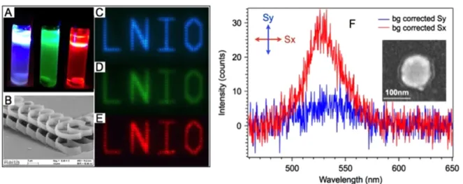
With QDs grafted by covalent bonds on the PETIA molecules, this type of polymerizable solution allows us to trap QDs in the integrated photopolymer structures. Figure10(A) shows a photo of photopolymerizable solution doped with three different QDs, ZnCdS/ZnS emitting at 460 nm, CdSe/ZnSeS emitting at 510 nm, and CdSe/CdS/ZnS emitting at 620 nm, respectively. Figure 10(B) is the SEM image of a high resolution far-field laser scanning lithography with the QD-containing photopolymerizable systems. Figure 10(B) was obtained via two-photon absorption photopolymerization.
Fluorescence images of the three different types of QD-doped photopolymer structures are shown in figures 10(C), (D) and (E), respectively.
On a 100 nm diameter gold nanodisk, two-lobe photo-polymer nanostructures were integrated via two-photon polymerization to trap QDs that emit at 510 nm in the active medium in the near-field of the nanoparticle. Figure 10(F) illustrates the resulting background corrected fluorescence spectra under incident polarizations along (Sx) and perpendi-cular (Sy) to the polymer nanostructures. As is shown in the SEM image (inset of figure 10(F)), photopolymer nanos-tructures were oriented along the X-axis. The polarization-dependentfluorescence indicates a 6-times higher intensity of Sx than Sy, which is due to the spatial overlap of the QD-doped active medium and the plasmonic near-field of the gold nanodisk. The optical anisotropy of the NE has been con-siderably enhanced by substituting organic dye to QDs.
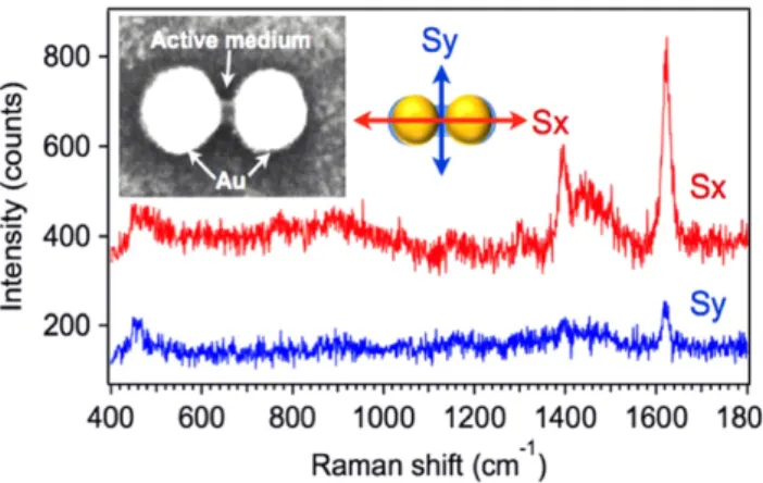
Another example is to position dye molecules in the gap of nanodimers via the photopolymerization process. It should be stressed that other thanfluorescence spectroscopy, surface-enhanced Raman scattering (SERS) can be used as well to probe the presence of molecules at the plasmonic hot spots.
Figure 10.(A) Photopolymerizable solutions doped with different QDs that emit at 460 nm, 510 nm, and 620 nm (from left to right), respectively. (B) SEM image of photopolymer structures fabricated by laser scanning photo-lithography with the QD-containing
polymerizable solutions. Fluorescence images of patterns of photopolymer structures doped by QDs emitting at (C) 460 nm (D) 510 nm and (E) 620 nm. (F) Background-correctedfluorescence spectra (two perpendicular incident polarization) from the green QDs that are embedded in the polymer lobes of a hybrid nano-emitter produced by two-photon plasmonic nano polymerization. The inset shows a SEM image representing the hybrid nano-emitter with polymer lobes oriented in the X-axis.
The MB molecule is known as a good SERS probe and was thus applied in this study. With incident light polarized along the interparticle axis of the nanodimer, MB molecules can be selectively positioned in the active medium formed in the nanodimer gap. MB molecules were initially embedded in the PPF to make it sensitive to the red light that corresponds to the wavelength of LSP resonance of the dimer. Figure 11 presents the polarization sensitivity of the SERS signal on a single gold nanodimer/photopolymer hybrid nanostructure with initial gap size of 10 nm. The inset shows a topographic SEM image that presents different contrasts on metal and photopolymer. As the spectra indicates, the characteristic SERS mode at 1620 cm−1is assigned to the stretching (C=C) ring of MB molecule. The SERS signal obtained with an incident polarization along the gap (Sx) is approximately 5 times higher than that perpendicular to the gap (Sy).
As forfigure10, this polarization sensitivity observed in figure11 is believed to result from a tunable spatial overlap between the plasmonic near-field and active molecules.
4.4. Application to nanophotochemistry
The studies carried out have revealed some very specific aspects regarding free-radical photopolymerization and asso-ciated chemistry that are worth being detailed. Using the plasmon-assisted configuration for local irradiation appears as a very interesting configuration for investigating nanophoto-polymerization. It is important to examine in detail the molecular pathway leading to photopolymerization (figure12). The absorption of a photon leads to the excited singlet state of the Eosin-Y, and then to its triplet state by intersystem crossing. From the triplet state, the dye can react with the amine to produce thefirst radical able to induce the free-radical polymerization of the methacrylate monomer. A trifunctional monomer is used so a rapid crosslinking of the polymer network is usually observed [69]. The right part of the scheme describes the inhibition processes that are due to the oxygen dissolved in the photopolymer. Oxygen can react with the triplet state of the dye, or with radicals, to create peroxide radicals that are not active for polymerization [70]. Usually, in free-radical polymerization, these later processes
are available for polymerization. In micro and nanofabrica-tion, such a molecular phenomenon is advantageously used to ensure a precise control of the polymerization volume. However, the presence of oxygen considerably affects the kinetics of photopolymerization and needs to be taken into account.
One major effect is on the threshold of polymerization, which depends on many chemical, physico-chemical, and photonic parameters [72]. From the triplet state, a protonated Eosin radical can be formed by reaction on MDEA. This radical is known to be ineffective for initiating polymeriza-tion. However, several reactions of this radical have to be considered [73]. First, it can react with another protonated Eosin radical to regenerate an Eosin molecule in the leuco form. The later form does not absorb at 532 nm, and the consequence of this reaction is the progressive bleaching of the Eosin formulation. Second, the protonated Eosin radical can undergo a disproportion reaction with other radicals (deprotonated amine radical, peroxide radicals or free-radicals of the growing polymer chains), where in this case, the Eosin is regenerated to its fundamental state. Such processes regenerate the Eosin in an active state that can photosensitize again other MDEA reactions.
By measuring the lateral extent of the lobes deduced from differential images (such as shown in figure9(A)), we investigated the role of irradiation dose on the polymerization at the nanoscale. Interestingly, it was demonstrated that the exposure time and the irradiation power are not equivalent to each other in terms of the spatial extent of the polymerization, although they correspond to the same energy (figure13). For a given dose, the lateral extent of polymerization is more pronounced for lower intensity, which is different from pre-vious studies of microscale photofabrication [72].
The behavior observed at the microscale [72] was inter-preted as follows. The inhibition role of oxygen is more pronounced at low intensity (i.e. long irradiation times) because the consumption of oxygen is slow enough to allow for a continual replenishment from the surroundings. The polymerization process starts only when the oxygen con-centration in the photopolymer droplet is low enough, which implies that replenishment increases the inhibition time. For higher incident power, the inhibition is less sensitive because oxygen diffusion has no time to proceed efficiently relative to the rate of new photons injected into the system. What we observed at the nanoscale reveals an opposite behavior to what was seen at the microscale.
Taking into account that the typical dye concentration is 0.5 wt%, and assuming a homogeneous distribution of Eosin-Y within the photopolymer solution, a volume of 10 × 10 × 10 nm3contains only four dye molecules. The same volume contains an average of 200 MDEA molecules and 6000 C=C double bonds. Considering this simple assessment, it appears that the limited reactant is the dye. The result is the curve in figure14, showing that low irradiation powers (e.g. long irradiation times) are beneficial for polymerization.
The dye diffusion turns out to be important to describe the photopolymerization at the nanoscale. Although in
Figure 11.Polarization-dependent SERS signals from MB mole-cules. Inset SEM image shows the topography of the hybrid nanodimer with MB-doped photopolymer integrated in the gap.
microscale photo-fabrication the dye diffusion is usually neglected, this parameter is of critical importance at the nanoscale. Dye diffusion in the monomer matrix is quite slow because of its high molecular weight and polarity. However, by reducing the scale of investigation and probing, it is possible to become sensitive to the dyes that diffuse in the irradiated volume. Furthermore, the diffusion process can be efficient since bleaching is negligible in the volume
corresponding to the optically enhanced region. Low intensity corresponds to higher irradiation times, which certainly increases the probability of the dye to diffuse in the near-field nanoscale volume. This discussion is specific to the nanoscale and is linked to the extremely limited number of molecules: at the microscale, the average number of molecules present in the irradiation volume (1000 × 1000 × 1000 nm3) is greater by a factor of 106and thus diffusion of the dye from the polymer droplet into the irradiated volume is not predominant. The limiting process at this length scale is the number of photons
Figure 12.Scheme illustrating the photoinduced polymerization of the methacrylate monomer, the inhibition processes, and the Eosin-Y regeneration pathways. Reprinted with permission from [71]. Copyright 2011 American Chemical Society.
Figure 13.Evolution of the spatial extent of the near-field
photopolymerization as a function of relative dose, at constant power (black curve) and constant irradiation time (red curve). Reprinted with permission from [71]. Copyright 2011 American Chemical Society.
Figure 14.Influence of the incident power (bottom axis) and the exposure time (top axis) on the spatial extent of polymerization. The incident dose was set at 7% of the threshold dose. Reprinted with permission from [71]. Copyright 2011 American Chemical Society.
given dose, competitive processes such as inhibition by oxygen are more important.
5. Conclusion and perspectives
In this review, we have highlighted recent advances in plas-mon-induced photopolymerization processes that create hybrid nanomaterials with modified optical properties. This is a highly multidisciplinaryfield requiring understanding of the complex range photochemical processes that impact the spa-tial features on the nanoscale hybrid nanostructures, as well as being motivated by complex interactions between molecules and metal nanostructures that produce novel optical effects. We have shown that plasmon-induced photopolymerization by metal nanoparticles produces important optical effects including anisotropic antenna behavior achieved with reso-nant interactions, as well as strong anisotropy in resoreso-nant effects, such as plasmon-enhanced emission processes.
Future work will be governed by advances in the ability to control the photopolymerization process at the nanoscale in order to optimize molecule–plasmon interactions for a parti-cular application. For example, control of the type of chro-mophore interaction with plasmons, such as strong or weak coupling, requires precise control over the spacing of the chromophores from the metal surface. For enhancement of molecular or quantum dot radiative effects, efficient routes to spacing the target chromophores at 5 nm from the surface should be found [73]. Conversely, strong coupling would generally benefit from a much closer, yet precise distance, to optimize strong coupling effects [74]. Both strong and weak coupling interactions would benefit from control of the orientation the molecular transition dipoles in the evanes-centfield.
The use of plasmon-induced photopolymerization could further significantly impact nanooptics if the fabrication processes can be optimized. These include tunable nanoan-tennas with greater efficiencies, optical gain and lasing, and highly efficient nonlinear nanostructures. All of these aspects are critical for miniaturization of next-generation optical devices. Hybridization at the nanoscale represents a highly plausible route towards these opportunities because of the novel optical properties that only hybrid nanomaterials can provide.
Acknowledgments
The authors would like to thank current and former colleagues that are (or have been) involved in this project in different ways (fruitful discussion, samples, previous achievements, participation in measurements, etc): P-M Adam, A Bouhelier, F Charra, C Ecoffet, C Fiorini, D Gérard, D Gosztola, L Huang, H Ibn El Ahrach, P K Jain, S Kostcheev, S Marguet, Y Qi, R D Schaller, S Telitel, A Vial and R Vincent. The authors thank the Partner University Funds program (PUF 2010) for partially supporting this work. X Z thanks the China
Materials was supported by the US. Department of Energy, Office of Science, Office of Basic Energy Sciences, under Contract No. DE-AC02-06CH11357. This work is also sup-ported by the French agency for research (ANR grant HAP-PLE) and by European community FEDER fund and the Region Champagne-Ardenne: Grants HYN-NOV and NANO’MAT.
References
[1] Maier S A 2007 Plasmonics Fundamental and Applications (Berlin: Springer)
[2] Bachelot R, Zhou X, Plain J, Adam P M, Baudrion A L, Gray S K and Wiederrecht G P 2014 Proc. of SPIE—The Int. Soc. Opti. Eng.9126 91260Z
[3] Koenderink A F 2010 Opt. Lett.35 4208–10 [4] Curto A G, Volpe G, Taminiau T H, Kreuzer M P,
Quidant R and Van Hulst N F 2010 Science329 930–3 [5] Willets K A and Van Duyne R P 2007 Annu. Rev. Phys. Chem.
58 267–97
[6] Atwater H , A and Polman A 2010 Nat. Mater.9 205–13 [7] Chen H, Mig T, Wang L, Sun L D, Wang J and Yan C H 2010
Nano Today5 494–505
[8] Acimovic S S, Kreuzer M P, Gonzalez M U and Quidant R 2009 ACS Nano3 1231–7
[9] Dintinger J, Klein S, Bustos F, Barnes W L and Ebbesen T W 2005 Phys. Rev. B71 035424
[10] Baudrion A L, Perron A, Veltri A, Bouhelier A, Adam P M and Bachelot R 2013 Nano Lett.13 282–6
[11] Salomon A, Gordon R J, Prior Y, Seideman T and Sukharev M 2012 Phys. Rev. Lett.109 073002
[12] Drexhage K H 1970 Sci. Am.222 108
[13] Chance R R, Prock A and Silbey R 1978 Adv. Chem. Phys.37
1–65
[14] Pockrand I, Brillante A and Mobius D 1980 Chem. Phys. Lett.
69 499–504
[15] Zhang J, Fu Y, Chowdury M H and Lakowicz J R 2007 J. Phys. Chem. C111 11784–92
[16] Ipe B I and Thomas K G 2004 J. Phys. Chem. B108 13265–72 [17] Jin S Y, DeMarco E, Pellin M J, Farha O K,
Wiederrecht G P and Hupp J T 2013 J. Phys. Chem. Lett.4
3527–33
[18] Wiederrecht G P, Wurtz G A and Hranisavljevic J 2004 Nano Lett.4 2121
[19] Wu X H, Gray S K and Pelton M 2010 Opt. Express18
23633–45
[20] Wurtz G A, Evans P R, Hendren W, Atkinson R, Dickson W, Pollard R J, Zayats A V, Harrison W and Bower C 2007 Nano Lett.7 1297–303
[21] Shah R A, Scherer N F, Pelton M and Gray S K 2013 Phys. Rev. B88 075411
[22] Govorov A O, Bryant G W, Zhang W, Skeini T, Lee J, Kotov N A, Slocik J M and Naik R R 2006 Nano Lett.6
984–94
[23] Peng S, McMahon J M, Schatz G C, Gray S K and Sun Y G 2010 Proc. Natl. Acad. Sci.107 14530–4
[24] Pramod P, Soumya C C and Thomas K G 2011 J. Phys. Chem. Lett.2 775–81
[25] Mukherjee S, Libisch F, Large N, Newmann O, Brown L V, Cheng J, Lassiter J B, Carter E A, Nordlander P and Halas N J 2013 Nano Lett.13 240–7
[26] Ueno K and Misawa H 2011 Photochem. Photobio. A221
[27] Watanabe K, Menzel D, Nilius N and Freund H J 2006 Chem. Rev.106 4301–20
[28] Gao S, Ueno K and Misawa H 2011 Acc. Chem. Res.44
251–60
[29] Suh J Y, Kim C H, Zhou W, Huntington M D, Co D T, Wasielewski M R and Odom T W 2012 Nano Lett.12
5769–74
[30] Hasobe T, Imahori H, Kamat P V, Ahn T K, Kim S K, Kim D, Fujimoto A, Hirakawa T and Fukuzumi S 2004 J. Am. Chem. Soc.127 1216–28
[31] Zhang H, Govorov A O and Bryant G W 2006 Phys. Rev. Lett.
97 146804
[32] Watanabe K, Menzel D, Nilius N and Freund H J 2006 Chem. Rev.106 4301–20
[33] Kamat P V 2002 J. Phys. Chem. B106 7729–44
[34] Chen C J and Osgood R M 1983 Phys. Rev. Lett.150 1705 [35] Ueno K and Misawa H 2013 J. Photochem. Photobio. C:
Photochem. Rev.15 31–52
[36] Hubert C et al 2005 Nano Lett.5 615–9
[37] Bian L L, Kumar J, Kim D Y, Williams J and Tripathy S K 1988 Appl. Phys. Lett.73 1817–9
[38] Juan M L, Plain J, Bachelot R, Vial A, Royer P, Gray S K, Montgomery J M and Wiederrecht G P 2009 J. Phys. Chem. A113 4647–51
[39] Hubert C et al 2008 J. Phys. Chem. C112 4111–6 [40] Galarreta B C, Rupar I, Young A and Lagugne-Labarthet F
2011 J. Phys. Chem. C115 15318–23
[41] Tsuboi Y, Shimizu R, Shoji T and Kitamura N 2009 J. Am. Chem. Soc.131 12623–7
[42] Nishi H, Asahi T and Kobatake S 2011 J. Phys. Chem. C115
4564–70
[43] Nie Z and Kumacheva E 2008 Nat. Mater.7 277–90 [44] Scott T F, Kowalski B A, Sullivan A C, Bowman C N and
McLeod R R 2009 Science324 913–7
[45] Li L, Gattass R R, Gershgoren E, Hwang H and Fourkas J T 2009 Science324 910–93
[46] Andrew T L, Tsai H Y and Menon R 2009 Science324 917–21 [47] Hell S W 2009 Nat. Methods6 24–32
[48] See (www.microchem.com/Prod-SU8_KMPR.htm)
[49] Srituravanich W, Fang N, Sun C, Luo Q and Zhang X 2004 Nano Lett.4 1085–8
[50] Srituravanich W, Durant S, Lee H, Sun C and Zhang X 2005 J. Vac. Sci. Technol. B23 2636–9
[51] Sundaramurthy A, Schuck P J, Conley N R, Fromm D P, Kino G S and Moerner W E 2006 Nano Lett.6 355–60
[52] Ueno K, Juodkazis S, Shibuya T, Yokota Y, Mizeikis V, Sasaki K and Misawa H 2008 J. Am. Chem. Soc.130
6928–9
[53] Deeb C et al 2010 ACS Nano4 4579–86 [54] Wiederrecht G P 2004 EPJ-Appl. Phys.28 3–18
[55] Douillard L, Charra F, Korczak Z, Bachelot R, Kostcheev S, Lerondel G, Adam P M and Royer P 2008 Nano Lett.8
935–8
[56] Kociak M and García de Abajo J 2012 MRS Bull.37 39–46 [57] Deeb C, Zhou X, Miller R, Gray S K, Marguet S, Plain J,
Wiederrecht G P and Bachelot R 2012 J. Phys. Chem. C.
116 24734−40
[58] Yi M, Zhang D, Wang P, Jiao X, Blair S, Wen X, Fu Q, Lu Y and Ming H 2011 Plasmonics6 515−9
[59] Sherry L J, Chang S-H, Schatz G C, Van Duyne R P, Wiley B J and Xia Y 2005 Nano Lett.5 2034−8 [60] Zhang S, Bao K, Halas N J, Xu H and Nordlander P 2011
Nano Lett.11 1657−63
[61] Scharte M, Porath R, Ohms T, Aeschlimann M, Krenn J R, Ditlbacher H, Aussenegg F R and Liebsch A 2001 Appl. Phys. B3 305–10
[62] Zhou X, Gérard D, Bouhelier A, Jain P K, Plain J, Soppera O, Royer P and Bachelot R 2011 J. Phys. Chem. Lett.2 7–11 [63] El Ahrach H I, Bachelot R, Vial A, Lerondel G, Plain J and
Royer P 2007 Phys. Rev. Lett.98 107402
[64] Kinkhabwala A, Yu Z, Fan S, Avlasevich Y, Mullen K and Moerner W E 2009 Nat. Photonics3 654–7
[65] Noginov M A, Zhu G, Belgrave A M, Bakker R, Shalaev V M, Narimanov E, Stout S, Herz E, Suteewong T and Wiesner U 2009 Nature460 1110–3
[66] Zhou X et al 2014 Appl. Phys. Lett.104 023114
[67] Derom S, Vincent R, G Bouhelier A and Colas des Francs G 2012 Europhys. Lett.98 47008
[68] Espanet A, Dos Santos G, Ecoffet C and Lougnot D J 1999 Appl. Surf. Sci.138-139 87–92
[69] Decker C and Jenkins A D 1985 Macromolecule18 1241–4 [70] Fouassier J P and Chesneau E 1991 Makromol. Chem.192
245–60
[71] Deeb C, Ecoffet C, Bachelot R, Plain J, Bouhelier A and Soppera O 2011 J. Am. Chem. Soc.133 10535–42 [72] Soppera O, Jradi S and Lougnot D J 2008 J. Polym. Sci. A46
3783–94
[73] Jin S, DeMarco E, Pellin M J, Farha O K, Wiederrecht G P and Hupp J T 2013 J. Phys. Chem. Lett.4 3527

![Figure 3. Molecular structure of diarylethene and spectra of open and closed forms. Reprinted with permission from [41]](https://thumb-eu.123doks.com/thumbv2/9libnet/5950588.124116/6.892.147.359.102.396/figure-molecular-structure-diarylethene-spectra-closed-reprinted-permission.webp)