Yazışma Adresi/Address for Correspondence: Dr. Sema Polat, Cukurova University Faculty of Medicine, Department of Anatomy, Adana, Turkey E-mail: [email protected]
Geliş tarihi/Received: 15.05.2019 Kabul tarihi/Accepted: 07.08.2019 Çevrimiçi yayın/Published online: 27.09.2019
ARAŞTIRMA / RESEARCH
Morphometric measurements of the internal acoustic meatus
Meatus acusticus internus’un morfometrik ölçümleri
Sema Polat
1, Ayşe Gül Uygur
1, Fatma Yasemin Öksüzler
2, Mahmut Öksüzler
3,
Ahmet Hilmi Yücel
11Cukurova University Faculty of Medicine, Department of Anatomy, Adana, Turkey
2Adana City Research and Training Hospital University of Health Sciences, Department of Radiology, Adana, Turkey 3Adana Medline Hospital, Department of Radiology, Adana, Turkey
Cukurova Medical Journal 2019;44(Suppl 1):419-426.
Abstract Öz
Purpose: This study was aimed to determine the
morphometry of internal acoustic meatus in healthy Turkish population aged between 18-60 years.
Materials and Methods: This study which was a
retrospective study, was carried out from the 142 healthy adult subjects (59 females; 83 males) aged 18-60 years. Subjects had undergone routine test in the Radiology Department.
Results: The groups were divided into four groups
according to age. The overall means of the measurements were: the internal acoustic meatus length 9.71 mm (R) and 9.92 mm (L); the internal acoustic meatus width 3.97 mm (R) and 3.95 mm (L); the internal acoustic meatus height 4.65 mm (R) and 4.66 mm (L) in females, respectively. The same dimensions were found 9.61 mm (R) and 9.87 mm (L); 4.18 mm (R) and 4.15 mm (L); 5.13 ±0.85 mm (R) and 5.13±0.90 mm (L) in males, respectively. Also, the ratio of IAM’width to height was calculated 0.860±0.125 and 0.825±0.137 in females and males in right side, respectively whereas in left side the corresponding value was 0.855±0.125 and 0.820±0.142 in females and males, respectively.
Conclusion: Knowledge of the normal dimensions of the
internal acoustic meatus is more important for determination of the clinical and tumor processes.
Amaç: Bu çalışma 18-60 yaş arası sağlıklı Türk
popülasyonunda meatus acusticus internus morfometrisini belirlemek amaçlandı.
Gereç ve Yöntem: Yaşları 18-60 arasında değişen yüz kırk
iki sağlıklı yetişkin kişilerle (59 kadın, 83 erkek) yürütülen bu çalışma retrospektif bir çalışmadır. Çalışmaya katılan kişiler Radioloji bölümüne rutin test yaptırmak için gelen kişilerdi.
Bulgular: Gruplar yaşa göre 4 gruba ayrıldı. Bütün
ölçümlerin ortalamaları kadınlarda sırasıyla meatus acusticus internus uzunluğu 9,71 mm (sağ) ve 9,92 mm (sol); meatus acusticus internus genişliği 3,97 mm (sağ) ve 3,95 mm (sol); meatus acusticus internus yükseklik 4,65 mm (sağ) ve 4,66 mm (sol) iken aynı ölçümler erkeklerde sırasıyla 9,61 mm (sağ) ve 9,87 mm (sol); 4,18 mm (sağ) ve 4,15 mm (sol); 5,13 ±0,85 mm (sağ) ve 5,13±0,90 mm (sol) olarak bulunmuştur. Aynı zamanda, sağ tarafta meatus acusticus internus genişliğinin yüksekliğe oranı kadınlarda ve erkeklerde sırasıyla 0.860±0.125 ve 0.825±0.137 olarak hesaplanırken, aynı ölçümler sol tarafta kadınlarda ve erkeklerde sırasıyla 0.855±0.125, ve 0.820±0.142 olarak bulunmuştur.
Sonuç: Meatus acusticus internus’un normal değerlerinin
bilinmesi klinik ve tümör gelişiminin değerlendirilmesi açısından oldukça önemlidir.
Keywords: Internal acoustic meatus measurements,
INTRODUCTION
Internal acoustic canal is named as internal auditory canal or meatus. The internal acoustic meatus (IAM) is a short and narrow bony canal. Auris interna is interconnected by this canal with fossa cranii posterior. The IAM lies laterally for nearly 1 cm within the temporal bone petrous part1-4. The IAM’s opening is located at the posteromedial part of the temporal bone and the internal acoustic meatus is closed by thin perforated plate of bone that separates this meatus from the internal ear2. The IAM has clinically important functions. Moreover, IAM is a closer to the cerebello pontine angle. Acoustic neuromas may develop IAM. Any tumors or traumas may compress to structure in this region and lead to loss of functions. So, the detailed anatomical and morphometric knowledge of auris interna is necessary for struggle with the tumors4-5. Also, the normal values of IAM provide the radiograph interpretation5. The more crucial structures such as facial nerve and vestibulocochlear cranial nerves, labyrinthine artery and vein pass through IAM3,6. In the studies of internal acoustic meatus’ morphometry and anatomy, the differences between races were reported3,5-7. Normal adult IAM volume, shape, and dimensions vary greatly, even between right and left side of same subject 6. Internal auditory canal might be affected from the eighth (8th) cranial nerve formation and deveopment7. Narrow internal acoustic canal or meatus were related with both unilateral and bilateral congenital sensorineural hearing loss, and cochlear nerve hypoplasia8,9. Dilated internal acoustic meatus indicates to expansion secondary to an acoustic neuroma or other occupying lesion8.
However, few studies about internal acousticus meatus or canal height, length and width measurements regarding age changes were found in Turkish population. Computerized tomography (CT), and Magnetic Resonance Imaging (MRI) are stated as very sensitive methods to investigate the Internal acoustic meatus or canal and anomalies7. Also, CT allows the correlation between radiological images and clinical data10.
Knowledge of the normal dimensions of the internal acoustic canal is more important for determination of the clinical and tumor processes. We aimed to evaluate the normal dimensions about internal acoustic meatus dependent on age and gender in Turkish healthy population.
MATERIALS AND METHODS
This study was carried out from the 142 healthy adult subjects (59 females; 83 males) aged 18-60 years. The study period extended from January 2018 and December 2018. All the test procedures were conducted after ethics committee approval. This study was a retrospective observational study which done in Department of Radiology at Medline Hospital in Turkey. All CT scans were obtained using a 64x2-slice multidetector CT (Siemens Somatom Definition AS, Siemens Healthcare). The mid-sagittal and axial image was used to obtain the Internal acoustic meatus length, width, and height. Image analyses were performed randomly by three observers [observer 1, a radiologist (MÖ) and observer 2, an anatomist (SP), and observer 3, an anatomist (AU)]. The three observers reviewed MR images. For intraobserver variability, internal acoustic meatus measurements were randomly performed by consensus in different sessions. Averages of the three measurements were used for final value of all measured region. Healthy adult subjects were selected by criteria of optimal health. The main exclusion criteria were having a history of brain or auris tumors and having pathology of auris.
This study was approved by the Institutional Review Ethics Committee at Cukurova University (2019/100-86).
Procedure of the tests
The data were divided into both two groups according to gender (healthy adult female and male subjects), and age groups (Group I, 18-30 years; Group II, 31-40 years; Group III, 41-50 years; and Group IV, 51-60 years). Estimations were expressed as millimeters. Measurements were performed bilaterally.
These parameters were as follows:
• The Internal Acoustic Meatus height: Vertical diameter of IAM at the middle part of the meatus6. Figure 3.
• The Internal Acoustic Meatus width: Horizontal diameter of IAM at the middle part of the meatus6. Figure 2.
• The Internal Acoustic Meatus length: The distance from the lateral margin of internal acoustic pore6. Figure 2.
421
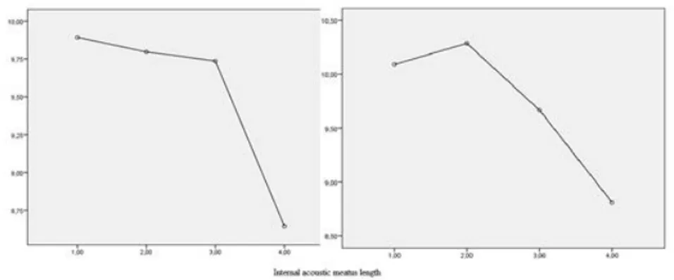
Figure 1. The showing as charts of internal acoustic meatus measurements. Figure 1a. Internal Acoustic Meatus Length (Right and left side, respectively).
Figure 1b. Internal Acoustic Meatus Width (Right and left side, respectively).
Figure 2. The width and length measurements of the internal acoustic meatus IAM: Internal acoustic meatus
Figure 3. The height measurements of the internal acoustic meatus IAM: Internal acoustic meatus.
Table 1. Internal acoustic meatus (IAM) parameters in healthy adult subjects
Parameters (mm) Female (n=59) Male (n=83) P*
IAM Length (Right) 9.71±1.46 (7.0-13.2) 9.61±1.58 (5.7-13.5) 0.708 IAM Length (Left) 9.92±1.45 (6.8-13.9) 9.87±1.64 (4.6-14.6) 0.865 IAM Width (Right) 3.97±0.6 (2.3-5.2) 4.18±0.74 (2.6-5.7) 0.079 IAM Width (Left) 3.95±0.61 (2.4-5.0) 4.15±0.71 (2.4-5.7) 0.085 IAM Height (Right) 4.65±0.69 (3.10-6.80) 5.13±0.85 (3.5-7.0) 0.001 IAM Height (Left) 4.66±0.69 (3.1-6.9) 5.13±0.90 (3.6-7.7) 0.001
*ANOVA test, SD; Standard Deviation, Min-Max; Minimum-Maximum. All values are given as Mean ± SD (Min.-Max.)
Table 2. Internal acoustic meatus (IAM) parameters in age groups. Parameters (mm) Group I (n=43) (18-30 years) Group II (n=49) (31-40 years) Group III (n=30) (41-50 years) Group IV (n=20) (51-60 years) Total (n=142) P* IAM length (Right) 9.89±1.45 (6.9-13.2) 9.80±1.67 (5.7-13.5) 9.74±1.38 (6.7-12.3) (6.7-11.40) 8.65±1.16 9.65±1.53 (5.7-13.5) 0.015 IAM length (Left) 10.09±1.54 (6.8-13.9) 10.29±1.62 (6.8-14.6) 9.67±1.50 (4.6-12.1) 8.81±0.99 (6.7-10.5) 9.89±1.56 (4.6-14.6) 0.002 IAM width (Right) 3.94±0.68 (2.6-5.1) 4.15±0.81 (2.3-5.7) 4.18±0.61 (3.1-5.7) 4.16±0.75 (3.0-5.2) 4.09±0.73 (2.3-5.7) 0.415 IAM width (Left) 3.95±0.68 (2.5-5.4) 4.15±0.75 (2.4-5.4) 4.12±0.60 (2.9-5.7) 4.01±0.56 (3.2-5.1) 4.06±0.68 (2.4-5.7) 0.475 IAM height (Right) 4.63±0.83 (3.2-6.9) 5.02±0.85 (3.1-6.9) 5.17±0.78 (3.9-7.0) 4.98±0.79 (3.5-6.2) 4.93±0.84 (3.2-6.9) 0.032 IAM height (Left) 4.71±0.84 (3.3-7.2) 4.87±0.77 (3.1-6.7) 5.33±0.99 (3.9-7.7) 4.94±0.65 (3.6-6.0) 4.93±0.85 (3.1-7.7) 0.019
*ANOVA test, SD; Standard Deviation, Min-Max; Minimum-Maximum; All values are given as Mean ± SD (Min.-Max.) Statistical analysis
The SPSS 21.0 program was used for statistical analysis of the measurement results. From these measurements, means, standard deviations (SD), minimum and maximum values were calculated; In all statistical analyses; p value under 0.05 was considered statistically significant. Furthermore, according to show normal distribution of the data ANOVA from
parametric tests were used to determine significance degree between sex and different age groups. Also, Paired Samples T Test were used to evaluate the differences between right and left side.
RESULTS
The aspect of the Internal Acoustic Meatus measurement charts according to gender in sagittal
423
and axial CT is shown in Figure 1. The values of minimum, maximum, mean and standard deviation of internal acoustic meatus measured in 142 healthy subjects (59 females and 83 males) are shown in Table 1 and 2. There were found significant difference in internal acoustic meatus height measurements between females and males (Table 1). Moreover, there were found significant difference between age groups in four parameters such as internal acoustic meatus length, and height in different age groups. There were no significant difference in internal acoustic meatus width measurements [p=0.415 (R); p=0.475 (L)] in different age groups (Table 2). In right and left sides, there were significant difference in internal acoustic meatus length (p=0.001). However, internal acoustic meatus width
(p=0.352) and height (p=0.953) were no showed significant difference.
When analyzed values, there were decrease after the age of 51 years in internal acoustic meatus length, width and height both genders. Also, the mean ratio of IAM’s width to height was calculated as 0.839±0.133 in right side, and 0.835±0.136 in left side. This ratio of right side was found as 0.864±0.154 in 18-30 years; 0.831±0.129 in 31-40 years; 0.815±0.107 in 41-50 years; and 0.842±0.129 in 51-60 years, respectively. Additionally, in left side the same ratio was measured as 0.851±0.155 in 18-30 years; 0.858±0.138 in 31-40 years; 0.786±0.120 in 41-50 years; and 0.815±0.841-50 in 51-60 years, respectively.
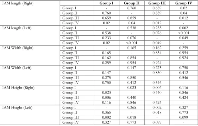
Table 3. Internal Acoustic Meatus (IAM) parameters between Groups
IAM length (Right) Group I Group II Group III Group IV
Group 1 - 0.760 0.659 0.02
Group II 0.760 - 0.859 0.04
Group III 0.659 0.859 - 0.012
Group IV 0.02 0.04 0.012 -
IAM length (Left) Group 1 - 0.538 0.233 0.002
Group II 0.538 - 0.076 <0.001
Group III 0.233 0.076 - 0.049
Group IV 0.02 <0.001 0.049 -
IAM Width (Right) Group 1 - 0.165 0.162 0.259
Group II 0.165 - 0.854 0.954
Group III 0.162 0.854 - 0.924
Group IV 0.259 0.954 0.924 -
IAM Width (Left) Group 1 - 0.147 0.275 0.750
Group II 0.147 - 0.850 0.412
Group III 0.275 0.850 - 0.546
Group IV 0.750 0.412 0.546 -
IAM Height (Right) Group I - 0.023 0.006 0.116
Group II 0.023 - 0.440 0.846
Group III 0.006 0.440 - 0.424
Group IV 0.116 0.846 0.424 -
IAM Height (Left) Group I - 0.365 0.002 0.327
Group II 0.365 - 0.018 0.773
Group III 0.002 0.018 - 0.099
Group IV 0.327 0.773 0.099 -
No significant difference was no found in ratio of IAM’s width to height (p value, 0.448 in right side; 0.095 in left side). The same dimension was calculated 0.860±0.125 and 0.825±0.137 in females and males in right side, respectively (p=0.120) whereas in left side the corresponding value was 0.855±0.125 and 0.820±0.142 in females and males, respectively (p=0.135). Also, the significance degree of the
Internal Acoustic Meatus length, width and height measurements between four groups were shown in Table 3.
DISCUSSION
Morphometric evaluation of the internal acoustic meatus (IAM) are vital to occur anatomical basis for
microsurgery of the cerebellopontine angle and intracranial pathology such as acoustic neuroma11. Moreover, knowledge of normal values and anatomy of the IAM is essential in determination of temporal bone trauma, congenital anomalies, affecting the individual nerves, and ear diseases surgery6. The differences in size of the internal auditory meatus on two sides may be a marker of the some diseases such as tumor, or internal auditory stenosis. Also, the suitable width for the neurovascular bundle is regarded approximately 2 mm. If difference in diameter of IAM between two internal acoustic canal is more than 2 mm, it can be a marker of tumor or, if there is a loss of 3 mm and more in IAM height, it can be thought as internal auditory meatus stenosis4,11. Additionally, the IAM’s narrowness is associated with cochlear nerve hypoplasia9. Glastonburry et al stated that if IAM’ vertical or transverse diameter was less than 4 mm, IAM was regarded as abnormal12. Mishra et al reported that the variation in IAM’diameter between two sides should not exceed 1 or 2 mm13. In a Stjernholm and Moren’s study, if the internal acoustic canal diameter is more than 1.4 mm, cochlear nerve abnormality can be thougth14. Valvassory et al declared there were differences in internal auditory canal’size. It’s diameter might vary from 2-3 mm to 12 mm (mean 5 mm)15. Furthermore, there are some differences in IAM’s morphometric data due to different races, and regions and also IAM varies between right and left sides6,11. In a study of Papangelou performed with the exact casts of 242 randomly-selected paired normal human ears, the length of the internal auditory canal was larger in males than females. Also, the relation found length and sex, whereas the relation no found between length and age. The length of the IAM was developed in second decade (11-20 years)16. In this study, there were no found significant difference in internal acoustic meatus width (p=0.352) and height (p=0.953) in both sides and it means there are no pathology of our study population.
There are many studies on radiographs, dissected temporal bone, dry skulls, or on CT, MRI regarding the morphometry or volume of IAM3,4,6,9-12. However, there are a few studies regarding gender and age changes5,9.
In Farahani’s study performed with Iranian cadavers, the mean of the IAM height was found as 4.04 mm (ranged from 3 mm to 5 mm)6. Sakashita and Sando reported the same dimension as 4.8 mm (ranged between 3.2 mm to 6.5 mm)17. In a study performed
Mamatha et al by using 40 adult temporal dry skull and casting method, means of height of the IAC were found as 3.52 mm (ranged between 2.5 mm and 4.9 mm) and 3.44 mm (ranged between 2.5 mm and 5.1 mm) in right and left sides, respectively11. Prashaanthi and Krishnamoorthy studied with Indian cadavers and the same parameter was found as 3.8 mm and 3.6 mm in right and left side, respectively4. In Germany population, the height of IAM was reported as 3.95 mm on CT, 3.70 mm in skull7. In Japanese population, the height of IAM was measured 6.8 mm (ranged from 3.5 mm to 12.00 mm) and males’value was significantly larger than females5. In Turkish patients with chronic otitis the IAM length was 9.33 mm and 9.59 mm in right and left side18. In this study, the means of IAM’s height were 4.65 mm and 4.66 mm of females in right and left side, respectively. The same measurements were 5.13 mm and 5.13 mm of males in right and left side, respectively. We found significant differences in the mean height value of IAM of Iranians, Indians, and German with our population; having lower value than Japanese. having greater value than Turks. From this data, our results are more similar to Belgium and Italian healty group. The mean of the internal acoustic meatus width was 3.96 mm (ranged between 3 mm and 5 mm)6. In Indian dried skull, means of width of the IAC were 3.37 mm (ranged from 2.00 mm to 5.08 mm), and 3.47 mm (ranged from 2.2 mm to 4.8 mm), in right and left sides, respectively11. Also, Indian cadavers, the corresponding value was measured as 6.4 mm and 6.9 mm in right and left side4. Berlis et al measured the IAM width as 3.87 mm (ranged between 2 mm and 6 mm) on CT, 4.11 mm (ranged from 2.35 mm to 6.36 mm) in European skulls19. In Brazilian adults the IAC anteroposterior (AP) width was found as 4.47 mm10. In Erkoç et al’s study, the AP diameter of IAC were reported as 5.93 mm and 5.83 mm in Turkish males and females with MRI. Also, Erkoç et al was found no significant difference in IAM width between gender, and in third decade (20-30 years) the same measurement was reached maximum value, whereas the minimum value was obtained in fifth decade (41-50 years)9. In chronic otitis Turkish population the same measurement was reported as 4.00 mm and 4.34 mm in right and left side18. In Egyptian, the same parameter was found 5.44 mm in control group; 5.27 mm in normal group; 11.61 mm in Patulous; and 1.43 mm in stenotic group7. In this study, the means of IAM width of females were 3.97 mm and 3.95 mm in right and left side, respectively. In males the corresponding values were found as 4.18
425
mm and 4.15 mm in right and left side, respectively. The maximum value was reached in fourth and fifth decade, whereas the minimum value was obtained in 18-30 years.
Internal acoustic canal length was declared as 1 cm in anatomy books2,3,20. Internal acoustic meatus length were reported as 8.7 mm (minimum 4 mm and maximum 18 mm) by Kobayashi and Zusho5. Moreover, the inferior length of the IAM was found 8.39 mm, whereas the superior length of the IAM was 10.75 mm6. In Indians, the means superior length of the IAM were measured as 9.89 mm and 9.94 mm on right and left side, whereas inferior length of the IAM was 8.43 mm and 8.59 mm, in right and left side11. Marcues et al reported the same dimension as 9.84 mm in Brazilian adult patients undergone routine tests10. IAC’s length of Germans were measured on CT as axial and coronal projection (between 10.98 mm and 11.98 mm); and in skull (11.68 mm), respectively19. In Egyptian, the same parameter was found 6.95 mm in control group; 7 mm in normal group; 8.46 mm in Patulous; and 10.46 mm in stenotic group7. Additionally, according our IAM length values of females were measured as 9.71 mm and 9.92 mm in right and left side, respectively. In males the same measurement were found as 9.61 mm and 9.87 mm in right and left side, respectively. The ratio of the IAM’s width to height was 1.00 mm in Iran population6. The our dimension (female, 0.860±0.125; and male, 0.825±0.137) was found lower than Farahani et al’s study.
In conclusion, we think that knowledge of the IAM anatomy in healthy population is important for safe and accurate surgical interventions in otology. Especially, we believe that data obtained in this study may give crucial information for IAM morphometry, age related changes and gender differences. Also, it may help the otolaryngology surgeon during presurgical evaluation for having a succesful surgery and minimize the related problems. Additionally, this study presents a normative data of internal acoustic meatus in healthy Turkish population.
Yazar Katkıları: Çalışma konsepti/Tasarımı: SP, AGU, FYÖ, MÖ,
AHY; Veri toplama: SP, AGU, FYÖ, MÖ, AHY; Veri analizi ve yorumlama: SP, AGU, FYÖ, MÖ, AHY; Yazı taslağı: SP; İçeriğin eleştirel incelenmesi: SP, AGU, FYÖ, MÖ, AHY; Son onay ve sorumluluk: SP, AGU, FYÖ, MÖ, AHY; Teknik ve malzeme desteği: SP, AGU, FYÖ, MÖ, AHY; Süpervizyon:SP, AGU, FYÖ, MÖ, AHY; Fon sağlama (mevcut ise): yok.
Bilgilendirilmiş Onam: Katılımcılardan yazılı onam alınmıştır. Hakem Değerlendirmesi: Dış bağımsız.
Çıkar Çatışması: Yazarlar çıkar çatışması beyan etmemişlerdir. Finansal Destek: Yazarlar finansal destek beyan etmemişlerdir.
Author Contributions: Concept/Design :SP, AGU, FYÖ, MÖ, AHY; Data acquisition: SP, AGU, FYÖ, MÖ, AHY; Data analysis and interpretation: SP, AGU, FYÖ, MÖ, AHY; Drafting manuscript: SP; Critical revision of manuscript: SP, AGU, FYÖ, MÖ, AHY; Final approval and accountability: SP, AGU, FYÖ, MÖ, AHY; Technical or material support: SP, AGU, FYÖ, MÖ, AHY; Supervision: SP, AGU, FYÖ, MÖ, AHY; Securing funding (if available): n/a.
Informed Consent: Written consent was obtained from the
participants.
Peer-review: Externally peer-reviewed.
Conflict of Interest: Authors declared no conflict of interest. Financial Disclosure: Authors declared no financial support
REFERENCES
1. Stranding S. Gray’s Anatomy. The anatomical basis of clinical practice. 40th Ed. New York: Elsevier Limited: 2016.
2. Moore KL, Dalley AF. Clinically oriented anatomy. Fourth Ed. Canada: Lippincolt Williams&Wilkins;1999.
3. Özocak O, Unur E, Ülger H, Ekinci N, Aycan K, Acer N. Meatus acusticus internus’un morfometrisi ve varyasyonları. EÜ Sağlık Bilimleri Dergisi. 2004;13:1-7.
4. Prashaanthi N, Karpagam K. Morphometric analysis of internal acoustic meatus. Res J Phar Tech. 2016;9:1575-6.
5. Kobayashi H, Zusho H. Measurements of internal auditory meatus by polytomography. Br J Radiol. 1987;60:209-14.
6. Farahani RM, Nooranipour M, Nikakhtar KV. Anthropometry of internal acoustic meatus. Int. J. Morphol. 2007;25:861-5.
7. El Sadik AO, Shaaban MH. The relationship between the dimensions of the internal auditory canal and the anomalies of the vestibulocochlear nerve. Folia Morphol. 2017;76:178-85.
8. Manzari L, Scagnelli P. Large bilateral internal auditory meatus associated with bilateral superior semicircular canal dehiscence. Ear, Nose Throat J. 2013;92:25-33.
9. Erkoç MF, Ímamoglu H, Okur A, Gümüş C, Dogan M. Normative size evaluation of internal auditory canal with magnetic resonance imaging: review of 3786 patients. Folia Morphol. 2012;71:217–20. 10. Marques SR, Ajzen S, D´Ippolito G, Alonso L, Isotani
S, Lederman H. Morphometric analysis of the internal auditory canal by computed tomography imaging. Iran J Radiol. 2012;9:71-8.
11. Mamatha Y, Trisha KR, Kumar V. Anthropometry of internal acoustic meatus in dry adult human skull using casting method. Int J Anat Res. 2019;7:6113-18. 12. Glastonbury CM, Davidson HC, Ric Harnsberger H, Butler J, Kertesz TR, Shelton C. Imaging findings of cochlear nerve deficiency. Am J Neuroradiol. 2002;23:635-43.
13. Mishra LCAK, Mehta LCAK, Singh BH. Hypoplasia of internal acoustic meatus. Med J Armed Forces India. 2006;62:196-7.
14. Stjernholm C, Muren C. Dimensions of the cochlear nerve canal: A radioanatomic investigation. Acta Otolaryngol. 2002;122:43–8.
15. Valvassori GE, Morales FG, Palacios E, Dobben GE. MR of the normal and abnormal internal auditory canal. AJNR Am J Neuroradiology. 1988;9:115-9. 16. Papangelou L. Study of the human internal auditory
canal in relation to age and sex. J Laryngol Otol. 1975;89:79-89.
17. Sakashita T, Sando I. Postnatal development of the internalauditory canal studied bycomputer-aided
three-dimensional reconstruction and measurement. Ann Otol Rhinol Laryngol. 1995;104:469-75. 18. Akbal İ. Meatus acusticus internus boyutları ile orta
kulaktaki varyasyon ilişkilerinin incelenmesi. (Yükek lisans tezi). Afyon, Afyon Kocatepe Üniversitesi 2015.
19. Berlis A, Putz R, Schumacher M. Direct and CT measurements of canals and foramina of the skull base. Br J Radiol. 1992;65:653-61.
20. Yücel AH. Dere Anatomi Atlası ve Ders Kitabı. 7.baskı; Adana: Akademisyen Kitabevi; 2018.