CASE REPORT
Date submitted: May 11, 2016 • Date accepted: Aug 04, 2017 • Online publication date: June 30, 2017
Introduction
Surgically assisted rapid maxillary expansion (SARME) is a treatment modality used for the management of transver-se maxillary hypoplasia, especially in adolescents and young adults (1). Many factors such as congenital, developmental, iatrogenic and traumatic factors are responsible in the etiology. The cause of maxillary constrictions could be; oral breathing, infancy type swallowing and tongue thrust. Radiographic and clinical examination should be performed for the exact diagno-sis of this type of deformity. The clinical findings are unilateral or bilateral crossbite, narrow palatal vault, rotated, crowded and palatally or buccally displaced teeth and a narrow and V shaped maxillary arch form (2-4). The cause of crossbite in adult patients is either skeletal or dental. Dental crossbites are usually treated by only orthodontic management. However, crossbites of more than one teeth are suggested to be the result of skeletal problems (5). SARME can be defined as the use of orthodontic expansion appliance with maxillary surgery. It is used especially in patients who has completed their skele-tal maturation (6). The aim of this case report was to evaluate dentofacial effects of SARME treatment protocol.
Case Report
A 17-year-old male patient referred to our clinic with primary complaint of unaesthetic smile and severe maxillary crowding. The patient had an oral breathing and he was snoring due to the nasal obstruction. It was suggested that all these compla-ints were the result of transverse maxillary constriction. Extra-oral examination revealed that the patient had a doligocepha-lic head type on the frontal plane and a respiratory face. On the sagittal plane, the patient had a convex profile due to the clockwise rotation of the mandible and the lower anterior face height was increased also. The smile analysis revealed an increased occurence of buccal corridors as well as unesthetic incisor view (Fig. 1). Panoromic radiographic analysis showed that the lower left molar was extracted (Fig. 2). The patient’s intraoral examination revealed; 1mm overbite on frontal pla-ne, 4mm overjet on the sagittal plapla-ne, both molar associations were Angle Class II, bilateral posterior crossbite on the trans-verse plane in addition to V shaped upper arch form. Model analysis revealed tooth/arch size discrepancy 18mm on upper arch and 4mm on lower arch. According to the lateral cephalo-metric analysis, skeletal Class II relationship was seen due to mandibular retrognathia (Table I)
Treatment Strategy
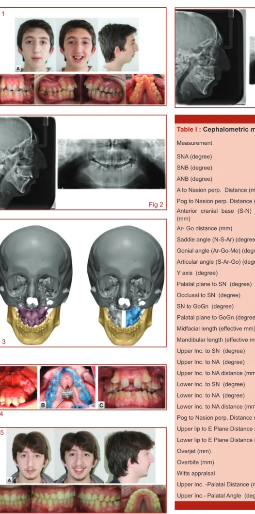
Computed tomographic (CT) images were segmented by using Mimics software (Materialise, Leuven, Belgium) for plan-ning of the treatment strategy (Fig. 3). The surgical planplan-ning was performed in SBÜ Gülhane industrial design and
manu-*Gülhane Diş Hekimliği Fakültesi Ortodonti Ad.Bşk.Lığı (Araştırma asistanı.) **Gülhane Diş Hekimliği Fakültesi Ortodonti Ad.Bşk.Lığı (Lisansüstü öğrenci.) ***Gülhane Diş Hekimliği Fakültesi Ortodonti Ad.Bşk.Lığı (Doçent.) ****Medipol Üniversitesi Hastanesi Ortodonti (Doçent)
*****Gülhane Diş Hekimliği Fakültesi Ağiz Diş Ve Çene Cerrahisi (Doçent)
Corresponding Author: Gökhan Serhat Duran
Araştırma asistanı. [email protected]
Cerrahi Destekli Üst Çene Genişletme Tedavisi
•
49 SUMMARYThe aim of this case report is to present an adult patient with a narrow maxilla and severe crowding treated with the help of surgically assisted maxillary expansion. A 17-year-old male patient referred to our clinic for the primary complaint of unaesthetic smile and severe upper anterior crowding. Extraoral examination revealed convex profile. Intraoral examination revealed narrow maxilla, posterior cross bite, V shaped upper arch form and severe crowding. The treatment was initiated with corticotomy operation for maxillary expansion. A tooth-borne acyrilic cap splint appliance with Hyrax screw was then fixed for distraction. Distraction protocol was started 7 days postoperatively by 2 turns a day (0.5 mm/ day). The distractor was left in place for an additional 3 months for stabilization and reossification after the required space had been achieved. Orthodontic fixed treatment started within 4 weeks after removal of the appliance. Computed tomography scans of the patient were performed to help the planning of the treatment 1 month before the orthodontic treatment. The data reconstructed using the mimics software (materialise, Leuven, Belgium) for surgical planning. surgically assisted maxillary expansion is a successful treatment modality for adult patients requiring palatal expansion. The total treatment period was 24 months and the results were acceptable.
Key words: SARME, Surgical assisted rapid maxillary expansion ,palatal expansion.
ÖZET
Maksiller darlığı ve şiddetli çapraşıklığı olan hastanın cerrahi destekli üst çene genişletme tedavisi (SARME) : Olgu sunumu
Bu olgu sunumunun amacı dar üst çeneye ve şiddetli yer darlığına sahip yetişkin hastanın cerrahi destekli üst çene genişletilmesi yardımı ile tedavisinin sunulmasıdır. 17 yaşında erkek hasta estetik olmayan gülümseme ve şiddetli üst ön çapraşıklık ile kliniğimize başvurdu. Ağız dışı incelemede konvex bir profil olduğu gözlendi. Ağız içi incelemede dar bir maksilla, arka bölgede çapraz kapanış, V şekilli üst ark formu ve şiddetli çapraşıklık gözlendi. Tedavi cerrahi operasyon ile başladı. Hyrax vida içeren diş destekli akrilik cap splint distraksiyon apareyi uygulandı. Distraksiyon protokolüne operasyon sonrası 7 günlük bekleme sonrası günde 2 tur (0.5mm/ gün) olacak şekilde başlandı. Gerekli yer elde edildikten sonra stabilizasyon ve yeni kemik oluşumunun beklenmesi amacıyla distraktör operasyon sonrası 3 ay ağızda bırakıldı. Apareyin çıkartılmasını takiben 4 hafta sonra sabit tedaviye başlandı. Tedavi planlaması amacıyla hastanın bilgisayarlı tomografi görüntüleri tedaviden 1 ay önce alındı. Analiz amaçlı, veriler mimics (materialise, Leuven, Belgium) yazılımı ile yeniden oluşturuldu. Cerrahi destekli üst çene genişletme tedavisi palatal ekspansiyon ihtiyacı olan yetişkin hastalarda başarılı bir tedavi yöntemidir. Toplam tedavi 24 ay sürmüştür ve kabul edilebilir sonuçlar elde edilmiştir.
Anahtar Kelimeler: SARME, Cerrahi destekli üst çene genişletme tedavisi,erişkin
ortodonti,üst çene darlığı.
Gökhan Serhat Duran(*), Alaattin Tekeli(**), Serkan Görgülü(***), Sıla Mermut Gökçe(****),
Hasan Ayberk Altuğ(*****)
Treatment of severe crowding and a narrow maxilla with
surgically assisted rapid maxillary expansion (SARME) : A case
report
Gülhane Med J 2017;59: 49-52 © Gülhane Medical Faculty 2017 doi: 10.5455/Gulhane.228324
50
•
June 2017 • Gulhane Med J Duran et. al. Table I : Cephalometric measurementsMeasurement Pre-treatment Post-treatment SNA (degree) 68.1 69.4
SNB (degree) 66 65.6
ANB (degree) 2.1 3.8
A to Nasion perp. Distance (mm) -4 -4.7 Pog to Nasion perp. Distance (mm) -11.2 -12.9 Anterior cranial base (S-N) distance
(mm) 71.9 72.1
Ar- Go distance (mm) 50.3 52.1 Saddle angle (N-S-Ar) (degree) 128.2 129.9 Gonial angle (Ar-Go-Me) (degree) 129.2 131 Articular angle (S-Ar-Go) (degree) 142.1 145.2
Y axis (degree) 64.3 65
Palatal plane to SN (degree) 6.3 6.9 Occlusal to SN (degree) 19.9 20.3 SN to GoGn (degree) 44.6 42.3 Palatal plane to GoGn (degree) 38.3 40.5 Midfacial length (effective mm) 78.1 82.3 Mandibular length (effective mm) 109.8 110.9 Upper Inc. to SN (degree) 96 94.7 Upper Inc. to NA (degree) 27.9 24.8 Upper Inc. to NA distance (mm) 8.5 7.4 Lower Inc. to SN (degree) 97.2 97.6 Lower Inc. to NA (degree) 27.7 29.3 Lower Inc. to NA distance (mm) 10 7.5 Pog to Nasion perp. Distance (mm) 2.9 1.9 Upper lip to E Plane Distance (mm) -3.7 -3 Lower lip to E Plane Distance (mm) -2.1 -1.8
Overjet (mm) 4.1 3.4
Overbite (mm) 1.2 2.7
Witts appraisal 7.9 6.3
Upper Inc. -Palatal Distance (mm) 44.1 37.4 Upper Inc.- Palatal Angle (degree) 100.8 102.6
Fig 1 Fig 2 Fig 3 Fig 4 Fig 5 Fig 6
Cerrahi Destekli Üst Çene Genişletme Tedavisi
•
51 facturing center. An acrylic cap splint type expansionapplian-ce and surgically assisted corticotomy were planned for the maxillary narrowing in a patient who has completed his growth and development. In addition to extraction of the upper third molars, due to the need for more arch perimeter, the pull of the vestibular positioned canine teeth and giving a form of canines to the first premolars were planned.
Treatment period
Cephalometric, panoromic radiographs and CT images for surgical planning in addition to the patient’s pretreatment intra-oral and extraintra-oral photographs were taken. Upper third molars were extracted for presurgical preparation. The treatment was started with surgically assisted expansion, after corticotomy for the treatment of transverse direction insufficiency. Acrylic cap splint type expansion appliance was used intraorally after sub-total Lefort I osteotomy. One-week latent period after the surgi-cal procedure, the activation was started. The activation of the appliance was performed by turning 0.5mm/day or twice daily. After ensuring an adequate expansion, the appliance’s screw was stabilized with a steel wire and the consolidation period was started (Fig. 4). Three months after the waiting period, retention was continued via an acrylic plate supported trans-palatal arch assistance and stantart orthodontic treatment was started (22 slot, Roth type). Upper canines were extracted and the leveling stage was started via 0,12 NiTi archwire. Stripping (1,5mm) was performed at the lower anterior arch area. The molars were mesialized with NiTi closed coil springs on the thick arch wires for the closure of the gap at the right lower posterior area. The standart orthodontic treatment ended at 20 months. Thermoplastic and fixed retainer was applied to avo-id posttreatment relapse. Intraoral and extraoral photographs were taken (Fig. 5) and cephalometric and panaromic images were obtained at the end of the treatment period (Fig. 6).
Discussion
The maxillary hypoplasia due to the insufficient growth in transverse direction, negatively effects the individuals both functionally and also esthetically. It is suggested that, rapid maxillary transverse expansion is the most commonly used technique in the management of malocclusion due to both dental and skeletal irregularities and the most appropriate time for this procedure is between the ages 8 and 15 (7). In the adult population, due to the reduced elastical capacity of bones with increasing age, the management of malocclusion is limited only with dentoalveolar structure and maxillary ex-pansion could not be obtained due to this skeletal resistance (8). SARME became a popular management protocol in the treatment of transverse disharmony of upper maxillary arches in adult patients (9) .
The patient was 17 years-old, thus surgery assisted expan-sion with corticotomy was planned as the patient was in the end of his growth and development period. The purpose of all surgical procedures for the rapid expansion of maxilla in the adult patients is to obtain the best sutural opening and transverse skeletal expansion by minimum surgical procedu-re. We started the maxillary expansion in our patient after a latent period of 7 days from the surgery. It is suggested that 5-7 days are satisfactory after surgery for capillary healing in the bone cavity, thus maxillary expansion was started 5-7 days after surgery (6). It is reported that, the daily expansion amount of the screw could be modified, taking into the considerations of the factors such as the age of the patient, the state of the occlusion, the amount of expansion planned and the toleran-ce to the appliantoleran-ce used for expansion (10) .In our patient, a
satisfactory expansion was obtained without a discomfort or a problem, by turning the screw for a quarter tour two times daily. Clinically, the most important change seen during SAR-ME is the formation of diastema between the upper incisors. It is reported that SARME causes a manifest increase ment in the intermolar width (6). In our patient, we have also seen the formation of diastema between the upper incisors and an increase in the width of the intermolar width.
It is suggested in the literature that orthopedic forces during the expansion period resulted in important dimensional incre-ases of dentoalveolar structures, base of maxilla and width of the nasal cavity by causing an opening in midpalatal suture .It is known that SNA angle increases as a result of forward movement after the rapid expansion of maxilla by an expan-sion appliance (7, 11). In our patient an increase of 1.3° in the SNA angle (Table 1) In various studies decrease in the SNB and an increase in the ANB angles were detected. In this case report, we have seen a decrease of 0.4° in SNB angle and an increase of 1.7° in ANB angle, (Table 1). We obtained accep-table molar relationship, transverse maxillary hypoplasia was corrected by maxillary expansion. The smile esthetics and the crowding were corrected. As a conclusion, SARME is a suc-cessful method for the management of maxillary narrowing in patients with a completed growth and development.
References
1. Glassman, A.S., Nahigian S.J., Medway J.M., Arono-witz H.I. Conservative surgical orthodontic adult ra-pid palatal expansion: Sixteen cases. Am J Orthod. 1984;86:207-213.
2. Rossi, R.C., Rossi N.J., Rossi N.J.C., Yamashita H.K., Pignatari S.S.N. Dentofacial characteristics of oral bre-athers in different ages: A retrospective case-control study. Prog Orthod. 2015;16:23.
3. McNamara, J.A., Lione R., Franchi L., Angelieri F., Cevi-danes L.H.S., Darendeliler M.A., et al. The role of rapid maxillary expansion in the promotion of oral and general health. Prog Orthod. 2015;16:33.
4. Dahan, J.S., Lelong O. Effects of bite raising and occ-lusal awareness on tongue thrust in untreated children. Am J Orthod Dentofacial Orthop. 2003;124:165-172. 5. Andrade, A.d.S., Gameiro G.H., Derossi M., Gavião
M.B.D. Posterior crossbite and functional changes. A systematic review. Angle Orthod. 2009;79:380-386. 6. Koudstaal, M.J., Poort L.J., van der Wal K.G.H.,
Wolvi-us E.B., Prahl-Andersen B., Schulten A.J.M. Surgically assisted rapid maxillary expansion (sarme): A review of the literature. Int J Oral Maxillofac Surg. 2005;34:709-714.
7. Bucci, R., D'Antò V., Rongo R., Valletta R., Martina R., Michelotti A. Dental and skeletal effects of palatal ex-pansion techniques: A systematic review of the current evidence from systematic reviews and meta-analyses. J Oral Rehabil. 2016;43:543-564.
8. Angelieri, F., Cevidanes L.H.S., Franchi L., Gonçalves J.R., Benavides E., McNamara J.A. Midpalatal suture maturation: Classification method for individual assess-ment before rapid maxillary expansion. Am J Orthod Dentofacial Orthop. 2013;144:759-769.
52
•
June 2017 • Gulhane Med J Duran et. al.pansion: A literature review. Am J Orthod Dentofacial Orthop. 2008;133:290-302.
10. Sygouros, A., Motro M., Ugurlu F., Acar A. Surgically as-sisted rapid maxillary expansion: Cone-beam computed tomography evaluation of different surgical techniques and their effects on the maxillary dentoskeletal comp-lex. Am J Orthod Dentofacial Orthop. 2014;146:748-757.
11. Machado-Júnior, A.-J., Zancanella E., Crespo A.-N. Rapid maxillary expansion and obstructive sleep apnea: A review and meta-analysis. Med Oral Patol Oral Cir Bucal. 2016:0.