71
Case Report
Selective Retreatment of a Three-Canalled Mandibular Premolar using Cone Beam Computed Tomography: 5-Year Follow-up
Neslihan Yılmaz Çırakoğlu1
1Department of Endodontics, Faculty of Dentistry, Karabük University, Karabük, Turkey
Abstract: Root canal retreatment is traditionally considered an “all or none” treatment approach. However, the use of three-dimensional imaging such as cone-beam computed tomographic imaging allows for a more accurate evaluation of the periapical status of individual roots associated with multi-rooted teeth. This information has introduced a novel and conservative treatment alternative for previously endodontically treated teeth with multiple roots presenting with post-treatment disease. This new approach is termed selective root retreatment. This case report presents the selective retreatment by using CBCT and 5-year follow-up of the lower second premolar tooth, which has been previously treated with canal treatment but has recurrent infection symptoms. The patient was followed up for 6 months, 1 year and 5 years. At control appointments, the patient was asymptomatic and radiographic evaluation showed that periapical infection had healed. This case report shows that the selective retreatment method can be successfully applied to multi-rooted teeth with symptoms of recurrent infection after root canal treatment.
Keywords: Cone beam computed tomography; multi-rooted tooth; reinfection; selective retreatment
Address of Correspondence: Neslihan Yılmaz Çırakoğ[email protected],
0000-0002-8316-3133, Department of Endodontics, Faculty of Dentistry, Karabük University, Kastamonu Yolu, Demir Çelik Kampüsü, Kılavuzlar/Karabük Merkez, Turkey.
1. Introduction
The fundamental aims of root canal therapy are properly preparing and disinfectioning of all pulp spaces and its wholly filling it in three-dimensions with an biocompatible obturation material to avoid reinfection of the tooth (Schilder, 1967; Schilder, 1974). Initial root canal treatment has been stated to result in a desirable consequence between 86% and 98% of the time (Friedman et al., 2003, Friedman and Mor, 2004). This shows that the endodontically treated teeth that developed post-treatment pathosis or presenting persistence of disease were dramatically rate. Previous studies informed failure percentages of 14%– 16% for initial root canal treatment (Torabinejad et al., 2007). Causes of failure are based on persistent intraradicular infection residing in initially untreated canals, dentinal tubules, or the complicated root canal
72
system (Nair, 2006; Peters et al., 2004). The extraradicular reasons of endodontic lack of success attribute periapical actinomycosis (Tronstad et al., 1990), a foreign body reaction induced by extruded endodontic materials (Nair et al., 1990), an accumulation of endogenous cholesterol crystals in the apical tissues (Nair, 1999) and an unresolved cystic lesion (Ramachandran Nair et al., 1996). Treatment alternatives for the endodontically treated teeth with post-treatment disease are restricted. They comprise non-surgical retreatment, surgical treatment (including intentional replantation), or a combination of both procedures. Root canal retreatment is a non‐surgical process that includes removal of root canal filling materials from the root canal, and after that cleaning, shaping and filling of the canals. Root canal retreatment is conventionally considered an “all or none” treatment modality (Roda, 2006). However, the use of three-dimensional imaging such as cone-beam computed tomographic visualization permits for a more correct assessment of the periapical condition of individual roots interrelated with multi-rooted teeth. This data has asserted a modernistic and conservative treatment option for initially endodontically treated teeth with multiple roots exhibiting with post-treatment disease. This new procedure is entitled selective root retreatment. New generation imaging permits the clinician to manage presumable treatment determination in terms of the presence or absence of periapical disease of individual roots as opposed to guessing about the tooth completely (Nudera, 2015). In that way, retreatment could be restricted to a single root or roots evidently indicating periapical pathosis while leaving the root(s) with no apparent or noticed pathosis untouched. The aim of this case report is to present the selective retreatment by using CBCT and 5-year follow-up of the lower second premolar tooth, which has been previously treated with canal treatment but has recurrent infection symptoms.
2. Description of the Case
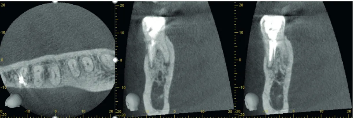
A 38-year-old male patient with a non-contributory medical history was referred to our clinic with the complaint of swelling and pain the previous two weeks in the lower left mandibular region. Extraoral examination demonstrated swelling in the left mental area. Clinical and radiographical examination presented that there was a root canal treatment in the second mandibular premolar tooth, but there was a lesion in the periapical area. It is thought that there might have been a missing extra canal. In the intraoral examination, the tooth was tender to percussion and palpation tests and exhibited no mobility. Probing depths were within normal limits for the tooth. Also, cone-beam computed tomography sections were taken to determine the exact diagnosis and lesion borders. As a result of the examined sections, it was determined that the second mandibular premolar tooth, which is generally single-rooted and single canals, had three canals. Two root canals were treated and a root canal was missed out. The periapical lesion originated from the missing root canal (Figure 1).
73 Figure 1. CBCT sections that taken from the tooth.
The treatment of the tooth was completed in two appointments. At the first visit, under local anesthesia, an access cavity was opened by applying rubber-dam to the tooth (Figure 2a). The root canal, which was missed out previously, was localized by endodontic sond without any intervention to other root canal fillings. Working length was determined using a radiograph with stainless steel hand #10 K file and electronic apex locator (Root ZX mini; J Morita Co, Kyoto, Japan) (Figure 2b). Cleaning and shaping of the canal was performed with rotary nickel-titanium system (ProTaper Next, Dentsply Maillefer, Ballaigues, Switzerland) up to X2. During the instrumentation, the canal was irrigated with copious amounts of 2.5% sodium hypochlorite. After the final irrigation (5.25% sodium hypochlorite, 17% ethylene diamine tetra acetic acid and 2% chlorhexidine) the canal was dried with paper cons, and dressed with a calcium hydroxide paste (Calasept, Nordiska Dental, Angelholm, Sweden), and the access was sealed with temporary filling material (Cavit W, 3M Espe, Seefeld, Germany).
When the patient came for control two weeks later, the patient did not have any symptoms (pain, swelling). The medication paste was removed with irrigation solutions. Final preparation was made with the X2 file. After the final irrigation, the root canal was dries with paper cons and was filled with gutta-percha and AH Plus sealer (AH Plus, Dentsply, De Trey, UK) with cold lateral compaction method. Then the tooth was restored with flowable and bulk-composite filling material. (Figure 2c and 2d).
Figure 2. a) Clinical view of the tooth after access cavity b) Radiographical view of localized missing canal c) Radiographical view of master con d) Clinical view of the tooth after filling the canal and placing flowable composite
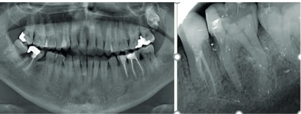
The patient was followed up for 6 months, 1 year and 5 years. At control appointments, the patient was asymptomatic and radiographic evaluation showed that periapical infection had healed (Figure 3 and 4).
74
Figure 3. After 1-year follow-up radiograps
Figure 4. After 5-year follow-up radiographs
3. Discussion
The presence of untreated root canals may be a reason for failure of endodontic therapy. Because of this, to achieve a satisfactory root canal therapy, the clinician should be aware of the multiple and complex variations that can occur during root formation and identify the different canal configurations (Parekh et al., 2011). The importance to have the knowledge of extra root canals and their locations with respect to the main respective canal depicts the success rate of their endodontic therapy (Maggiore et al., 2002; Vertucci and Haddix, 2011). Although mandibular 1st and 2nd premolar teeth are usually single-rooted and single-canalled, they can show variations in terms of the number of roots and canals. In the study of Prakash et al. two-rooted mandibular second premolars were found (Prakash et al., 2008). Vertucci and Francois 1986 reported that the mandibular first premolar had single canal at the apex in 74.0% of the teeth researched, two canals at the apex in 25.5%, and three canals at the apex in the rest 0.5% of the teeth. Only 12% of mandibular second premolars studied had a second or third canal. They also stated that the second premolar had one canal at the apex in 97.5% and two canals at the apex in only 2.5% of the teeth researched (Vertucci and Francois, 1986).
In 1991, Bram and Fleisher revealed a mandibular second premolar with four canals (Bram and Fleisher, 1991). Lotfi et al. mandibular second premolars with three canals and atypical orifices were studied. They found one distolingual and one distobuccal at the same level and one mesiolingual on the wall of the second premolar (Lotfi et al., 2008). Mandibular premolars with three canals were stated in other previous studies (Aguiar et al., 2010; Chan et al., 1992; Cleghorn et al., 2008; Poorni et al., 2010). As seen in
75 mentioned above studies, the incidence of second premolar teeth with atypical morphology with more
than one root and canal is considerably higher. As a result of that, these extra canals can be overlooked when performing canal treatment and this situation may cause post-treatment lesions.
Conservative treatment options are limited for teeth with recurrent or persistent infectional disease. These alternative approaches are the non-surgical retreatment (by removal of all root canal filling and restorations), surgical root treatment, or extraction. Although favourable results can be achieved with all these options, present technology may allow a more conservative and equally successful approach. It is generally recommended that all restorative and obturation materials be removed from all roots irrespective of the presence or absence of periapical pathosis in traditional root canal retreatment. However this treatment approach had some disadvantages. Weakening of the tooth structure through unnecessary removal of dentin and possible iatrogenic mistakes can generate difficulties for re-building after nonsurgical retreatment. Additionally, undesired heat generation can occur during post-removal and it may harm the tooth and its connecting periodontium (Davis et al., 2010). These risks could cause tooth loss. Besides these, removal of whole restorations is never practical and in the interest of patient. Patients usually refer to root canal retreatment with restorations considered to be short while ago builded. Patients are generally reluctant to undertake the financial and/or psychological pressure of a full restoration removal and reconstruction. Also, removal completely the existing restoration can cause loss of time for patients. Previously, treatment planning and finding permorfing has been confined by two-dimensional radiographic imaging. Cone-beam computed tomographic (CBCT) imaging has become a worthy device for the objective of endodontics (Patel, 2009). This improved imaging dramatically enhances the clinician’s facility to forseeable estimate root morphology, internal anatomy, and periapical pathosis before treatment (Barnett Abbott et al., 2011). This provides us to perform a differential diagnosis of which root arises the periapical pathosis, and selective retreatment could be managed more accurately.
As a conclusion, clinicians should be aware of root canal configuration malformations and be able to apply this knowledge in radiographic and clinical interpretation. It is the rationalist method to apply the selective retreatment method, which is the most conservative treatment approach, in teeth that developed post-treatment lesions due to the lack of internal anatomy knowledge. Meanwhile, CBCT can be used for precise diagnosis of the individual roots and canals. In this way, no attempts can be performed on root canal obturations that do not have pathosis and appear healthy. Also access cavity reformings may be required for unrestricted entry to complex anatomy and missing canals.
The main aim of this case study is to report a successful selective retreatment of a mandibular second premolar with three canals. In our case report, CBCT imaging confirmed the presence of an untreated root canal in the lower second premolar tooth. Owing to the fact that, there was no deficiency in filling and no periapical pathosis of other roots, no intervention was done to them. Thus, weakening of the roots and whole tooth is prevented without the unnecessary additional interventions.
Conflict of Interest
76
References
Aguiar, C., Mendes, D., Camara, A., Figueiredo, J. (2010). Endodontic treatment of a mandibular second premolar with three root canals. J Contemp Dent Pract, 11(2), 078-084.
Barnett, F., Abbott, J., Hartwell, G. (2011). Cone beam-computed tomography in endodontics. Endodontics: colleagues for excellence. Chicago: American Association of Endodontists, 1-7.
Bram, S. M., Fleisher, R. (1991). Endodontic therapy in a mandibular second bicuspid with four canals. J Endod, 17(10), 513-515.
Chan, K., Yew, S. C., Chao, S. Y. (1992). Mandibular premolar with three root canals--two case reports. Int Endod J, 25(5), 261-264.
Cleghorn, B. M., Christie, W. H., Dong, C. C. (2008). Anomalous mandibular premolars: a mandibular first premolar with three roots and a mandibular second premolar with a C-shaped canal system. Int Endod J, 41(11), 1005-1014.
Davis, S., Gluskin, A. H., Livingood, P. M., Chambers, D. W. (2010). Analysis of temperature rise and the use of coolants in the dissipation of ultrasonic heat build-up during post removal. J Endod, 36(11), 1892-1896. Friedman, S., Abitbol, S., Lawrence, H. P. (2003). Treatment outcome in endodontics: the Toronto Study. Phase 1: initial treatment. J Endod, 29(12), 787-793.
Friedman, S., Mor, C. (2004). The success of endodontic therapy--healing and functionality. J Calif Dent Assoc, 32(6), 493-503.
Lotfi, M., Vosoughhosseini, S., Zand, V., Fatemi, A., Shyezadeh, V., Ranjkesh, B. (2008). A mandibular second premolar with three canals and atypical orifices. J Oral Sci, 50(3), 363-366.
Maggiore, F., Jou, Y. T., Kim, S. (2002). A six-canal maxillary first molar: case report. Int Endod J, 35(5), 486-491. Nair, P. N. (1999). Cholesterol as an aetiological agent in endodontic failures--a review. Aust Endod J, 25(1), 19-26.
Nair, P. N. (2006). On the causes of persistent apical periodontitis: a review. Int Endod J, 39(4), 249-281. Nair. P. N., Sjogren, U., Krey, G., Sundqvist, G. (1990). Therapy-resistant foreign body giant cell granuloma at the periapex of a root-filled human tooth. J Endod, 16(12), 589-595.
Nudera, W. J. (2015). Selective root retreatment: a novel approach. J Endod, 41(8), 1382-1388.
Parekh, V., Shah, N., Joshi, H. (2011). Root canal morphology and variations of mandibular premolars by clearing technique: an in vitro study. J Contemp Dent Pract, 12(4), 318-321.
Patel, S. (2009). New dimensions in endodontic imaging: part 2. Cone beam computed tomography. Int Endod J, 42(6), 463-475.
77 Peters, O. A., Barbakow, F., Peters, C. I. (2004). An analysis of endodontic treatment with three
nickel-titanium rotary root canal preparation techniques. Int Endod J, 37(12), 849-859.
Poorni, S., Karumaran, C. S., Indira, R. (2010) Mandibular first premolar with two roots and three canals. Aust Endod J, 36(1), 32-34.
Prakash, R., Nandini, S., Ballal, S., Kumar, S. N., Kandaswamy, D. (2008). Two-rooted mandibular second premolars: case report and survey. Indian J Dent Res, 19(1), 70-73.
Ramachandran Nair, P. N., Pajarola, G., Schroeder, H. E. (1996). Types and incidence of human periapical lesions obtained with extracted teeth. Oral Surg Oral Med Oral Pathol Oral Radiol Endod, 81(1), 93-102. Doi: 10.1016/s1079-2104(96)80156-9
Roda, R. S. (2006). Nonsurgical retreatment. Pathways of the Pulp, 944-1010.
Schilder, H. (1967). Filling root canals in three dimensions. Dent Clin North Am, 723-744. Schilder, H. (1974). Cleaning and shaping the root canal. Dent Clin North Am, 18(2), 269-296.
Torabinejad, M., Anderson, P., Bader, J. (2007). Outcomes of root canal treatment and restoration, implant-supported single crowns, fixed partial dentures, and extraction without replacement: a systematic review. J Prosthet Dent, 98(4), 285-311.
Tronstad, L., Barnett, F., Cervone, F. (1990). Periapical bacterial plaque in teeth refractory to endodontic treatment. Endod Dent Traumatol, 6(2), 73-77.
Vertucci, F. A., Francois, K. J. (1986). Endodontic therapy of a mandibular second premolar: a case report with clinical correlations. Fla Dent J, 57(1), 25-27.
Vertucci, F. J., Haddix, J. E. (2011). Tooth morphology and access cavity preparation. Cohen’s Pathways of the Pulp, Elsevier, 136-222.