Abstract:
We aimed to assess the relationship between age, pulpal blood flow (PBF), and orthodontic treatment outcomes. Decreased blood supply to pulp cells commonly occurs with age and can change the response of pulp to orthodontic tooth movement. This study was conducted in 28 human subjects divided into 2 groups according to age. A laser Doppler flowmeter was used to record blood flow to the teeth prior to and during the course of orthodontic treatment (days 1, 3, and 7; week 3; and month 1). Data were analyzed using Wilcoxon signed-rank and Mann-Whitney U tests. Mean PBF values were significantly higher in the young group compared to the old group at all time points (P < 0.001). The decreased PBF in response to tooth movement was more severe in the old group and was also of longer duration. Pulp in younger patients had significantly higher blood flow values compared to that in older patients at baseline and throughout the course of the study.Keywords: pulpal blood flow; laser Doppler flowmeter; aging; tooth movement; human dental pulp.
Introduction
Aging affects the vascular system, causing changes in structure and function (1). Like other parts of the
body, dental pulp shows age-related changes, and these changes have been extensively studied. Apical deposi-tion of secondary dentin and cementum are shown to increase with age (2) and tend to narrow the originally wide open root apex. Accordingly, blood, lymphatic, and nerve supplies passing through the apical foramen to the pulp can be compromised.
Pulpal blood flow (PBF) measurement provides the most accurate and reliable assessment of pulp status (3). Laser Doppler flowmetry (LDF) is a well-documented, noninvasive technique providing direct, objective measurements of blood circulation (3); however, it has several drawbacks, the most significant being the possi-bility of signal contamination. According to one study, LDF signals from human teeth do not necessarily indicate PBF, as signals obtained from pulp can be confounded by signal contamination from the periodontium and other neighboring tissue (4). Despite this limitation, LDF is widely used to monitor pulpal reaction to orthodontic procedures and may also be used to monitor age-related changes in PBF.
To our knowledge, only one prior LDF study has reported the effects of aging on human pulp hemody-namics. Those authors associated the extremely small PBF signal identified in elderly subjects with a reduction in pulp volume (5). However, the effect of orthodontic treatment on pulpal vasculature and blood flow changes in older subjects has not been studied using LDF. Therefore, this study aimed to examine PBF responses to orthodontic tooth movement using LDF, compare the responses of younger and older pulp, and obtain informa-tion about age-related changes in PBF. We hypothesized that the application of orthodontic force would produce changes in dental pulp circulation that would be more severe and last longer in older than in younger pulp. Journal of Oral Science, Vol. 60, No. 3, 446-452, 2018
Original
Effect of age on pulpal blood flow in human teeth during
orthodontic movement
Seyda Ersahan
1)and Fidan A. Sabuncuoglu
2)1)
Department of Endodontics, Faculty of Dentistry, Istanbul Medipol University, Istanbul, Turkey
2)Department of Orthodontics, Gulhane Dentistry Faculty, Health Sciences University, Ankara, Turkey(Received September 5, 2017; Accepted November 18, 2017)
Correspondence to Dr. Seyda Ersahan, Department of Endodontics, Faculty of Dentistry, Istanbul Medipol University, Birlik Mahallesi, Bahceler Cad. No:5, Esenler/Istanbul, 34230, Turkey
E-mail: [email protected]
Color figures can be viewed in the online issue at J-STAGE. doi.org/10.2334/josnusd.17-0316
Materials and Methods
SubjectsClinical and radiographic examinations were performed on 85 patients requiring initial placement of a fixed orth-odontic appliance following approval from the Hospital Research Ethics Committee (B.10.4.ISM.4.06.68.49/582). Of patients, 28 were selected according to the following criteria: 1) age 18-55-years; 2) Class I skeletal pattern with only moderate crowding in the upper anterior segment (<4 mm); 3) clinically healthy upper central and lateral incisors (free of caries, restorations, defects, attri-tion, and discoloration) and periodontal tissue (normal gingival appearance, gingival sulcus depth of <2 mm, no symptomatic mobility); 4) radiographically healthy upper central and lateral incisors (visible pulp chamber and root canals) and normal periapical tissue (6) with no obliterated root canals, pulp stones (denticles), or diffuse calcifications; 5) no history of trauma or previous orthodontic treatment; and 6) no history of smoking or systemic vascular disease or evidence of hypertension or use of cardiovascular medication. In order to better eval-uate PBF differences between young and old subjects, the patients were arranged into “young” and “old” groups. Of 33 patients meeting the above criteria, the 14 oldest patients were included in the old group (age range: 42-55 years; mean age: 47.6 years), the 14 youngest in the young group (age range: 18-25 years; mean age: 20.3 years), and the remaining 5 individuals (age range: 25-40 years) were excluded from the study. Age range of subjects in the young group was fairly narrow (18-25 years) in order to reduce variations related to increases in dentin
deposition (7), decreases in pulp chamber size, (8) and reductions in PBF (5) that occur with increasing age. In each patient, the left central and lateral incisors (“experi-mental teeth”) underwent alignment and leveling as part of fixed orthodontic treatment, while the orthodontically untreated right central and lateral incisors were used as “control teeth” (Fig. 1). Informed consent was obtained from all individual participants included in the study.
Orthodontic treatment
Orthodontic treatment was planned on a case-by-case basis. Leveling was performed using 0.014-inch round nickel-titanium (NiTi) wires (Round Sentalloy Accuform, Dentsply GAC International, NY, USA). Preadjusted edgewise appliances with 0.022” × 0.028” slot twin brackets (Roth prescription, Gemini Metal Brackets; 3M Unitek Corporation, Monrovia, CA, USA) were direct-bonded (Concise, Dental Express, Kent, UK) to all maxillary teeth between and including the second premolars, except for the right central and lateral (control) incisors. Straight-wire orthodontic bands were cemented (Ketac, Baxter Dental, Watford, UK) onto the first molar teeth, and round NiTi wires were placed in the bracket slots and tied with stainless steel ligatures. All intervention and follow-up was performed by the second author of the study, who is a qualified orthodontist.
Laser Doppler flowmetry
PBF was recorded with a laser Doppler flowmeter (Peri-Flux System 4001 with a 632.8 nm laser and straight type 416 probe, 2 mm diameter; Perimed, Järfälla, Sweden).
(n =28) (n =28) (n =28) (n =28) Lost to follow up n=0
28 left incisors were included in the data
analysis
28 right incisors were included in the data
analysis
28 left incisors were included in the data
analysis
28 right incisors were included in the data
analysis
Prior to each measurement, the flow meter was calibrated according to the manufacturer’s instructions, and the obtained flux values were recorded in arbitrary perfusion units (PUs).
Recording Procedures
For each group, LDF measurements were recorded for 56 anterior teeth (28 experimental and 28 control) just prior to orthodontic bracket bonding (T0) and at 24 h (T1), 3 days (T2), 7 days (T3), 3 weeks (T4), and 1 month (T5) following the application of orthodontic force. Each tooth was provided a custom-fabricated splint of self-curing acrylic resin to achieve accuracy and reproducibility of measurements. A small hole was placed in the splint through which the probes could be positioned (Fig. 2). The probe site location used on all teeth in the study was approximately 2 mm coronal to the level of the free gingival margin, at the mesiodistal center of the tooth, and perpendicular to the buccal surface. The archwire and brackets were temporarily removed prior to LDF measurement, and an opaque, heavy-gauge rubber dam and a total of four splints (one per tooth) were positioned in the patient’s mouth. After the patient rested in the dental chair for 10 minutes, data were taken continuously until 2 min of stable data values were registered on the flowmeter screen. The measurements were performed at the same position by the same operator. Once a constant reading was obtained, the splints and rubber dam were removed and the archwire and brackets were repositioned on the teeth.
For each measurement session, the mean PU for each tooth was calculated based on the phase of stable values, excluding peaks attributable to movement artifacts. LDF data were transferred to a computer connected to the RS-232 serial port of the flowmeter using the system’s own software (PeriSoft for Windows, Perimed) and stored for analysis at a later date.
Statistical Analysis
Statistical analysis was performed using MedCalc Statis-tical Software, Version 13.0 (MedCalc Software BVBA, Ostend, Belgium; http://www.medcalc.org; 2014). PBF changes within and between groups were assessed by Wilcoxon signed-rank and Mann-Whitney U tests, respectively, and statistical significance was set at P < 0.05.
Results
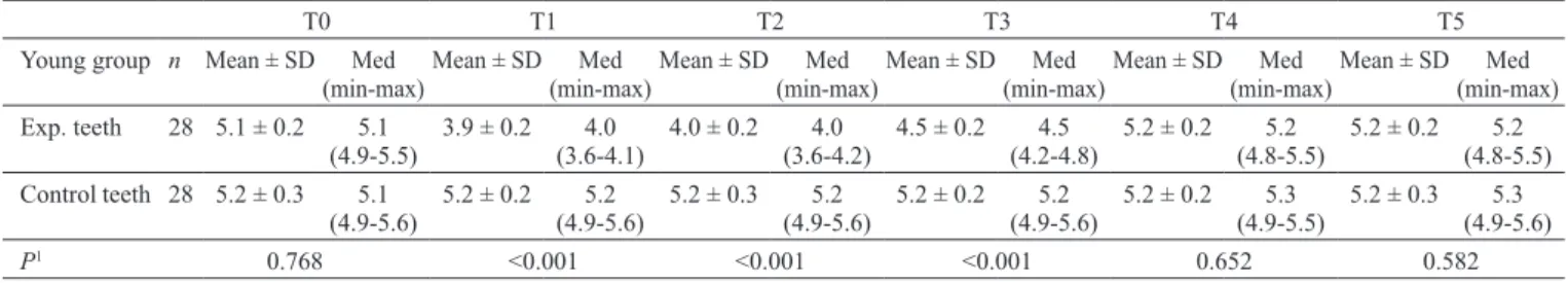
Mean PBF values for the young and old groups at all times tested are shown in Tables 1 and 2. Mean baseline PBF measured just prior to the adhesion of orthodontic appliances was 5.2 (± 0.2) PU for the young group and 3.6 (± 0.3) PU for the old group (Mann-Whitney U test,
P < 0.001) (Fig. 3). Mean PBF values were significantly
higher in the young compared to the old group at all time points (P < 0.001). Baseline-adjusted PBF changes for both groups are shown in Fig. 4 and 5.
Orthodontic tooth movement caused a significant reduction in PBF in the experimental teeth in both groups (Friedman test, P < 0.001), while PBF did not change in the control teeth over the course of the study (Friedman test, P = 0.981 and P = 0.972, respectively for the young and old groups). Experimental teeth in the Young group had significantly higher PBF at T0 prior to orthodontic appliance insertion (5.1 ± 0.2) when compared to T1 (3.9 ± 0.2), T2 (4.0 ± 0.2), and T3 (4.5 ± 0.2) (P < 0.001); however, there was no statistically significant difference in PBF between T0 and T4 (Wilcoxon signed-rank test,
P = 0.327) or between T0 and T5 (Wilcoxon signed-rank
test, P = 0.448). In the experimental teeth, PBF values in the old group decreased significantly at T1 (1.7 ± 0.1) when compared to baseline values (3.6 ± 0.3) and remained suppressed throughout the observation period (P < 0.001). Although there was a significant increase in mean PBF values from T3 (1.8 ± 0.1) to T4 (2.1 ± 0.1, P Fig. 2 Diagram of the experimental setup and custom acrylic splint for LDF
< 0.001), PBF had not returned to baseline levels at the end of the study.
Discussion
This study evaluated PBF response to orthodontic tooth movement in subjects of different ages. We used human subjects in order to examine orthodontic forces under simulated therapy over a 4-week period. Tissue reaction to orthodontic therapy may vary according to
the type of movement, dimensions of force, mechanical method used, and observation time. In the present study, all subjects had a Class I skeletal pattern and moderate crowding. To eliminate discrepancies in horizontal, vertical, and rotational positioning, orthodontic therapy was initiated with leveling using 0.014-inch NiTi wires. Differences in patient response, root-surface area, and frictional losses within the appliance make it difficult to accurately determine the appropriate force level to use with fixed appliances in clinical situations (9). This study aimed to reduce the effects of the first two vari-ables using the contralateral incisors of each subject as the experimental and control teeth. In order to compare early and late responses to orthodontic tooth movement, LDF was used to measure flux values over a 1-month period. Measuring flux values at 24 hours and 3 days made it possible to detect gradual signs of inflammation due to orthodontic force, whereas those taken at 7 days, 3 weeks, and 1 month were designed to capture possible chronic changes (10). Although we were not able to measure the force magnitude delivered during clinical alignment and leveling, a previous study estimated 0.014-inch NiTi wires to deliver 0.7-1.0 N of force at 1.5 mm of deflection at all sites in vitro (11). Given that light forces in the range of 0.5-1 N are considered adequate for achieving orthodontic movement, leveling, which involves a combination of labiolingual and mesiodistal tipping as well as intrusion and extrusion, is assumed to pose less risk to pulp microcirculation compared to other types of tooth movement, especially pure intrusion (10).
We found that age significantly affected PBF values.
T0 T1 T2 T3 T4 T5
Old group n Mean ± SD Med
(min-max)Mean ± SD(min-max)Med Mean ± SD(min-max)Med Mean ± SD(min-max)Med Mean ± SD(min-max)Med Mean ± SD(min-max)Med Exp. teeth 28 3.6 ± 0.3 3.6
(2.9-4.1) 1.7 ± 0.1 (1.5-1.9)1.7 1.7 ± 0.1 (1.5-1.9)1.7 1.8 ± 0.1 (1.5-1.9)1.8 2.1 ± 0.1 (1.9-2.2)2.1 2.1 ± 0.1 (1.9-2.2)2.1 Control teeth 28 3.6 ± 0.3 3.6
(2.9-4.1) 3.6 ± 0.3 (2.9-4.1)3.6 3.6 ± 0.3 (2.9-4.1)3.6 3.6 ± 0.3 (2.9-4.1)3.6 3.6 ± 0.3 (2.9-4.1)3.6 3.6 ± 0.2 (3.2-4.0)3.6
P 0.8431 <0.0012 <0.0011 <0.0012 <0.0012 <0.0012
1Student t test, 2 Mann-Whitney U test.
Fig. 3 Scattering representing the relationship between baseline
PBF (PU, at T0) and subjects’ ages (years) (Group 1: experimental teeth values for the young group, Group 2: control teeth values for the young group, Group 3: experimental teeth values for the old group, and Group 4: control teeth values for the old group)
Mean PBF values varied significantly prior to orth-odontic treatment and throughout the remainder of the study between the groups. PBF in the young group was nearly 1.5 times that in the old group at baseline and more than twice that in the old group at all other time points (T1-T5). The finding that baseline PBF decreases with age is in agreement with previous histological studies demonstrating an increase in age accompanied by a decrease in the number of blood vessels (12) as well as a reduction in the size and volume of pulp due to an increase in calcified tissue (8,13).Ikawa et al. (5) also reported age-related decreases in PBF and described the limitations and difficulties associated with measuring PBF using LDF in subjects aged 60-80 years. They (5) found that the magnitude of PBF signal in the subjects was very small and close to the limit of resolution of the measurement device (<1 PU, n = 5), which they attrib-uted to an age-related increase in the amount of calcified tissue surrounding the pulp. In the present study, while initial radiographs showed a greater volume of enamel and dentin in old compared to young pulp, LDF accu-rately measured PBF in older subjects throughout the course of the study (with a minimal flux signal of 1.5 PU). In other words, calcified tissue thickness did not pose a challenge to the LDF measurements in this study, which is in accordance with Vongsavan and Matthews (14), who showed that LDF was capable of passing through enamel and dentin thicknesses of 2-3.5 mm. Additionally, Ikawa et al. (15) demonstrated laser light penetrating to root depths of up to 6 mm. Apart from differences in enamel and dentin volume, differences in the their optical properties may also affect scattering and filtering of light in human teeth. Although we did not include subjects with discolored teeth, further studies
evaluating optical properties of dental tissue in relation to PBF data are needed.
Most previous studies examining age-related changes in dental pulp were conducted using either histomorpho-logical or immunohistomorpho-logical techniques (5,16-20). Shroff (16) pointed out that the volume of the pulp cavity becomes progressively smaller with advancing age due to continuous deposition of dentin onto the cavity walls, and that this volume decrease results in a decrease in the demand for blood as well as well as in the number of pulp cells. A histological study by Daud et al. (20) showed a change in pulp cell morphology and a reduction in cell (especially odontoblast) density with age. In their histomorphometric analysis, Mitsiadis et al. (18) claimed that pulp volume gradually decreases with age due to continuous dentin matrix production by odontoblasts, and that this age-related reduction in pulp-chamber size is associated with the apoptotic elimination of odontoblasts. Likewise, microarray analysis confirmed a decrease in age corresponding with vitality of the pulp-dentin complex as demonstrated by the low expression of genes encoding for transcription regulation and the high expression of genes involved in apoptotic processes (19). However, growth factor expression in older dental pulp confirms ongoing reparative processes throughout the entire life of the tooth (19). Kishi et al. (17) discovered a triple-layer vascular network in the superficial layer of young cat pulp on scanning electron microscopy compared to a coarse, single-layer capillary network in old cat pulp; they also reported fewer blood vessels in old compared to young cat pulp.
Only one published study has investigated the age-related changes in dental pulp using LDF (5). In that study, Ikawa et al. (5) examined 22 subjects ranging Fig. 4 Baseline-adjusted PBF changes for the young group Fig. 5 Baseline-adjusted PBF changes for the old group
by 1 second of cold stimulation (5). Control PBF read-ings were significantly lower in younger compared to older subjects, which is in line with our findings, and the magnitude of reduction (%) decreased significantly following cold stimulation with increased participant age (5). They reported relatively low signals in patients aged between 20 and 40 years (approximately 3 PU). However, it is difficult to compare their findings with those of the present study due to the numerous factors affecting LDF readings including tooth type, isolation device (resin cap or rigid acrylic splint, with or without rubber dam), and laser output.
We found that orthodontic tooth movement signifi-cantly affected PBF values. LDF measurements showed that PBF decreased significantly in the experimental teeth in both age groups following the application of orthodontic force, whereas the control teeth showed no significant changes in PBF over the course of the study, indicating that the changes in PBF were unrelated to repeated measurement, flowmeter calibration, or test sensitivity in subjects undergoing orthodontic treatment. The reduction in PBF after the application of force, especially at T1 and T2, is possibly due to constriction in the vessels entering and leaving the apical foramen as a result of dental dislocation. The significant decrease in PBF values observed initially following the application of orthodontic force was eventually followed by a pattern of gradual recovery and a return to baseline levels by week 3 among the younger participants in our study. Thus, we conclude that orthodontic movement produces a transient reduction in PBF in young subjects, and this finding is supported by several other studies (21,22-26). Using LDF, McDonald and Pitt Ford (22) found a temporary decrease in PBF when light, continuous tipping forces were applied to maxillary canines; however, the study did not provide means with standard deviations (although values for control canines could be approximated as 13 PU). Similarly, five other studies (21,23-26) reported intrusive orthodontic forces temporarily reducing PBF (from baseline values ranging from 6.38 to 11.74 PU).
effect of leveling forces on PBF has largely been ignored. Furthermore, with the exception of Barwick and Ramsay (27), the other mentioned studies were conducted with relatively young individuals (aged 10-31 years) whose dental anatomy, including degree of mineralization and ratio of mineralized tissue to dental pulp, differed from that of older adults (21,22-26,28), which may have affected LDF recordings, e.g., larger tubules and less mineralized tissue may facilitate light penetration into teeth. Although Barwick and Ramsay (27) included participants ranging between ages 25 and 49 years, their sample size was small (n = 8), and they did not evaluate PBF values in relation to age.
We found that mean PBF significantly decreased in the old group following the application of orthodontic force and remained suppressed during the first weeks of treat-ment. Despite a subsequent marked increase in PBF after 3 weeks and 1 month, PBF in the experimental teeth in the old group had not returned to baseline levels at the end of the study, and it is not known whether PBF in these teeth would reach baseline values over time. Further research is required to determine whether blood flow in aged pulp returns to normal over time, remains suppressed, or decreases further with continued orthodontic force. The present study also found that initial decrease in PBF following the application of orthodontic force (T1) was more severe in older (52.7%) versus younger pulp (25%) (Mann-Whitney U test, P < 0.001). Thus, our hypothesis, that dental pulp circulation is affected by the application of orthodontic force and that PBF changes in response to orthodontic force are more severe and long lasting in older than younger pulp, is accepted. The more severe the decrease in blood flow of older versus younger pulp subjected to orthodontic force may be associated with age-related arteriosclerotic changes in tooth pulp reported by various histological studies (12,20).
In conclusion, this study found that pulpal blood flow values in younger patients were significantly higher than in older patients at baseline and throughout the course of the study. Furthermore, PBF showed a more pronounced
decrease during the initial leveling phase of orthodontic treatment in older compared to younger patients.
Conflict of interest
The authors declare that they have no conflict of interest.
References
1. Marín J (1995) Age-related changes in vascular responses: a review. Mech Ageing Dev 79, 71-114.
2. Morse DR (1991) Age-related changes of the dental pulp complex and their relationship to systemic aging. Oral Surg Oral Med Oral Pathol 72, 721-745.
3. Fratkin RD, Kenny DJ, Johnston DH (1999) Evaluation of a laser Doppler flowmeter to assess blood flow in human primary incisor teeth. Pediatr Dent 21, 53-56.
4. Soo-ampon S, Vongsavan N, Soo-ampon M, Chuckpaiwong S, Matthews B (2003) The sources of laser Doppler blood-flow signals recorded from human teeth. Arch Oral Biol 48, 353-360.
5. Ikawa M, Komatsu H, Ikawa K, Mayanagi H, Shimauchi H (2003) Age-related changes in the human pulpal blood flow measured by laser Doppler flowmetry. Dent Traumatol 19, 36-40.
6. Hartmann A, Azérad J, Boucher Y (1996) Environmental effects on laser Doppler pulpal blood-flow measurements in man. Arch Oral Biol 41, 333-339.
7. Woods MA, Robinson QC, Harris EF (1990) Age-progressive changes in pulp widths and root lengths during adulthood: a study of American blacks and whites. Gerodontology 9, 41-50.
8. Philippas GG, Applebaum E (1966) Age factor in secondary dentin formation. J Dent Res 45, 778-789.
9. Drenker E (1988) Calculating continuous archwire forces. Angle Orthod 58, 59-70.
10. Ramazanzadeh BA, Sahhafian AA, Mohtasham N, Hassanzadeh N, Jahanbin A, Shakeri MT (2009) Histological changes in human dental pulp following application of intrusive and extrusive orthodontics forces. J Oral Sci 51, 109-115.
11. Hemingway R, Williams RL, Hunt JA, Rudge SJ (2001) The influence of bracket type on the force delivery of Ni-Ti archwires. Eur J Orthod 23, 233-241.
12. Bernick S (1967) Age changes in the blood supply to human teeth. J Dent Res 46, 544-550.
13. Morse DR (1991) Age-related changes of the dental pulp complex and their relationship to systemic aging. Oral Surg Oral Med Oral Pathol 72, 721-745.
14. Vongsavan N, Matthews B (1993) Experiments on extracted teeth into the validity of using laser Doppler techniques for recording pulpal blood flow. Arch Oral Biol 38, 431-439. 15. Ikawa M, Vongsavan N, Horiuchi H (1999) Scattering of
laser light directed onto the labial surface of extracted human upper central incisors. J Endod 25, 483-485.
16. Shroff FR (1953) The physiologic pathology of changes in the dental pulp. I. Senile pulp atrophy. Oral Surg Oral Med Oral Pathol 6, 1455-1460.
17. Kishi Y, Shimozato N, Takahashi K (1989) Vascular archi-tecture of cat pulp using corrosive resin cast under scanning electron microscope. J Endod 15, 478-483.
18. Mitsiadis TA, De Bari C, About I (2008) Apoptosis in devel-opmental and repair-related human tooth remodeling: a view from the inside. Exp Cell Res 314, 869-877.
19. Tranasi M, Sberna MT, Zizzari V, D’Apolito G, Mastrangelo F, Salini L et al. (2009) Microarray evaluation of age-related changes in human dental pulp. J Endod 35, 1211-1217. 20. Daud S, Nambiar P, Hossain MZ, Rahman MR, Bakri MM
(2016) Changes in cell density and morphology of selected cells of the ageing human dental pulp. Gerodontology 33, 315-321.
21. Ikawa M, Fujiwara M, Horiuchi H, Shimauchi H (2001) The effect of short-term tooth intrusion on human pulpal blood flow measured by laser Doppler flowmetry. Arch Oral Biol 46, 781-787.
22. McDonald F, Pitt Ford TR (1994) Blood flow changes in permanent maxillary canines during retraction. Eur J Orthod 16, 1-9.
23. Brodin P, Linge L, Aars H (1996) Instant assessment of pulpal blood flow after orthodontic force application. J Orofac Orthop 57, 306-309.
24. Sano Y, Ikawa M, Sugawara J, Horiuchi H, Mitani H (2002) The effect of continuous intrusive force on human pulpal blood flow. Eur J Orthod 24, 159-166.
25. Sabuncuoglu FA, Ersahan S (2014) Changes in maxillary incisor dental pulp blood flow during intrusion by mini-implants. Acta Odontol Scand 72, 489-496.
26. Sabuncuoglu FA, Ersahan S (2014) Changes in maxillary molar pulp blood flow during orthodontic intrusion. Aust Orthod J 30, 152-160.
27. Barwick PJ, Ramsay DS (1996) Effect of brief intrusive force on human pulpal blood flow. Am J Orthod Dentofacial Orthop 110, 273-279.
28. Babacan H, Doruk C, Bicakci AA (2010) Pulpal blood flow changes due to rapid maxillary expansion. Angle Orthod 80, 1136-1140.