Address for Correspondence: Serdar Balcı e.mail: [email protected]
©Copyright 2016 by the Turkish-German Gynecological Education and Research Foundation - Available online at www.jtgga.org DOI: 10.5152/jtgga.2016.16040
Introduction
Preeclampsia and intrauterine growth restriction (IUGR), as well as related perinatal death or preterm birth before the 32nd week of pregnancy, are very closely related with the underly-ing potential placental pathology. In the last 25 years, many studies have been conducted regarding the early recogni-tion of placental insufficiency. Doppler flow measurements of uterine and umbilical arteries, as well as maternal serum alpha fetoprotein (AFP) and human chorionic gonadotropin (hCG) levels, during the first and second trimesters of preg-nancy are most frequently studied to predict placental insuf-ficiency and its effects on the fetus (1).
Uterine artery Doppler velocimetry results have demonstrated that hemodynamic changes detectable in the uterine artery as early as in the first trimester of pregnancy are associated with an increased risk of preeclampsia and IUGR (2-4). This association can also be demonstrated in the second and third trimesters (5-7).
Several studies have recently shown that low serum levels of pregnancy-associated plasma protein A (PAPP-A) might be as-sociated with poor pregnancy and poor neonatal outcomes (1, 8, 9). PAPP-A, which was first purified from the serums of pregnant women in 1974, is a member of the metzincin family of metalloproteinases (10, 11). PAPP-A is an insulin-like growth factor-binding protein (IGFBP)-specifically, a protease. IGFBPs bind to insulin-like growth factor (IGF) 1 and 2 and disconnect these proteins from cell surface receptors; thus, low levels of serum PAPP-A are associated with low levels of bioactive IGF (11).
A low level of maternal serum PAPP-A is an important sign of early placental insufficiency in the first three months of preg-nancy; however, the effects on the fetus reaches recognizable levels in the second trimester. Nevertheless, growth restriction determined in the second trimester is also directly associated with poor pregnancy and poor neonatal outcomes. Detailed ul-trasonography for fetal biometric measurements (FBMs) and Doppler flow measurements that would be performed in the second trimester in patients with low levels of serum PAPP-A, Objective: To determine predictive values of maternal serum PAPP-A (msPAPP-A) levels, uterine artery Doppler velocimetry, and fetal biomet-ric measurements (FBMs) for poor pregnancy and poor neonatal outcomes.
Material and Methods: This prospective cohort study was conducted on singleton pregnancies followed until delivery. Pregnancy and neo-natal outcomes were evaluated with respect to the msPAPP-A level at the 11th–14th weeks, uterine artery Doppler velocimetry at the 15th–18th
weeks, and FBMs at the 20th–24th and 28th–32nd weeks of pregnancy.
Results: One hundred fifty-eight women constituted the study group; 17 (10.75%) of them had at least one poor pregnancy outcome. The cut-off point of 0.72 multiple of the median (MoM) for the PAPP-A level achieved a sensitivity of 82.4% and a specificity of 29.8% for poor pregnancy out-comes. The mean birth weight was significantly lower in the subgroup with a higher mean pulsatility index of uterine arteries (UAPImean≥1.19) (p=0.025) as well as in the subgroup with a higher mean resistance index of uterine arteries (UARImean≥0.62) (p=0.013). When the subgroup of pregnant women under the risk of early-onset IUGR according to FBMs was compared to the low-risk group, statistically significant differ-ences were seen in terms of pregnancy outcomes (p=0.045) and birth weight (p=0.011).
Conclusion: Maternal serum PAPP-A level and FBMs could be used for predicting pregnancy outcomes, while uterine artery Doppler velocim-etry and FBMs could be used for predicting neonatal outcomes, specifically the birth weight. (J Turk Ger Gynecol Assoc 2016; 17: 143-9)
Keywords: Pregnancy-associated plasma protein-a, ultrasonography, Doppler, color, fetal biometry, pregnancy outcome, fetal development
Received: 7 March, 2016 Accepted: 21 July, 2016
Predictive values of maternal serum PAPP-A level,
uterine artery Doppler velocimetry, and fetal
biometric measurements for poor pregnancy and
poor neonatal outcomes in pregnant women
Serdar Balcı
Department of Gynecology and Obstetrics, Başkent University School of Medicine, Ankara, Turkey
which is measured routinely within the scope of first-trimester aneuploidy screening at the 11th–14th weeks of pregnancy may be beneficial in predicting poor pregnancy and poor neonatal out-comes and in taking necessary measures (12, 13).
The present study aimed to determine the predictive value of maternal serum PAPP-A levels measured in the first trimester, uterine artery Doppler velocimetry performed during the sec-ond trimester, and FBMs in the secsec-ond and third trimesters for poor pregnancy and poor neonatal outcomes in pregnant women.
Material and Methods
This prospective cohort study was conducted in a single uni-versity-based pregnancy clinic in the Department of Gynecol-ogy and Obstetrics in Turkey between July 2013 and July 2015, with approval of the hospital ethics committee. The singleton pregnant women, who presented to our pregnancy clinic, were enrolled into the study from September 2013 to May 2015 after their informed consents were obtained. The study complied with the Declaration of Helsinki and ethical standards. Pregnant women were excluded from the final analyses if they missed follow-up visits or had complicated pregnancies, such as abor-tion or iatrogenic terminaabor-tion of the pregnancy.
The women included in the study were followed up after the confirmation of pregnancy until delivery. Initially, all the study participants underwent the first-trimester aneuploidy screen-ing test at the 11th–14th weeks of pregnancy, including ma-ternal serum PAPP-A, β-hCG, and nuchal translucency mea-surements. Subsequently, color pulsed Doppler ultrasound examination of the bilateral uterine arteries was performed at the 15th–18th weeks of pregnancy. The women with diastolic notches, unilateral or bilateral, in the uterine artery Doppler waveforms were recorded. The pulsatility index (PI) and resis-tance index (RI) of both uterine arteries were measured and their arithmetic means were calculated. FBMs, including fetal biparietal diameter (BPD), head circumference (HC), femur length (FL), and abdominal circumference (AC), were mea-sured at the 20th–24th and 28th–32nd weeks of pregnancy, and the HC/AC ratio was calculated. Poor pregnancy outcomes, such as pregnancy-induced hypertension, preeclampsia, and pre-term birth, and poor neonatal outcomes as well as birthweight measured at delivery were noted. Women were divided into two groups according to the pregnancy or neonatal outcomes: the group with and the group without poor outcomes. These two groups were compared with each other with respect to the test results specified above, in order to determine the pre-dictive values of the analyses.
All the clinic-demographic characteristics and test results were recorded in a prospectively formed electronic database. All the ultrasonographic measurements were performed by the same physician, using color Doppler ultrasound (Voluson 730 Expert, General Electric Healthcare; Chicago, USA) and the data were assessed by a single researcher.
Pregnant women were considered to be under risk of early-on-set IUGR when the estimated gestational age according to the ultrasonographic FBMs at the 20th–24th and 28th–32nd weeks of
pregnancy was more than one-week behind the gestational age calculated according to the last menstrual period (LMP) and when HC/AC ratio was over 1.15 and 1.10 at the 20th–24th and 28th–32nd weeks of pregnancy, respectively (12, 13). This group was compared with the group of pregnant women in whom the estimated gestational age according to the FBMs was consistent with the gestational age calculated according to the LMP, with respect to the test results as well as pregnancy outcomes, in-cluding the birth weight.
Pregnancy-induced hypertension (PIH) was diagnosed if the arterial blood pressure of the patient was 140/90 mmHg and higher, whereas the diagnosis of preeclampsia was made if a high blood pressure was accompanied by proteinuria ≥300 mg in 24 hours (14). Birth before the 37th week of pregnancy was considered as a preterm birth. The threat of preterm labor was diagnosed when there were regular uterine contractions (4 per 20 minutes) without any cervical dilatation before the 37th week of pregnancy. Preterm premature rupture of the membranes (PPROM) was defined as rupture of the membranes before the onset of labor before the 37th week of pregnancy. Low birth weight was defined as a birth weight <2500 g.
A prospective cohort study design was chosen to conduct the study in order to minimize a potential bias that could result from the nature of the trial. In addition, ultrasonographic measure-ments and the data assessment were performed by two differ-ent researchers.
Data were analyzed by Chi-square, Mann–Whitney U test, and Student t-test using SPSS (Statistical Package for Social Scienc-es version 15.0, SPSS Inc.; Chicago, USA) program. A value of p<0.05 was considered as statistically significant. The param-eters assumed to predict pregnancy outcomes were evaluated by receiver operating characteristic (ROC) curves.
Results
A total of 175 singleton pregnant women were enrolled in the study. Of these pregnancies, three were terminated due to missed abortion and one was terminated due to Trisomy 21. Thirteen patients were lost to follow-up at different phases of the study. The remaining 158 patients formed the study group. Table 1 summarizes the clinicodemographic characteristics of the study participants. Over the course of the follow-up period, a total of 17 (10.75%) pregnant women were determined to have at least one of the poor pregnancy outcomes (Table 2).
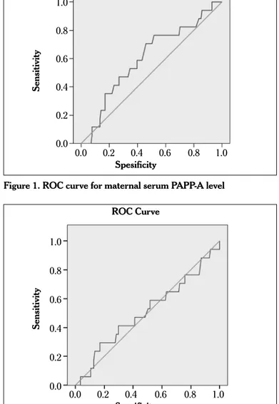
An ROC curve generated for the first-trimester maternal serum PAPP-A values is presented in Figure 1. Considering the cut-off value as a 0.72 multiple of the median (MoM), (Standard Error, SE: 0.039) (95% CI 0.646–0.798), the sensitivity, specificity, posi-tive predicposi-tive value (PPV), and negaposi-tive predicposi-tive value (NPV) achieved for the poor pregnancy outcomes were 82.4%, 29.8%, 67%, and 56%, respectively.
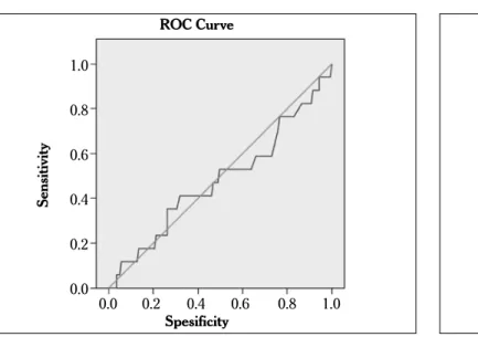
In the ROC curve analysis of the arithmetic mean of PI of the right and left uterine arteries (UAPImean), the cut-off value was considered to be 1.08 with 58.8% sensitivity, 48.2% specificity, 76% PPV, and 55% NPV for poor pregnancy outcomes (Standard Error, SE: 0.039) (95% CI 0.646–0.798) (Figure 2). Pregnancy out-comes were compared between the two groups formed based
on this cut-off value; however no statistically significant differ-ence was determined (p=0.582) (Table 3). After calculating the UAPImean for each pregnant woman, the mean value of the entire group was calculated as 1.18. The entire group was divided into two subgroups according to the UAPImean value: ≤1.18 or ≥1.19. There was no statistically significant difference between the two subgroups in terms of poor pregnancy out-comes (p=0.723), but the mean birth weights showed a signifi-cant difference (p=0.025), being lower in the subgroup with an UAPImean of ≥1.19 than that of the subgroup with an UAPIme-an ≤1.18 (Table 4).
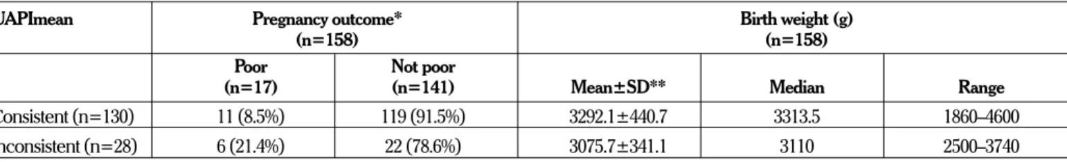
The mean RI (UARImean) value of both uterine arteries was calculated for each pregnant woman, and then an ROC curve was drawn (Figure 3). The sensitivity, specificity, PPV, and NPV values for the poor pregnancy outcomes were 58.8%, 34%, 36%, and 47%, respectively, when the cut-off value was taken as 0.582 (Standard Error, SE: 0.039) (95% CI 0.646–0.798). After
calculat-Table 1. Clinicodemographic characteristics of the study
participants (n=158)
Characteristics Descriptive values Additional (mean±SD or n) explanation
Age 27.9±4.4 years Range: 17–41
Weight 62.8±10.1 kg Range: 42–108
Smoking 8 women (5.06%) 2–8 cigarettes day
First pregnancy 64 women (40.5%)
-Previous poor pregnancy 18 women (11.4%) PIH, Preeclampsia, GDM,
outcome Preterm birth, PPROM,
IUGR, Trisomy 21, Fetal anomaly Concomitant disease 13 women (8.2%) Hypo-hyperthyroidism,
FMF, Psoriasis, Cardiac valve diseases, Chronic
HT, Asthma, Thalassemia carrier
Medications 7 women (4.4%) Levothyroxine,
Colchicine, Alfa methyldopa
PIH: pregnancy-induced hypertension; GDM: gestational diabetes mellitus; PPROM: preterm premature rupture of the membranes; IUGR: intrauterine growth restriction; FMF: familial Mediterranean fever; HT: hypertension
Table 2. Numeric and proportional distribution of poor
pregnancy outcomes determined during follow-up
Outcomes Number of patients Percentage (%)
(n=17) (n=158)
PIH/Preeclampsia 5 (3/2) 3.16
Threat of preterm labor 1 0.63
PPROM 4 2.53
Preterm birth <37th week 14 8.86
Preterm birth <32nd week -
-Need for neonatal intensive care 7 4.43
Low birth weight 7 4.43
Placental abruption (ablatio - -
placentae)
PIH: pregnancy-induced hypertension; PPROM: preterm premature rupture of the membranes
Table 3. Comparison of pregnancy outcomes between two
subgroups formed according to the cut-off value of
UA-PImean determined by the ROC curve analysis
Number of patients Percentage (%)
Outcomes (n=17) (n=158)
<1.08 (n=75) 7 (9.3%) 68 (90.7%)
≥1.08 (n=83) 10 (12%) 73 (88%)
∗Chi-square test: p=0.582.
UAPImean: arithmetic mean of the pulsatility indices of the right and left uterine arteries; ROC curve: receiver operating characteristic curve
Figure 1. ROC curve for maternal serum PAPP-A level ROC Curve Spesificity 1.0 0.8 0.6 0.4 0.2 0.0 Sensitivity 0.0 0.2 0.4 0.6 0.8 1.0
Figure 2. ROC curve for UAPImean ROC Curve Spesificity Sensitivity 0.0 0.2 0.4 0.6 0.8 1.0 1.0 0.8 0.6 0.4 0.2 0.0
ing the UARImean for each pregnant woman, the mean value of the entire group was calculated as 0.62 and the entire group was divided into two subgroups according to the UARImean val-ues: ≥0.62 or <0.62. No statistically significant difference was determined between the two subgroups in terms of pregnancy outcomes (p=0.797), while the mean birth weight was signifi-cantly higher in the subgroup with UARImean <0.62 than that of the subgroup with an UARImean of ≥0.62 (p=0.013) (Table 5). There was no significant difference between the women with unilateral uterine artery notch and the women with bilateral uterine artery notch in terms of pregnancy outcomes.
Receiver operating characteristic curves were drawn for HC/AC ratio measured at the 20th–24th and 28th–32nd weeks of pregnancy (Figure 4, 5, respectively). Taking the cut-off value of HC/AC ratio as 1.135 for the 20th–24th weeks of pregnancy (Standard Error, SE: 0.039) (95% CI 0.646–0.798), the sensitivity, specificity, PPV, and NPV for poor pregnancy outcomes were 58.8%, 49.6%, 56%, and 49%, respectively. The cut-off value of HC/AC ratio for the 28th–32nd weeks of pregnancy was taken as 1.075 (Standard Er-ror, SE: 0.039) (95% CI 0.646–0.798) with 52.9% sensitivity, 41.1% specificity, 59% PPV, and 67% NPV.
Pregnant women considered to be under the risk of early-onset IUGR (inconsistent FBMs subgroup), as specified in the Material and Methods section, were compared with the pregnant wom-en with whom the estimated gestational age according to the
fetal biometric measurement was consistent with the gestation-al age cgestation-alculated according to the last menstrugestation-al period (consis-tent FBMs subgroup), in terms of the pregnancy outcomes and birth weight (Table 6). Statistically significant differences were
Table 4. Comparison of pregnancy outcomes and birth weight between two subgroups formed according to the
UA-PImean of the entire group
Pregnancy outcome* Birth weight (g)
(n=158) (n=158)
Poor Not poor
UAPImean (n=17) (n=141) Mean±SD** Median Range
≤1.18 (n=90) 9 (10%) 81 (90%) 3320.2±411.1 3350 2400–4105
≥1.19 (n=68) 8 (11.8%) 60 (88.2%) 3165.7±445.8 3145 1860–4600
*Chi-square test: p=0.723 for comparison of the pregnancy outcomes between the two subgroups. **T-test: p=0.025 for comparison of the mean birth weight between the two subgroups.
UAPImean: arithmetic mean of the pulsatility indices of the right and left uterine arteries; SD: standard deviation
Figure 3. ROC curve for UARImean ROC Curve Spesificity Sensitivity 0.0 0.2 0.4 0.6 0.8 1.0 1.0 0.8 0.6 0.4 0.2 0.0
Figure 5. ROC curve for HC/AC ratio measured at the 28th–32nd
weeks of pregnancy ROC Curve Spesificity Sensitivity 0.0 0.2 0.4 0.6 0.8 1.0 1.0 0.8 0.6 0.4 0.2 0.0
Figure 4. ROC curve for HC/AC ratio measured at the 20th–24th
weeks of pregnancy ROC Curve Spesificity Sensitivity 0.0 0.2 0.4 0.6 0.8 1.0 1.0 0.8 0.6 0.4 0.2 0.0
determined between the two subgroups in terms of pregnancy outcomes (p=0.045) and birth weight (p=0.011).
Discussion
Impaired placentation is one of the most important causes of poor pregnancy outcomes for mother and/or the baby. Re-searches on the etiology of unpleasant outcomes of pregnancy, such as preeclampsia, IUGR, and preterm birth, highlight the importance of normal placental development. Markers that could indicate these changes prior to the onset of disorder will provide us a chance to take early preventive measures and even to prevent this in the future. For this reason, the world of perina-tology has carried out many studies, particularly in recent years, on numerous placental biochemical markers, various Doppler ultrasound techniques and parameters, and various ultrasono-graphic methods to evaluate fetal and placental development in order to determine placental defects (1, 8, 10, 11). We aimed to determine the predictive value of the maternal serum PAPP-A level of the first trimester, uterine artery Doppler velocimetry of the second trimester, and FBMs of the second and third tri-mester for poor pregnancy and poor neonatal outcomes which would probably occur due to placentation defect in pregnant women, and we found that the early prediction of unfavorable maternal and unfavorable neonatal outcomes of placental in-sufficiency might be possible using some test results, such as FBMs or an assessment of the HC/AC ratio, even though there is no method that can be used alone as a screening test.
The association between low hCG (<0.5 MoM) and PAPP-A (<0.4 MoM) levels, which are measured on the 10th–14th weeks of pregnancy, and the complications of pregnancy has been demonstrated previously (15, 16). In the present study as well, poor pregnancy outcomes could be predicted with 82.4% sen-sitivity and 29.8% specificity when the cut-off value was taken as 0.72 MoM in the ROC curve drawn for PAPP-A, which was measured in the 11th–14th weeks of pregnancy.
Doppler ultrasonography of the uterine arteries is the other method used for the early prediction of pathological placenta-tion and can be performed at various weeks of pregnancy. In the present study, a Doppler ultrasound of uterine arteries was performed on the 15th–18th weeks of pregnancy, the mean PI value of both sides was calculated (UAPImean), and then an ROC curve was drawn. We achieved a sensitivity of 58.8% and a specificity of 48.2% for poor pregnancy outcomes when the cut-off value was taken as 1.08. The average value of the entire group of pregnant women for UAPImean was found to be 1.18±0.40, with no significant difference determined between pregnancy outcomes when the group was divided into two based on this value. However, a statistically significant difference was deter-mined in terms of the mean birth weight (p=0.025). The results, thus, indicate that increased uterine artery PI might cause a de-crease in birth weight. Cooper et al. (17) conducted a study in 229 pregnant women and underlined the value of an increased mean uterine artery PI measured at the 22nd week of pregnancy in predicting preterm birth, the small for gestational age (SGA), and a low birth weight, particularly in pregnant women with a PAPP-A value lower than 0.4 MoM. Pilalis et al. (18) evaluated 878 pregnant women on the 11th–14th weeks of pregnancy and emphasized that each of the Doppler ultrasound of uterine arter-ies and the PAPP-A value is an independent factor for predicting SGA and that the combination of both is more effective for pre-diction. The present study, showing an inverse relation between uterine artery PI and birth weight, differs from the other studies in that uterine artery PI has been evaluated within a different gestational age period of pregnancy. Nevertheless, it is neces-sary to underline that a normal PI does not exclude obstetric complications, as was demonstrated in almost all studies. RI is another parameter assessed by Doppler ultrasound of the uterine arteries. In the present study, the mean RI of the uterine arteries (UARImean) was calculated for each pregnant woman and then an ROC curve was drawn. We were able to predict poor pregnancy outcome with 58.8% sensitivity and 34% speci-ficity when the cut-off value was taken as 0.582. The average
Table 6. Comparison of pregnancy outcomes and birth weight between two subgroups formed according to fetal biometric
measurements
UAPImean Pregnancy outcome* Birth weight (g)
(n=158) (n=158)
Poor Not poor
(n=17) (n=141) Mean±SD** Median Range
Consistent (n=130) 11 (8.5%) 119 (91.5%) 3292.1±440.7 3313.5 1860–4600
Inconsistent (n=28) 6 (21.4%) 22 (78.6%) 3075.7±341.1 3110 2500–3740
*Chi-square test: p=0.045 for comparison of the pregnancy outcomes between the two Subgroups. **Mann–Whitney U test: p=0.011 for comparison of the mean birth weight between the two Subgroups.
Table 5. Comparison of pregnancy outcomes and birth
weight between two subgroups formed according to the
UARImean of the entire group
Pregnancy outcome* Birth weight (g)** (n=158) (n=158)
Poor Not poor
UARImean (n=17) (n=141) Mean±SD**
<0.62 (n=79) 8 (10.1%) 71 (89.1) 3338.3±413.5 ≥0.62 (n=79) 9 (11.4%) 70 (%88.6) 3169.1±435.8
*Chi-square test: p=0.797 for comparison of the pregnancy outcomes between the two subgroups.
**T-test: p=0.013 for comparison of the mean birth weight between the two subgroups.
UARImean: arithmetic mean of the resistance indices of the right and left uterine arteries
value of UARImean was calculated for the whole group and the group was divided into two according to this value (0.62±0.10). We failed to determine a significant difference between the groups in terms of obstetric complications; however, there was significant difference between the mean birth weights of the groups (p=0.013). This result suggests that increased uterine artery RI, as well as increased PI, might cause a decrease in birth weight. The literature includes studies propounding that increased uterine artery RI and uterine notch can predict pre-eclampsia and SGA (19, 20). In these studies, which were per-formed in large population groups and usually in the 18th–24th weeks of pregnancy, the sensitivity reached 63–95% when RI was above the 95th percentile or when the limit values of 0.56– 0.58 were used for RI (21, 22). The RI found in the present study is close to those determined in earlier studies, although the cut-off value 0.582 was measured in a different gestational age pe-riod of pregnancy. The present study found no significant differ-ence between the women with unilateral uterine artery notch and the women with bilateral uterine artery notch in terms of pregnancy outcomes.
Ultrasonographic biometry has always been an important tool in monitoring fetal development. In the present study, the pregnant women with whom estimated gestational age that was calculated based on ultrasonographic FBMs performed at the 20th–24th and 28th–32nd weeks of pregnancy was more than 1 week behind the gestational age calculated according to the last menstrual period and the pregnant women with an HC/ AC ratio over 1.15 and 1.10 at the 20th–24th and 28th–32nd weeks of pregnancy, respectively, were considered to be under a risk of early onset fetal growth restriction. This high-risk group was compared with the other group of pregnant women and statistically significant differences were determined in terms of pregnancy outcomes (p=0.045) and mean birth weight (p=0.011). In the literature, studies on this subject are not so many, excluding two separate studies conducted by Fox et al. (12, 13) suggesting an association between a HC/AC ratio >90th percentile and poor pregnancy outcomes and the study con-ducted by Colley et al. (23) determining a weak correlation between the HC/AC ratio and the ponderal index. In addition to the literature information, the present study indicated that it is possible to obtain significant results in terms of pregnancy outcomes and birth weight in the light of precise FBMs and an assessment of the HC/AC ratio (23, 24).
Our study has some limitations. First, power calculation was not performed, and we had a small sample size in terms of the sub-group with poor pregnancy outcomes. Second, we did not com-bine the results of the tests to analyze the predictive values of the combinations of current biochemical and ultrasonographic methods.
In conclusion, we aimed to determine the predictive value of maternal serum PAPP-A levels, uterine artery Doppler velocim-etry, and FBMs for poor pregnancy and poor neonatal outcomes in pregnant women and found that serum PAPP-A levels and FBMs could be used for predicting pregnancy outcomes, while uterine artery Doppler velocimetry and FBMs could be used for predicting neonatal outcomes, specifically the birth weight. Therefore, an early prediction of unfavorable maternal and
neo-natal outcomes of placental insufficiency might be possible us-ing some test results, such as FBMs or by the assessment of the HC/AC ratio, even though there is no method that can be used alone as a screening test. Therefore, different combinations of current biochemical and ultrasonographic methods may be necessary. It is clear that a screening test including certain com-binations of biochemical and ultrasonographic markers with certain cut-off values, which will be created in multicenter stud-ies with larger study populations, is needed in the future in order to predict obstetric complications earlier.
Ethics Committee Approval: Ethics committee approval was received
for this study from the ethics committee of Dokuz Eylül University School of Medicine.
Informed Consent: Written informed consent was obtained from
pa-tients who participated in this study.
Peer-review: Externally peer-reviewed.
Conflict of Interest: No conflict of interest was declared by the authors. Financial Disclosure: The authors declared that this study has received
no financial support.
References
1. Smith GC, Shah I, Crossley JA, Aitken DA, Pell JP, Nelson SM, et al. Pregnancy-associated plasma protein A and alpha-fetoprotein and prediction of adverse perinatal outcome. Obstet Gynecol 2006; 107: 161-6. [Crossref]
2. Van den Elzen HJ, Cohen-Overbeek TE, Grobbee DE, Quartero RW, Wladimiroff JW. Early uterine artery Doppler velocimetry and the outcome of pregnancy in women aged 35 years and older. Ultra-sound Obstet Gynecol 1995; 5: 328-33. [Crossref]
3. Martin AM, Bindra R, Curcio P, Cicero S, Nicolaides KH. Screen-ing for pre-eclampsia and fetal growth restriction by uterine artery Doppler at 11-14 weeks of gestation. Ultrasound Obstet Gynecol 2001; 18: 583-6. [Crossref]
4. Dugoff L, Lynch AM, Cioffi-Ragan D, Hobbins JC, Schultz LK, Malone FD, et al. First trimester uterine artery Doppler abnormalities pre-dict subsequent intrauterine growth restriction. Am J Obstet Gyne-col 2005; 193: 1208-12. [Crossref]
5. Campbell S, Diaz-Recasens J, Griffin DR, Cohen-Overbeek TE, Pearce JM, Willson K, et al. New doppler technique for assessing uteroplacental blood flow. Lancet 1983; 1: 675-7.[Crossref]
6. Harrington K, Cooper D, Lees C, Hecher K, Campbell S. Doppler ul-trasound of the uterine arteries: the importance of bilateral notch-ing in the prediction of pre-eclampsia, placental abruption or deliv-ery of a small-for-gestational-age baby. Ultrasound Obstet Gynecol 1996; 7: 182-8. [Crossref]
7. Steel SA, Pearce JM, McParland P, Chamberlain GV. Early Doppler ultrasound screening in prediction of hypertensive disorders of pregnancy. Lancet 1990; 335: 1548-51. [Crossref]
8. Huang T, Hoffman B, Meschino W, Kingdom J, Okun N. Predic-tion of adverse pregnancy outcomes by combinaPredic-tions of first and second trimester biochemistry markers used in the routine pre-natal screening of Down syndrome. Prenat Diagn 2010; 30: 471-7.
[Crossref]
9. Salvig JD, Kirkegaard I, Winding TN, Henriksen TB, Torring N, Uldb-jerg N. Low PAPP-A in the first trimester is associated with reduced fetal growth rate prior to gestational week 20. Prenat Diagn 2010; 30: 503-8. [Crossref]
10. Berry E, Aitken DA, Crossley JA, Macri JN, Connor JM. Screening for Down’s syndrome: changes in marker levels and detection rates between first and second trimesters. Br J Obstet Gynaecol 1997; 104: 811-7. [Crossref]
11. Consuegra-Sanchez L, Fredericks S, Kaski JC. Pregnancy-associ-ated plasma protein A: Has this biomarker crossed the boundary from research to clinical practice? Drug News Perspect 2009; 22: 341-8. [Crossref]
12. Fox NS, Shalom D, Chasen ST. Second-trimester fetal growth as a predictor of poor obstetric and neonatal outcome in patients with low first-trimester serum pregnancy-associated plasma protein-A and a euploid fetus. Ultrasound Obstet Gynecol 2009; 33: 34-8.
[Crossref]
13. Fox NS, Huang M, Chasen ST. Second-trimester fetal growth and the risk of poor obstetric and neonatal outcomes. Ultrasound Obstet Gynecol 2008; 32: 61-5. [Crossref]
14. ACOG Committee on Obstetric Practice. Diagnosis and manage-ment of preeclampsia and eclampsia. Number 33, January 2002. American College of Obstetricians and Gynecologists. Int J Gynae-col Obstet 2002; 77: 67-75.
15. Morris RK, Cnossen JS, Langejans M, Robson SC, Kleijnen J, Ter Riet G, et al. Serum screening with Down’s syndrome markers to predict pre-eclampsia and small for gestational age: systematic review and meta-analysis. BMC Pregnancy Childbirth 2008; 8: 33.
[Crossref]
16. Gagnon A, Wilson RD, Audibert F, Allen VM, Blight C, Brock JA, et al. Obstetrical complications associated with abnormal maternal serum markers analytes. J Obstet Gynaecol Can 2008; 30: 918-49.
[Crossref]
17. Cooper S, Johnson JA, Metcalfe A, Pollard J, Simrose R, Connors G, et al. The predictive value of 18 and 22 week uterine artery Doppler
in patients with low first trimester maternal serum PAPP-A. Prenat Diagn 2009; 29: 245-8. [Crossref]
18. Pilalis A, Souka AP, Antsaklis P, Basayiannis K, Benardis P, Haido-poulos D, et al. Screening for pre-eclampsia and small for gesta-tional age fetuses at the 11-14 weeks scan by uterine artery Dop-plers. Acta Obstet Gynecol Scand 2007; 86: 530-4. [Crossref]
19. Arduini D, Rizzo G. Normal values of Pulsatility Index from fetal ves-sels: a cross-sectional study on 1556 healthy fetuses. J Perinat Med 1990; 18: 165-72. [Crossref]
20. Dane B, Dane C, Kiray M, Cetin A, Koldas M, Erginbas M. Correlation between first-trimester maternal serum markers, second-trimester uterine artery doppler indices and pregnancy outcome. Gynecol Obstet Invest 2010; 70: 126-31. [Crossref]
21. Fonseca E, Yu CK, Singh M, Papageorghiou AT, Nicolaides KH. Re-lationship between second-trimester uterine artery Doppler and spontaneous early preterm delivery. Ultrasound Obstet Gynecol 2006; 27: 301-5. [Crossref]
22. Jacobson SL, Imhof R, Manning N, Mannion V, Little D, Rey E, et al. The value of Doppler assessment of the uteroplacental circulation in predicting preeclampsia or intrauterine growth retardation. Am J Obstet Gynecol 1990; 162: 110-4. [Crossref]
23. Colley NV, Tremble JM, Henson GL, Cole TJ. Head circumference/ abdominalcircumference ratio, ponderal index and fetal malnutri-tion. Should head circumference/abdominal circumference ratio be abandoned? Br J Obstet Gynaecol 1991; 98: 524-7. [Crossref]
24. Chambers SE, Hoskins PR, Haddad NG, Johnstone FD, McDicken WN, Muir BB. A comparison of fetal abdominal circumference measurements and Doppler ultrasound in the prediction of small-for-dates babies and fetal compromise. Br J Obstet Gynaecol 1989; 96: 803-8. [Crossref]