Summary
The aim of the study was to evaluate acute phase response via Haptoglobin and serum amyloid-A concentrations in dairy cows naturally infected with Anaplasma marginale. The second aim of the study was to determine the changes in clinical, hematological and biochemical parameters in dairy cows naturally infected Anaplasma marginale. A total of 40 dairy cattle suffering from bovine anaplasmosis were included to the study from a dairy cattle herd. A total of 10 healthy dairy cattle were selected for control group. Analysis of acute phase proteins, hematologic analysis and biochemical analysis was performed in this study. Serum haptoglobin and serum amyloid-A concentrations significantly increased in cattle infected with Anaplasma marginale compared to healthy cattle. All cattle in infected group demonstrated clinical signs of anaplasmosis. Significantly decreased red blood cell count, packed cell volume, and hemoglobin concentration were observed in infected cattle compared to the control group. Serum aspartate aminotransferase, alkaline phosphatase, creatinine and bilirubin concentrations were significantly increased in infected cattle compared with the control group. In conclusion, the changes of biochemical and hematological parameters may be indicate of anemia and tissue damage in cattle with anaplasmosis. Serum haptoglobin and serum amyloid-A concentrations could be usefull in evaluate of acute phase response in cattle infected with Anaplasma marginale.
Keywords: Anaplasmosis, Haptoglobin, Serum amyloid-A, Dairy cows
Anaplasma marginale İle Doğal Enfekte Sütçü İneklerde Akut Faz
Proteinler ile Klinik, Hematolojik ve Biyokimyasal Parametrelerin
Değerlendirilmesi
Özet
Bu çalışmanın amacı Anaplasma marginale ile doğal enfekte sığırlarda haptoglobin ve serum amyloid-A konsantrasyonlarında meydana gelen değişimleri belirleyerek akut faz cevabı değerlendirmektir. Çalışmanın ikinci amacı ise anaplazmozisli ineklerde gözlenen klinik, hematolojik ve biyokimyasal değişimleri belirlemektir. Araştırmaya anaplasmosis tanısı konulmuş 40 sütçü inek ve sağlıklı 10 sütçü inek dahil edildi. Çalışmada akut faz proteinlerin ölçümü, hematolojik ve biyokimyasal parametrelerin analizleri yapıldı. Enfekte gruptaki ineklerin serum haptoglobin ve serum amyloid-A konsantrasyonları kontrol grubundaki sığırlardan önemli ölçüde yüksek bulundu. Anaplasma marginale ile enfekte olan gruptaki tüm hayvanlar da anaplazmozisin klinik semptomları tespit edildi. Enfekte ineklerdeki serum aspartat aminotransferaz, alkalen fosfataz, kreatinin ve bilirubin konsatrasyonları kontrol grubuna göre önemli oranda yüksek tespit edilirken eritrosit sayısı, hematokrit değeri ve hemoglobin konsantrasyonu ise önemli oranda düşük olarak belirlendi. Sonuç olarak, anaplasmosisli sığırlarda, hematojik ve biyokimyasal değişiklikler aneminin ve doku hasarının bir göstergesi olabilir. Anaplasma marginale ile enfekte sığırlarda gelişen akut faz cevabın değerlendirilmesinde serum haptoglobin ve serum amyloid-A konsantrasyonları kullanılabilir.
Anahtar sözcükler: Anaplasmosis, Haptoglobin, Serum amyloid-A, İnek
Acute Phase Proteins, Clinical, Hematological and
Biochemical Parameters in Dairy Cows Naturally Infected with
Anaplasma Marginale
Alparslan COŞKUN *
Özlem DERİNBAY EKİCİ **
Hasan GÜZELBEKTEŞ *** Uğur AYDOĞDU *** İsmail ŞEN ***
* ** ***
Cumhuriyet University, Faculty of Veterinary Medicine, Department of Internal Medicine, TR-58140 Sivas - TURKEY Selcuk University, Faculty of Veterinary Medicine, Department of Parasitology, TR-42075 Konya - TURKEY
Selcuk University, Faculty of Veterinary Medicine, Department of Internal Medicine, TR-42075 Konya - TURKEY
Makale Kodu (Article Code): KVFD-2011-5822
İletişim (Correspondence)
+90 346 2191812INTRODUCTION
Anaplasma marginale is a rickettsial organism that
causes bovine anaplasmosis in cattle in tropical and sub-tropical areas throughout the world. The disease is a major constraint to cattle production in many countries and can be seen at any age1,2. To confirm the diagnosis,
laboratory tests such as light microscopic evaluation of Giemsa-stained blood smears or serological/molecular diagnostic procedures are required. Infected erythrocytes are not always detectable in stained blood smears during the persistent infection 3,4 so, a variety of serologic tests
are used for the detection of specific antibodies against anaplasmosis 5. A competitive ELISA (cELISA) has been
used to diagnose A. marginale infection in cattle 6. This
test is used to serologically detect both acute and chronic
Anaplasma infections in cattle 6-8.
The early protection mechanism of the host against infection, trauma or other tissue damage comprises a set of reactions known as the acute phase response (APR). During the APR, the serum concentration of the acute phase proteins (APP) changes dramatically 9-12. These
proteins are synthesized mainly in the liver. The secretion of APPs is regulated by proinflamatory cytokines such as interleukin-6 (IL-6), tumor necrosis factor-α, and IL-1β 10,12.
Serum haptoglobin (Hp) and serum amyloid-A (SAA) are the major APP in cattle 10-14. Many studies have indicated
the significance of serum Hp and SAA as clinically useful parameters in cattle various conditions 9-12,15-17.
The symptoms of clinical disease are fever, anemia, icterus, weight loss, abortion, and lethargy. Severity and death rate increase with advancing age 2 A. marginale
infection causes fever and mild to marked hemolytic anemia. After infection, parasitemia increases until the hemolytic crisis, frequently with more than 50% of RBCs infected 18. The number of infected erythrocytes increases
drastically and phagocytosis by reticulo-endothelial cells of parasitized erythrocytes lead to development of hemolytic anemia and icterus 19. Serum biochemical parameters such
as aspartate aminotransferase (AST), gamma-glutamyl transferase (GGT), concentrations are indicators of hepatic function. Hematological and biochemical alterations are the indicators of severity of disease 20. Hornok et al.21 have
suggest that biochemical values indicated pathological changes in the liver and in muscles, but not in the kidney in cattle with anaplasmosis. Anaplasmosis may induce elevation of AST and alkaline phosphatase (ALP) concentration in cattle 22.
The aim of the study was to evaluate acute phase response via acute phase proteins in dairy cows naturally infected with anaplasmosis. The second aim of the study was to determine the change in clinical, hematological and biochemical parameters in dairy cows naturally infected with anaplasmosis.
MATERIAL and METHODS
Animals
This study was performed in southern Turkey. A total of 40 dairy cattle (Holstein) suffering from bovine anaplasmosis were included to the study from a dairy cattle herd. All animals, which had symptoms of fever, weakness, lack of appetite, and decreasing milk yields, were females ranging from 2-5 years of age. All animals were in the first lactaction period. The affected cattle were selected on the basis of clinical signs and presence of anaplasma inclusion bodies in blood smears. These animals were treated with oxytetracycline (10 mg/kg, IM, Tenaline®LA, Ceva-Dif/ TURKEY), which was repeated after 48 h. The control group of this study was composed of 10 dairy cattle (Holstein). Control animals were selected based on clinical examination, results of cELISA and absence of anaplasma in blood smears.
Blood Smear Examination
Thin blood smears were prepared from smears of each examined animal. The smears were fixed with methyl alcohol, stained with 10% Giemsa, washed under regular tap water, and dried at room temperature. Giemsa-stained thin blood smears were examined under a light microscope with immersion-oil objective.
Blood Sample Collection
Blood samples were taken from the vena jugularis to measure serological, biochemical, and hematological parameters. An aliquot of blood was placed into an EDTA-containing plastic tube for routine hematologic examination, and another aliquot of blood was placed into glass tubes for determination of SAA, Hp, and serum biochemical analysis and cELISA. The tubes were centrifuged after clotting, and the serum was harvested and stored at -20°C until analyzed.
Competitive-ELISA Test
The cELISA currently used for diagnosis of bovine anaplasmosis employs monoclonal antibody ANAF16C1, which recognizes MSP5 in A. marginale. The cELISA test was performed according to the test procedure of the manufacturer (Anaplasma antibody test kit, cELISA, VMRD, Inc., USA).
Acute Phase Protein Measurement
Hp concentrations in serum were determined using a sandwich ELISA previously used for the analysis of Hp levels in cattle 15,16,23. Serum samples were diluted according
to the manufacturer’s instructions (Life Diagnostics Inc., West Chester, PA, USA). Optical density of the samples was measured by use of a microplate reader (MWGt Lambda Scan 200, Biotek Instrument Inc., USA) at 450 nm using
630 nm as the reference. The manufacturer of this assay reported a limit of detection in bovine serum of 0.25 mg/L. Cutpoints for serum haptoglobin of >150 mg/L or >500 mg/L have been recommended to identify an acute phase response in postparturient dairy cows, and >670 mg/L was recommended to identify cattle with traumatic reticuloperitonitis 16,23,24. Estimated values for sensitivity
and specificity (versus clinical examination as the gold standard) for a cutpoint of serum [haptoglobin] of >150 mg/L are 0.83 and 0.58, respectively 25.
SAA concentrations in serum were measured with a commercially available ELISA kit (Tridelta Development Ltd., Maynooth, Co. Kildare, Ireland), and serum samples were diluted according to the manufacturer’s instructions. Optical density of the samples was measured by use of a microplate reader (MWGt Lambda Scan 200, Biotek Instrument Inc., USA) at 450 nm using 630 nm as the reference. The manufacturer of this assay reported a limit of detection in bovine serum of 0.3 mg/L and a reference range of 9-150 mg/L. Cutpoints for SAA of >9 mg/L or >600 mg/L have been recommended to identify an acute phase response in cattle 25,26. Estimated values for sensitivity
and specificity (versus clinical examination as the gold standard) for a cutpoint of >600 mg/L are 0.79 and 0.60, respectively 25.
Hematological and Biochemical Analysis
Hematologic analysis was performed using an automated hematology cell counter (Medonic CA 530 VET,
Sweden). The levels of GGT, AST, ALP, blood urea nitrogen (BUN), total protein (TP), total bilirubin, glucose, creatinine kinase and creatinine were measured using an automatic analyzer (BT 3000plus, Biotecnica Instruments SpA, Italy).
Statistical Analysis
Data are expressed asmeans ± SE. The level of statistical significance was set at P<0.05. A statistical software program (SPSS 10.0) was used for statistical analysis. Comparisons of values between the two groups were analysed with the independent sample t test.
RESULTS
All cattle in infected group demonstrated clinical signs of anaplasmosis. Anaplasmosis was confirmed by smear test for the presence of A. marginale in blood cells. The organisms, of approximately 0.5-10 µm and they were located peripherally in erythrocytes. Anaplasma marginale antibodies were found in 37 (92.5%) of 40 dairy cows. Clinical findings in all cattle with anaplasmosis included fever (>40°C), pale mucous membrane, lack of appetite, and decrease in milk yields. Most cattle demonstrated icterus, lethargy, weakness, weight loss and depression. Three cattle presented severe anemia, dehydration, and sternal recumbency and died within 3 days. Control animals were negative for antibodies to A. marginale by cELISA.
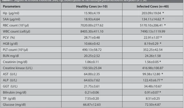
Table 1. The levels of SAA, Hp, biochemical and hematological parameters in infected and healthy cows (X ± Sx) Tablo1. Sağlıklı ve enfekte ineklerde SAA, Hp, biokimyasal ve hematolojik parametrelerin seviyeleri (X ± Sx)
Parameters Healthy Cows (n=10) Infected Cows (n=40)
Hp (µg/ml) 15.90±4.19 203.09±19.04 * SAA (µg/ml) 18.93±4.64 134.11±14.62 * RBC count (103/µl) 7020.00±277.62 5170.10±206.41 * WBC count (cell/µl) 8405.30±411.10 7490.13±1119.99 PCV (%) 28.71±0.48 22.91±1.07 * HGB (g/dl) 10.66±0.42 8.19±0.29 * PLT count (103/µl) 490.13±58.72 352.25±42.54 BUN mg/dl 20.25±2.52 24.26±1.58 Creatinin (mg/dl) 1.06±0.11 1.56±0.05 * Creatine kinase (U/L) 150.50±25.04 416.98±100.87 AST (U/L) 64.00±2.35 99.38±12.80 * ALP (U/L) 64.63±7.62 122.43±6.77 * GGT (U/L) 21.75±3.61 34.48±10.67 Bilirubin (mg/dl) 0.21±0.06 0.91±0.07 * TP (g/dl) 7.55±0.20 8.51±0.25 Glucose (mg/dl) 66.87±12.63 72.50±4.87
Haptoglobin (Hp), Serum Amyloid-A (SAA), Red blood cell count (RBC), White blood cell count (WBC), Packed cell volume (PCV), Hemoglobin (HGB), Plateleyt (PLT), Gamma-glutamyl transferase (GGT), Aspartate aminotransferase (AST), Blood urea nitrogen (BUN), Total protein (TP), Alkaline phosphatase (ALP)
The levels of SAA, Hp, and serum biochemical and hematological parameters in the infected animals and control group are presented in Table 1. Hp and SAA concentrations significantly differed between healthy cattle and A. marginale-infected cattle. While Hp and SAA concentrations in A. marginale-infected cattle were
203.09±19.04 µg/mL, 134.11±14.62 µg/mL respectively, Hp and SAA concentrations in healthy cattle was 15.90±4.19
µg/mL, 18.93±4.64 µg/mL respectively (Table 1). Significantly decreased RBC, PCV and HGB concentration were observed in infected cattle compared to the control group. Other values were within normal range (Table 1). Serum AST, ALP, creatinine and bilirubin concentrations were significantly increased in infected cattle compared with the control group. Serum GGT, creatine kinase, total protein, and blood urea nitrogen levels were increased in infected cattle compared to the control group, but with no statistically significant difference (Table 1).
DISCUSSION
Anaplasmosis, formerly known as gall sickness, traditionally refers to a disease of ruminants caused by obligate intra-erythrocytic ricketsia of the genus Anaplasma 27. Producers in endemic areas often suspect
anaplasmosis based on a history of previous disease out- breaks in that locality. Birdane et al. have indicated of a local outbreak in Turkey 8. Clinical outbreaks occur most
frequently during warm, wet seasons when vector-borne transmission is more prevalent. Naïve cattle in non-endemic areas may become infected with anaplasmosis following the introduction of a carrier animal from an endemic area 2,28. This study was performed on dairy cows
living in a southern region of Turkey that is warm and moist. Mosquitoes are observed commonly in this area.
Haptoglobin, an alpha-globulin constituent, is a major APP in numerous species of production and companion animals. In ruminants, its circulating level is negligible in healthy animals but increases over 100-fold upon immune stimulation 10,29. Murata et al.10 defines major
APPs as those proteins whose levels increase 10 to 100 times over the basal levels as a result of a stimulus such as inflammation or trauma. We observed more than a 10-fold increase in the Hp levels in infected cattle compared to healthy cattle (Table 1). In the meantime, SAA levels showed also appoximetely 10-fold increase in the infected cattle compared to healthy cattle (Table 1). Skinner et al.30
stated that Hp concentrations indicate mild inflammation when higher than 200 µg/mL, severe inflammation at 400 µg/mL, and extended pathological lesions at levels of 1-2 mg/dL. In the current study, the Hp concentration (203.09±19.04 µg/mL) increased in in cows natural infected with A. marginale (Table 1). The condition shown
that mild inflammation developed in cows natural infected with A. marginale. Haptoglobin binds free hemoglobin,
which is toxic and proinflammatory, in the plasma and thus reduces the oxidative damage associated with hemolysis, whereas SAA mainly modulates the immune response. SAA is a valuable APP in diagnosing cattle with inflammation 10.
An effect of free hemoglobin in serum samples towards reduction of measured Hp concentration has also been found 31,32. In contrast to those results, Nazifi et al.33-35
observed increased in cattle infected theileriosis and anaplasmosis. Our present sudy is consistent with Nazifi’s findings, with Hp concentration increased in A.
marginale-infected cattle compared to healthy cattle.
This significant increase indicates an inflammation in cows with anaplasmosis. Nazifi et al.35 indicated that SAA
demonstrated more obvious changes than Hp during different levels of parasitemia in A. marginale-infected cattle. However, the current study demonstrated that both Hp and SAA concentrations could be good indicators of inflamation in cattle with A. marginale-associated parasitemia. In inflammation following Anaplasma infection causes to important stimulation of the synthesis of APP. Thus, the evaluation of acute phase response in cattle with A. marginale is important for the determination of inflammation.
All animals in this study had high rectal temperature (>40°C), mild icterus, reduced milk yield, and restlessness. Three of 40 anaplasmosis-infected cattle died after oxytetracycline treatment following diagnosis. Clinical symptoms progressed from mild to severe anaplasmosis in cattle, suggesting that carrier cows in advanced pregnancy and/or lactation may relapse and develop signs of acute infection. Such events may be related to immunosuppression associated with the periparturient period in cows 36,37. Peracute anaplasmosis, characterized
by a high mortality rate within a few hours of clinical signs developing, is most frequently encountered in purebred animals and high-producing dairy cows 2,38. In the current
study, peracute anaplasmosis was not observed in any cattle. The severity of anaplasmosis may have been due to immunosuppression associated with the postparturient status of the cattle.
After erythrocytic infection is detected, the number of infected erythrocytes increases geometrically. Bovine anaplasmosis often results in development of mild to severe anemia and icterus without hemoglobinemia or hemoglobinuria, which arises from phagocytosis of these infected erythrocytes by bovine reticuloendothelial cells 1,2.
The decrease in RBC, HGB, and PCV resulted in anemia in the infected group (Table 1). Hematologycal analysis indicated pronounced decrease in platelets, but this value was not statistically significant. Other values were within normal range. Decrease in RBC, PCV, and Hb may be indicative of the severity of anemia as parasitemia progressed.
Serum AST, GGT, and ALT concentrations are indicators of hepatic function 20. In the present study, an increase in
AST, ALP and GGT concentration was observed in infected cattle compared with healthy cattle, indicating hepatic dysfunction. The rise of serum AST and cireatine kinase concentration in cattle may have been caused by musculer trauma as a result of recumbency due to anaplasmosis. The increased serum bilirubin concentration may be attributable to hemolysis of parasitized erythrocytes 39.
Furthermore, increased serum bilirubin concentration has been reported to result from hemolytic anemia and hepatic dysfunction 40,41. Biochemical analysis revealed
pronounced elevation of GGT, creatine kinase, total protein, and blood urea nitrogen plasma levels, but these increases were not statistically significant. Non-significant increase of total protein, and blood urea nitrogen levels in infected cattle may have resulted from dehydration observed in 5 dairy cows.
In conclusion, serum Hp and SAA concentrations could be also usefull in evaluate of acute phase response in cattle with Anaplasma marginale-associated parasitemia. Changes of biochemical and hematological parameters may be indicate of anemia and tissue damage in cattle with Anaplasma marginale. These parameters may be helpful to understanding the disease pathogenesis and could be used as tools for diagnosis.
REFERENCES
1. Kocan KM, De La Fuente J, Blouin EF, Garcia-Garcia JC: Anaplasma
marginale (Rickettsiales: Anaplasmataceae): Recent advances in defining host-pathogen adaptations of a tick-borne rickettsia. Parasitology, 129, 285-300, 2004.
2. Kocan KM, De La Fuente J, Blouin EF, Coetzee JF, Ewing SA: The
natural history of Anaplasma marginale. Vet Parasitol, 167, 95-107, 2010.
3. Mcelwain TF: Bovine anaplasmosis. In, Manual of Standards for
Diagnostic Tests and Vaccines. pp. 399-411, Office International des Epizooties, Paris, 2000.
4. Waal DTD: Anaplasmosis control and diagnosis in South Africa. Ann N
Y Acad Sci, 916, 474-483, 2000.
5. Goff WL, Stiller D, Roeder RA, Johnson LW, Falk D, Gorham JR, Mcguire TC: Comparison of a DNA probe, complement-fixation and
indirect immunofluorescence tests for diagnosing Anaplasma marginale in suspected carrier cattle. Vet Microbiol, 24, 381-390, 1990.
6. Torioni De Echaide S, Knowles DP, Mcguire TC, Palmer GH, Suarez CE, Mcelwain TF: Detection of cattle naturally infected with Anaplasma
marginale in a region of endemicity by nested PCR and a competitive enzyme-linked immunosorbent assay using recombinant major surface protein. J Clin Microbiol, 36, 777-782, 1998.
7. Knowles D, Torioni De Echaide S, Palmer G, Mcguire T, Stiller D, Mcelwain T: Antibody against an Anaplasma marginale MSP5 epitope
common to tick and erythrocyte stages identifies persistently infected cattle. J Clin Microbiol, 34, 2225-2230, 1996.
8. Birdane FM, Sevinc F, Derinbay O: Anaplasma marginale infections in
dairy cattle: Clinical disease with high seroprevalence. Bull Vet Inst Pulawy, 50, 467-470, 2006.
9. Ganheim C, Alenius S, Persson WK: Acute phase proteins as indicators
of calf herd health. Vet J, 173, 645-651, 2007.
10. Murata H, Shimada N, Yoshioka M: Current research on acute phase
proteins in veterinary diagnosis: An overview. Vet J, 168, 28-40, 2004.
11. Orro T, Jacobsen S, Lepage JP, Niewold T, Alasuutari S, Soveri T:
Temporal changes in serum concentrations of acute phase proteins in
newborn dairy calves. Vet J, 176, 182-187, 2008.
12. Yoshioka M, Watanabe A, Shimada N, Muratha H, Yokomizo Y, Nakajima Y: Regulation of haptoglobin secretion by recombinant
bovine cytokines in primary cultured bovine hepatocytes. Domest Anim
Endocrinol, 234, 425-433, 2002.
13. Eckersall PD, Conner JG: Bovine and canine acute phase proteins.
Vet Res Commun, 12, 169-178, 1988.
14. Eckersall PD: Acute phase proteins as monitoring tools in farm
animals. 13th Intertional Conference Production Diseases in Farm Animals.
July 29th - August 4th, Leipzig, Germany, 2007.
15. Coskun A, Sen I: Acute phase response and clinical changes in calves
with lipopolysaccharide induced endotoxemia. Eurasian J Vet Sci, 28 (1): 21-26 2012.
16. Guzelbektes H, Sen I, Ok M, Constable PD, Boydak M, Coskun A:
Serum amiloid A and haptoglobin concentrations and liver fat percentage in lactacting dairy cows with abomasal displacement. J Vet Int Med, 24, 213-219, 2010.
17. Çitil M: Puerperal enfeksiyonlu ve abomasum deplasmanlı ineklerde
serum amiloid-A ve haptoglobin düzeyleri. Kafkas Univ Vet Fak Derg, 9 (2):
147-151, 2003.
18. Allison RW, Meınkoth JH: Anemia caused by Rickettsia, Mycoplasma,
and Protozoa erythrocytes. In, Weiss DJ, Wardrop KJ (Eds): Schalm’s Veterinary Hematology. 6th ed., pp.199-210, Blackwell Publishing Ltd, 2010.
19. De UK, Dey S, Banerjee Ps, Sahoo M: Correlations among Anaplasma marginale parasitemia and markers of oxidative stress in crossbred calves. Trop Anim Health, DOI 10.1007/s11250-011-9938-6
20. Turgut K: Veteriner Klinik Laboratuvar Teşhis. s. 157-158, Bahçıvanlar
Basımsanayi, Konya, 2000.
21. Hornok S, Elek V, de la Fuente J, Naranjo V, Farkas R, Majoros G, Földvari G: First serological and molecular evidence on the endemicity
of Anaplasma ovis and A. marginale in Hungary. Vet Microbiol, 122, 316-322, 2007.
22. Allen PC, Kuttler KL, Amerault BS: Clinical chemistry of anaplasmosis:
Blood chemical changes in infected mature cows. Am J Vet Res, 42, 322-325, 1981.
23. Hirvonen J, Pyorala S: Acute-phase response in dairy cows with
surgically-treated abdominal disorders. Vet J, 155, 3-62, 1998.
24. Stengarde L, Traven M, Emanuelson U, Holtenius K, Hultgren J, Niskanen R: Metabolic profiles in five high-producing Swedish dairy
herds with a history of abomasal displacement and ketosis. Acta Vet
Scand, 50, 31, 2008.
25. Humblet M, Guyot H, Boudry B, Mbayahi F, Hanzen C, Rollin F, Godeu JM: Relationship between haptoglobin, serum amyloid A, and
clinical status in a survey of dairyherds during a 6-month period. Vet Clin
Path, 35, 188-193, 2006.
26. Horadagoda NU, Knox KMG, Gibbs HA, Reid SWJ, Horadagoda A, Edwards SER, Eckersall PD: Acutephase proteins in cattle: Discrimination
between acute and chronic inflammation. Vet Rec, 44, 437-441, 1999.
27. Aubry P, Geale DW: Review of bovine Anaplasmosis. Transbound
Emerg Dis, 58, 1-30, 2011.
28. Smith RD, Hungerford LL, Armstrong CT: Epidemiologic
investigation and control of an epizootic of anaplasmosis in cattle in winter. JAVMA, 195, 476-480, 1989.
29. Conner JG, Eckersall PD, Doherty M, Douglas TA: Acute phase
response and mastitis in the cow. Res Vet Sci, 41, 126-128, 1986.
30. Skinner JG, Brown RA, Roberts L: Bovine haptoglobin response in
clinically defined field conditions. Vet Rec, 16, 147-149, 1991.
31. Eckersall P D, Duthie S, Safi S, Moffat D, Horagoda NU, Doyle S, Parton R, Bennett D, Fitzpatrick JL: An automated biochemical assay
for haptoglobin: Prevention of interference from albumin. Comp Hematol
Int, 9, 117-124, 1999.
32. Petersen HH, Nielsen J P, Jensen A L, Heegaard P M H: Evaluation
of an enzyme linked immunosorbent assay (ELISA) for determination of porcine haptoglobin. J Vet Med A, 48, 513-523, 2001.
33. Nazifi S, Razavi SM, Esmailnejad Z, Gheisari H: Study on acute phase
proteins (haptoglobin, serum amyloid A, fibrinogen, and ceruloplasmin) changes and their diagnostic values in bovine tropical theileriosis.
Parasitol Res, 105, 41-46, 2009.
34. Nazifi S, Khoshvaghti A, Gheisari HR: Evaluation of serum and milk
amyloid A in some inflammatory diseases of cattle. IJVR, 9, 222-226, 2008.
35. Nazifi S, Razavi SM, Kaviani F, Rakhshandehroo E: Acute phase
response in cattle infected with Anaplasma marginale. Vet Microbiol Doi:10.1016/j.vetmic.2011.08.024.
36. Jones EW, Brock WE: Bovine anaplasmosis: Its diagnosis, treatment,
and control. JAVMA, 149, 1624-1633, 1966.
37. Kehrli ME, Nonnecke BJ, Roth JA: Alterations in bovine neutrophil
function during the periparturient period. Am J Vet Res, 50, 207-214, 1989.
38. Ristic M: Anaplasmosis. In, Weinman D, Ristic M (Eds): Infectious
Blood Diseases of Man and Animals. Vol. II., pp. 478-542, Academic Press, New York, London, 1968.
39. Sandhu GS, Grewal AS, Singh A, Kondal JK, Singh J, Brar RS:
Haematological and biochemical studies on experimental Theileria
annulata infection in crossbred calves. Vet Res Commun, 22, 347-354,
1998.
40. Khan IA, Khan A, Hussain A, Riaz A, Aziz A: Hemato-biochemical
alterations in cross bred cattle affected with bovine theileriosis in semi arid zone. Pak Vet J, 31, 137-140, 2011.
41. Omer OH, El-Malik KH, Magzoub M, Mahmoud OM, Haroun EM, Hawas A, Omar HM: Biochemical profiles in Friesian cattle naturally
infected with Theileria annulata in Saudi Arabia. Vet Res Commun, 27, 15-25, 2003.