Experimental (X-ray Diffraction and FT-IR) and Quantum
Chemical Studies (HF and DFT) of Ethyl
3-hydroxy-7-methyl-
3-(3-methyl-3-phenylcyclobutyl)-5-phenyl-3,5-dihydro-5H-thiazolo [3,2-a]pyrimidine-6-carboxylate
Etil
3-hidroksi-7-metil-3-(3-metil-3-fenilsiklobütil)-5-fenil-3,5dihidro-5H-tiyazolo [3,2-a] pirimidin-6-karboksilat’ın Deneysel (X-ışını
kırınımı ve FT-IR) ve Kuantum Kimyasal (HF ve DFT) Çalışmaları
Research ArticleFatih Şen1*, İbrahim Yılmaz2, Muharrem Dinçer3, Alaaddin Cukurovalı4
1Bozok University, Sorgun Vocational School, Department of Opticianry, Yozgat, Turkey.
2Karamanoğlu Mehmetbey University, Faculty of Science, Department of Chemistry, Karaman, Turkey. 3Ondokuz Mayıs University, Arts and Sciences Faculty, Department of Physics, Samsun, Turkey. 4Firat University, Sciences Faculty, Department of Chemistry, Elazig, Turkey.
Ö Z
E
til 3-hidroksi-7-metil-3-(3-metil-3-fenilsiklobütil)-5-fenil-3,5-dihidro-5H-tiyazolo [3,2-a] pirimidin-6-karboksilat başlıklı bileşik sentezlenmiştir ve deneysel ve kuantum kimyasal yöntemlerle karakterize edilmiştir. Bileşiğin kristal yapısı tek-kristal X-ışını kırınımı yöntemi ile aydınlatılmış ve katı halde titreşimsel spektrumu (FT-IR) 400-4000 cm-1 aralığında gözlenmiştir. X-ışını kırınımından elde edilen moleküler geometritemel halde Yoğunluk Fonksiyoneli Teorisi (YFT) yöntemi 6-31G(d) baz seti ile optimize edilmiştir. Optimize geometriden elde edilen bağ uzunlukları, bağ açıları, torsion açıları ve titreşimsel işaretlemeleri deneysel dataları ile karşılaştırılmıştır. Yapısal ve titreşimsel işaretlemeler deneysel ölçümlerle iyi bir uyum içindedir. Optimize geometriye ait sınır moleküler orbital ve Mulliken yük analizi teorik hesaplama sonuçları kullanılarak incelenmiştir.
Anahtar Kelimeler
Tiazol[3,2-α]-pirimidin, X-ışını kırınımı, IR spektroskopisi, Yoğunluk Fonksiyoneli Teorisi, HOMO ve LUMO, Mulliken yükü.
A B S T R A C T
T
he title compound, ethyl 3-hydroxy-7-methyl-3-(3-methyl-3-phenylcyclobutyl)-5-phenyl-3,5-dihydro-5H-thiazolo[3,2-α]pyrimidine-6-carboxylate, was synthesized and characterized by experimental and quantum chemical methods. The crystal structure of compound was brought to light by single crystal X-ray diffraction method and its vibrational spectrum (FT-IR) in solid state was observed in the region 4000-400 cm-1. Themolecular geometry was those obtained from the X-ray structure determination was optimized using Hartree-Fock and Density Functional Theory (DFT/B3LYP) method with the 6-31G(d) basis set in ground state. From the optimized geometry of the molecule, geometric parameters (bond lengths, bond angles, torsion angles) and vibrational assignments of the compound have been theoretically calculated and compared with the experi-mental data. The obtained structural and vibrational results are well in agreement with the experiexperi-mental mea-surement. The frontier molecular orbital (FMO) and Mulliken population analysis of the optimized geometries were investigated by theoretical calculation results.
Key Words
Thiazolo[3,2-α]-pyrimidine, X-ray diffraction, IR spectroscopy, Density Functional Theory, HOMO and LUMO, Mulliken Charge.
Article History:Received: Jun 11, 2016; Revised: Jun 28, 2016; Accepted: Sep 20, 2016; Available Online: Apr 1, 2016. DOI: 10.15671/HJBC.2017.150
Correspondence to: F. Şen, Bozok University, Sorgun Vocational School, Department of Opticianry, Yozgat, Turkey.
INTRODUCTION
C
yclobutane is an organic compound formed by the four carbon atoms. Although itself is of no commercial or biological significance, it is known that the more complex derivatives are important in biology and biotechnology [1]. Also, 3-substituted cyclobutane carboxylic acid derivatives exhibit significant pharmacological activities such as inflammatory and anti-depressant activity [2]. Thiazole, or 1,3-thiazole, is a heterocyclic compound that contains both sulfur and nitrogen atoms. Thiazole derivatives show a diverse range of biological activities such as antimicrobial [3-5], anti-inflammatory [6,7], antitumor [8,9], anticonvulsant [10] and antifungal [11,12], etc. It is known that the thiazolo[3,2-a]pyrimidine derivatives possess a variety of biological and [13,14], antinociceptive [15], antibacterial [16-18], antiviral [19,20], antihypertensive [21], antibiofilm [22,23], antitubercular [17,18], antivirulence [24], antioxidant [25], etc. In the light of the effects given above, the compounds containing cyclobutane, thiazole and thiazolo[3,2-a] pyrimidine functionalities in one molecule have seen to be important.Determination of the structural and spectroscopic properties of compounds using both experimental techniques and theoretical methods has been attracted interest for many years. To the best of our knowledge, the title compound is a novel compound firstly synthesized in our laboratories by us and there is no any information present in literature about its molecular and vibrational spectroscopic properties. The aim of the present work was to describe and characterize the molecular structure and vibrational frequencies of the compound, both experimentally and theoretically. In the experimental study, the
molecule was prepared and characterized by single-crystal X-ray diffraction and FT-IR methods. In theoretical study, the molecular structure of the title compound in the ground state have been calculated using the Hartree-Fock (HF) and Density Functional Theory (DFT) (B3LYP) with 6-31G(d) basis set. The calculated geometric parameters (bond lengths, bond angles, torsion angles), and vibrational assignments compared with their experimental data.
MATERIALS AND METHODS Synthesis and Characterization
To a solution of 4-phenyl-2-thioxo-1,2,3,4-tetrahydro-pyrimidine-5-carboxylic acid ethyl ester (2.6233 g, 10 mmol) in 50 mL of ethanol, a solution of 1-methyl-1-phenyl-3-(2-chloro-1-oxoethyl) cyclobutane (2.2271 g, 10 mmol) in 20 mL of absolute ethanol was added, stirred 3 h (TLC), made alkaline with an aqueous solution of NH3 (5%), brilliant yellow precipitate separated by suction and crystallized from EtOH (Scheme 1). Yield: 63%, melting point: 427 K.
General Remarks
All chemicals were of reagent grade and used as commercially purchased without further purification. The melting point was determined by Gallenkamp melting point apparatus. The FT-IR spectrum of the tittle compound was recorded in the range 4000-400 cm-1 using a Mattson 1000 FT-IR spectrometer with KBr pellets.
Crystal Structures Determination and Refinement
The single-crystal X-ray data were collected on a STOE IPDS II image plate diffractometer at 296 K. Graphite-monochromated Mo Kα radiation (λ = 0.71073 Å) and the w-scan technique were used. The structure of title compound was solved by
N N H S H CH3 COOC2H5 H3C C O H2C Cl + S N N COOC2H5 CH3 OH H3C
direct methods using SHELXS-97 [26] and refined through the full-matrix least-squares method using SHELXL-97 [27], implemented in the WinGX [28] program suite. Non-hydrogen atoms were refined with anisotropic displacement parameters. All H atoms were located in a difference Fourier map and were refined isotropically. Data collection: Stoe X-AREA [29], cell refinement: Stoe X-AREA [29], data reduction: Stoe X-RED [29]. The general-purpose crystallographic tool PLATON [30] was used for the structure analysis and presentation of the results. The structure was refined to Rint= 0.053 with 3810 observed reflections using I > 2σ(I) threshold. The ORTEP-3 program for Windows was used for preparation of figures. Details of the data collection conditions and the parameters of the refinement process are given in Table 1.
Details of the Theoretical Calculation
All the calculations were carried out by using Gaussian 03 package [31] and Gauss-View molecular visualization software [32] on the personal computer without restricting any
symmetry for the title compound. For modeling, the initial guess of the compound was first obtained from the X-ray coordinates and this structure was optimized by Hartree-Fock (HF) and Density Functional Theory (DFT)/B3LYP methods with and 6-31G(d) as basis set. From the optimized geometry of the molecule, geometric parameters (bond lengths, bond angles, torsion angles) and theoretical harmonic frequencies of the title compound have been calculated and compared with the experimental data. Besides, frontier molecular orbitals (FMOs) and Mulliken population analysis of the title compound were investigated by theoretical calculations.
RESULTS AND DISCUSSION Structural Description
The title compound, methyl 5-(4-hydroxy-3- methoxyphenyl)-2-(4-methoxybenzylidene)-7- methyl-3-oxo-2,3-dihydro-5H-thiazolo[3,2-a]-pyrimidine-6-carboxylate, with the crystal data shown in Figure 1. X-ray diffraction analysis has revealed that the title compound crystallizes in monoclinic system with space group P 21/c. The unit cell dimensions are a= 6.5335 (3) Å, b= 19.7921 (7) Å, c= 19.3274 (8) Å and V= 2437.50 (17) Å3.
The molecular structure of compound has ’’C1’’ symmetry. It is composed of the cyclobutane,
phenyl rings, and an ester group in addition to the heterocycles such as a thiazole, and a pyrimidine rings. The dihedral angles between the cyclobutane plane A (C7-C10), the thiazole plane B (S1/N1/C12-C14), and the pyrimidine plane C (N1/ N2/C14-C16) are 86.10° (A/B), 13.32° (B/C) and 82.48° (A/C).
The cyclobutane ring was adopted butterfly conformation. The bond lengths between the carbon atoms are average 1.546 Å and bond angles formed by the three carbons are average 89.27° for the cyclobutane ring in the compound. When these values are compared with the previously reported cyclobutane derivatives [33,34], it is seen that there are no significant differences.
In the thiazole ring, the S1—C13 and S1—C14 bond
Table 1. Crystal data collection and structure refinement parameters for the title compound.
Data collection
Diffractometer STOE IPDS-2 Wavelength (Å) 0.71073 Mo Kα θ range for data collection (º) 1.5 ≤ θ ≤ 27.3 Index ranges
hmin, hmax -7, 8
kmin, kmax -24, 24
lmin, lmax -24, 24
Measurement method ω taraması Reflections collected 12583 Independent reflections 4964 Observed reflections [I > 2σ(I)] 3810
Absorption correction Integrasyon (X−RED32) Tmin, Tmax 0.905, 0.954
Rint 0.053
Refinement
Refinement method SHELXL-97
Parameters 298
R[F2 > 2σ(F2)] 0.046
wR(F2) 0.129
GooF = S 1.06
lengths are 1.790 (2) and 1.7361 (18) Å, respectively. These values are close to the standard value for an S—Csp2 single bond (1.76 Å) [35]. The thiazole ring is planar with a maximum deviation of 0.0794 Å. Thiazole ring are not coplanar with the pyrimidine
ring. The average angle between the plane of the thiazole ring and the plane of the pyrimidine ring is 13.32°. Structural parameters of the pyrimidine are as follows: average C═N distance is 1.310 (2) Å, the C–N distance is 1.331 (2) Å. These parameters are comparable with the related pyrimidine derivatives reported in the literature [36-38].
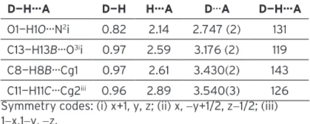
It does not observed π−π stacking (face-to-face) interactions in the crystal structure. There are, however, two D−H···A and two D−H···Cg(π-ring) (edge-to-face) interactions, details of which are given in Table 2 The hydroxyl atom O1 in the molecule at (x, y, z) acts as hydrogen-bond donor, via atom H1O, to pyrimidine ring atom N2 in the molecule at (x+1, y, z). The thiazole ring atom C13 in the molecule at (x, y, z) acts as hydrogen-bond donor, via atom H13B, to carboxyl atom O3 in the molecule at (x, −y+1/2, z−1/2). The cyclobutane atom C8 at (x,y,z) forms an intramolecular C—H···Cg(π-ring) contact, this time via atom H8B, with the centroid of the C22-C27 ring. Atom C14 at (x, y, z) forms an intermolecular C—H···Cg(π-ring) contact, via atom H11C, with the centroid of the C1-C6 ring of the molecule at (1-x,1-y, -z) (Figure 2).
The molecular structure of the title compound was also studied theoretically. The starting coordinates were those obtained from the X-ray structure determination. The molecular geometry
(X-ray coordinates) was optimized using Hartre-Fock (HF) and Density Functional Theory (DFT/ B3LYP) method with the 6-31G(d) basis set in ground state. The total energy, zero-point vibrational energy, entropy and dipole moment values obtained for optimized geometries are presented in Figure 3.
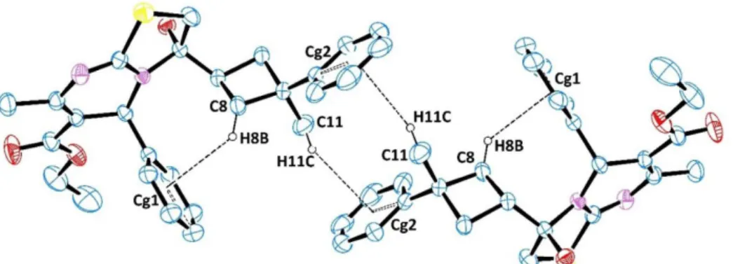
Conventionally, two methods were used in comparison the molecular structures. The first method is to calculate the correlation value (R2) between X-ray and theoretical structure. The optimized some geometrical parameters (bond lengths, bond angles and torsion angles) calculated by HF/6-31G(d) and DFT/B3LYP/6-31G(d) methods with corresponding to experimental values are listed in Table 3. We have calculated correlation value (R2) between these parameters. R2 values are 0.9962 and 0.9939 for bond lengths, 0.9985 and 0.9979 for bond angles, 0.9682 and 0.9658 for torsion angles at HF/6-31G(d) and DFT/ B3LYP/6-31G(d) levels, respectively. According to correlations values, the HF/6-31G(d) method gave accurate results for the bond lengths, bond
Figure 1 : A view of the title compound showing the atom-numbering scheme with the crystal data. Displacement ellipsoids are drawn at the 30% probability level.
Table 2. Hydrogen-bond geometry (Å, °) for the title compound.
D—H···A D—H H···A D···A D—H···A
O1—H1O···N2i 0.82 2.14 2.747 (2) 131
C13—H13B···O3ii 0.97 2.59 3.176 (2) 119
C8—H8B···Cg1 0.97 2.61 3.430(2) 143 C11—H11C···Cg2iii 0.96 2.89 3.540(3) 126
Symmetry codes: (i) x+1, y, z; (ii) x, −y+1/2, z−1/2; (iii) 1−x,1−y, −z.
Cg1: the centroid of the C22-C27 ring, Cg2: the centroid of the C1-C6 ring
angles, and torsion angles compared with the DFT/B3LYP/6-31G(d) method. Second method for globally comparing the structures obtained with the theoretical calculations is by overlapping the molecular skeleton with that obtained from X-ray diffraction, giving a RMSE of 0.497 Å for HF6-31G(d) and 0.483 Å for DFT/B3LYP/6-31G(d) calculations (Figure 4). According to these results, it may be concluded that the HF/6-31G(d) calculation well reproduce the geometry of the title compound. It was noted here that the experimental results belong to solid phase and theoretical calculations belong to gaseous phase. In the solid state, the existence of the crystal field along with the intermolecular interactions has connected the molecules together, which result in the differences of bond parameters between the calculated and experimental values [39].
Vibrational Spectra
The Fourier Transform Infrared Spectrum (FT-IR) of the title compound was recorded employing a “Mattson 1000 FT-IR spectrometer” using KBr
pellet technique in the range of 4000-400 cm-1. The theoretical IR spectra have been calculated with HF and DFT/B3LYP methods with the 6-31G(d) basis set in ground state. In order to compare the theoretical results with experimental values of those, scaling factor which are 0.8929 and 0.9626 for HF/6-31G(d) and DFT/B3LYP/6-31G(d) is applied to all of the calculated frequencies, respectively. The overlapped experimental and scaled theoretical FT-IR spectra of the title compound are given in Figure 5.
The title compound belongs to C1 point group symmetry and consists of 63 atoms. It has seen 183 normal modes of vibration using the formula 3N-6. The fundamental vibrational wavenumbers of compound calculated by HF/6-31G(d) and DFT/B3LYP/6-31G(d) methods. The vibrational bands assignments have been made by using Gauss View molecular visualization program [32]. Some observed and calculated frequencies in the infrared spectra of the title compound and their
Figure 2: The intra- and inter-molecular hydrogen bondings in the crystal structure
Figure 4. Atom-by-atom superimposing of the structures calculated [HF/6-31G(d) = red, DFT/B3LYP/6-31G(d) = blue] over the X-ray structure (black) for the title compound. Hydrogen atoms omitted for clarity.
Table 3. Some selected geometric parameters (Å, °).
Geometric Parameters [X–ray diffraction]Experimental Calculated
HF/6-31G(d) DFT/B3LYP/6-31G(d) Bond lengths (Å) C6—C7 1.506 (3) 1.518 1.517 C7—C11 1.528 (3) 1.535 1.539 C7—C8 1.545 (2) 1.555 1.566 C9—C12 1.525 (2) 1.531 1.536 C12—O1 1.401 (2) 1.403 1.427 C13—S1 1.790 (2) 1.810 1.832 S1—C14 1.7361 (18) 1.751 1.765 C14—N1 1.331 (2) 1.347 1.360 C14—N2 1.310 (2) 1.275 1.298 C16—C19 1.463 (3) 1.470 1.464 C19—O3 1.202 (2) 1.194 1.220 C19—O2 1.337 (3) 1.335 1.371 R2 0.9962 0.9939 Bond Angles (o) C7—C8—C9 89.66 (13) 89.08 89.08 C12—N1—C14 116.80 (13) 115.46 115.08 C13—S1—C14 92.18 (8) 91.87 91.58 N1—C14—N2 126.95 (16) 127.61 127.40 C16—C17—C22 111.74 (13) 111.50 111.78 O2—C19—O3 121.47 (19) 121.44 121.30 R2 0.9985 0.9979 Torsion Angles (o) C1—C6—C7—C10 48.2 (2) 39.74 38.77 C16—C17—C22—C27 61.2 (2) 70.83 70.85 C19—C16—C17—C22 75.35 (19) 76.39 78.10 N1—C17—C22—C23 119.91 (17) 129.59 129.33 R2 0.9682 0.9658
probable assignments are given in Table 4. As can be seen from the Table 4, an acceptable agreement exists between the experimental and theoretical vibrational frequencies. The correlation coefficients are 0.9908 and 0.9937 for HF/6-31G(d) and DFT/B3LYP/6-31G(d), respectively. According to these correlation values, DFT/B3LYP/6-31G(d) method gave accurate results compared with the HF/6-31G(d) method in determining vibrational spectra. Some experimentally obtained vibrational frequencies were supported by literature values.
O–H Vibrations
It is well known that the high frequency region above 3000 cm-1 is the characteristic region for the ready identification of C—H, O—H and N—H stretching vibrations. These vibrations are sensitive to hydrogen bondings. The non-hydrogen bonded or free hydroxyl group absorb strongly in 3550-3700 cm-1 region [40]. In the IR spectra of the compound, the absorption at 3327 cm-1 is assigned to the stretching vibrations of O—H bond that has been calculated at 3658 and 3587 cm-1 for HF/6-31G(d) and DFT/B3LYP/6-31G(d) levels, respectively. The difference between the experimental and theoretical values take its source from the O1—H1O···N2 intermolecular hydrogen bondings.
Vibrational modes: ν, stretching; α, scissoring; γ, rocking; ω, wagging; ω, twisting; θ, ring; breathing. Abbreviations: s, symmetric; as, asymmetric. Phenyl Ring Vibrations
It is known that the C—H and C═C stretching vibrations of the aromatic compounds usually appear in 2900-3150 and 1100-1500 cm-1 frequency ranges, respectively [41]. The C—H aromatic stretching modes were recorded at 3082 cm-1 experimentally and computed at 3014, 2995 and 2991 cm-1, respectively. The C═C stretching vibrations predicted at 1617, 1616, 1595 and 1590 cm-1 in the theoretical spectra show good agreement with the experimental FT-IR bands at 1600 cm-1.
Cyclobutane Vibrations
The cyclobutane ring of symmetric and asymmetric C—H2 vibration mode is known to monitor the 3100-3000 cm-1. The asymmetric stretching C—H2 observed at 2950 cm-1 in FT-IR spectrum. This value is not significantly different from those in the literature; 2960 [33], 2975 [34], 2929 and 2855 [42]. The symmetric stretching C—H2 observed at 2950 cm-1 in FT-IR spectrum and also these vibrations calculated at 2914, 2895 and 2974, 2961 cm-1 for HF/6-31G(d) and DFT/B3LYP/6-31G(d) levels, respectively. The, scissoring C—H2, wagging C—H2, twisting C—H2 and
Table 4. Comparison of the observed and calculated vibrational spectra of the compound (cm-1).
Assignment Experimental (Kbr) Calculated [6-31G(d)] HF DFT/B3LYP νO—H 3327 3658 3587 νsC—Haromatic - 3027 3092 νsC—Haromatic - 3023 3089 νasC—Haromatic 3082 3014 3085 νasC—H2thiazole - 3006 3063 νasC—Haromatic 3082 2995 3062 νasC—Haromatic 3082 2991 3057 νasC—H2cyclobutane 3023 2965 3029 νasC—H2cyclobutane 3023 - 3012 νsC—H2thiazole - 2953 3005 νasC—H2cyclobutane 3023 2939 -νC—H 2975 2921 2952 νsC—H2cyclobutane 2950 2914 2974 νsC—H2cyclobutane 2950 2895 2961 νC—Hcyclobutane 2912 2889 2940 νsC—H3 - 2869 2943 νsC—H3cyclobutane 2864 2857 2927 νC═Ocarboxyl 1694 1752 1705 νC═Caromatic 1600 1617 1602 νC═Caromatic 1600 1616 1599 νC═Npyrimidine + νC═Cpyrimidine 1674 1612 1574 νC═Caromatic 1600 1595 1486 νC═Caromatic 1600 1590 1490 νC═Npyrimidine + νC═Cpyrimidine 1495 1502 1495 αC—H2cyclobutane 1423 1482 1475 αC—H2cyclobutane 1423 1468 1443 αC—H2thiazole - 1462 1455 ωC—H - 1406 1385 νsC—H3cyclobutane 1370 1395 1382 ωC—H - 1381 1360 ωC—Hcyclobutane 1270 1265 1359 ωC—H2cyclobutane 1239 1235 1212 δC—H2thiazole - 1207 1185 ωC—Hpyrimidine - 1172 1165 δC—H2cyclobutane 1029 1035 1044 θcyclobutane 998 946 943 νS—CH2 766 805 794 R2 0.9908 0.9937
breathing vibrations observed at 1423, 1239 and 1029 cm-1 in FT-IR spectrum for cyclobutane ring.
HOMO and LUMO Analysis
The distributions of the HOMO and LUMO orbitals computed at the HF/6-31(d) and DFT/B3LYP/6-31G(d) levels for the title compound are shown in Figure 6. The calculations indicate that the title compound has 123 occupied molecular orbitals. HOMO and LUMO energies are -8.21 and 2.71 eV for HF/6-31G(d) level, -5.57 and -1.27 eV for DFT/ B3LYP/6-31G(d) level, respectively.
By using HOMO and LUMO energy values for a molecule, electronegativity, chemical hardness
and chemical softness can be calculated as follows:
χ=(I+A)/2(electronegativity) η=(I-A)/2 (chemical hardness)
S=1/2η (chemical softness)
where I and A are ionization potential and electron affinity;
I=-EHOMO A=-ELUMO
respectively [43]. The ionization potential (I), the electron affinity (A), the absolute electronegativity (χ), the absolute hardness (η) and softness (S) for molecule have been calculated at the same levels and the results are given in Table 5.
The value of the energy separation between the HOMO and LUMO is 10.92 eV for HF/6-31G(d) level and 4.30 eV for DFT/B3LYP/6-31G(d) level. These energy gaps indicate that the title structure
is quite stable.
Mulliken Population Analysis
The Mulliken charge distributions of the title compound were calculated using HF/6-31G(d)
Figure 6. The molecular orbital diagrams, energies and energy gaps for the optimized geometries.
Figure 7. The Mulliken charges of diagram for the title compound.
Table 5. The parameters related to frontier molecular orbitals. Parameters HF/6-31G(d) DFT/B3LYP/6-31G(d) I (eV) 8.21 5.57 A (eV) -2.71 1.27 χ (eV) 2.75 3.42 η (eV) 5.46 2.15 S(═eV-1) 0.092 0.233
and DFT/B3LYP/6–31G(d) levels. The calculated charge for all atoms has been given in Figure 7.
It is well-known that the Mulliken charges confirm the hydrogen bondings in the molecular structure of compound. For the title compound, the Mulliken charges corresponding to the atoms in hydrogen bondings are shown in Table 6. As can be seen in Table 6, these charges confirmed that the intra- and intermolecular hydrogen bondings. CONCLUSIONS
The title compound, ethyl 3-hydroxy-7-methyl- 3-(3-methyl-3-phenylcyclobutyl)-5-phenyl-
3,5-dihydro-5H-thiazolo[3,2-α]pyrimidine-6-carboxylate (C27H30N2O3S), was both
experimentally and theoretically investigated. The important conclusions to be obtained from this study are the following:
i. The compound was synthesized and brought to light its molecular structure by single-crystal X-ray diffraction method in our laboratory. The crystal structure and geometric parameters of the compound were obtained.
ii. The initial guess of the compound was first obtained from the X-ray coordinates was optimized by Hartree-Fock (HF) and Density Functional Theory (DFT)/B3LYP method with 6-31G(d) as basis set. The HF/6-31G(d) method gave accurate results compared with the DFT/ B3LYP/6-31G(d) method in determining molecular structure of the title compound.
iii. By using optimized geometries of molecule, theoretical IR spectra was calculated and compared with experimental FT-IR spectrum. The
DFT/B3LYP/6-31G(d) method is better than HF/6-31G(d) method for assigning vibrational modes of the title compound.
iv. The frontier molecular orbitals (HOMO and LUMO) and related to parameters were generated by theoretical results.
v. The calculated Mulliken charges confirm the hydrogen bonding in the crystal structure.
SUPPLEMENTARY MATERIAL
CCDC 929331 contains supplementary crystallographic data (excluding structure factors) for the structure reported in this article. These data can be obtained free of charge via http://www.ccdc.cam.ac.uk/data_request/cif, by e-mailing [email protected] or by contacting The Cambridge Crystallographic Data Centre, 12 Union
Road, Cambridge CB2 1EZ, UK; fax: +44 1223336033.
ACKNOWLEDGEMENTS
I wish to thank Prof. Dr. Orhan Büyükgüngör for his help with the data collection and acknowledge the Faculty of Arts and Sciences, Ondokuz Mayıs University, Turkey, for the use of the STOE IPDS II diffractometer.
This paper was orally presented at the International Conference on Natural Science and Engineering (ICNASE’16). Paper ID : ICNASE’16_408.
R e f e r e n c e s
1. Web-1: https://en.wikipedia.org/wiki/Cyclobutane, consulted 10 February 2016.
2. E.V. Dehmlow and S. Schmidt, Synthese von stereoisomeren 3-substituierten Cyclobutancarbonsäure-Derivaten, Liebigs Annalen der Chemie Liebigs, 5 (1990) 411-414. 3. P. Karegoudar, M.S. Karthikeyan, D.J. Prasad, M.
Mahalinga, B.S. Hollaa, and N.S. Kumari, Synthesis of some novel 2,4-disubstituted thiazoles as possible antimicrobial agents, European Journal of Medicinal Chemistry, 43 (2008) 261-267.
Table 6. Mulliken charges of the atoms in hydrogen bondings for the title compound.
HF/6-31G(d) DFT/B3LYP/6-31G(d) D—H•••A D H A D H A O1—H1O···N2 -0.739 0.449 -0.674 -0.614 0.399 -0.538 C13—H13B···O3 -0.543 0.224 -0.665 -0.496 0.215 -0.519 C8—H8B···Cg1 -0.332 0.179 -1.111 -0.308 0.146 -0.983 C11—H11C···Cg2 -0.453 0.172 -1.054 -0.451 0.139 -0.895
4. O. Bozdağ-Dündar, Ö. Özgen, A. Menteşe, N. Altanlar, O. Atlı, E. Kendi and R. Ertan, Synthesis and antimicrobial activity of some new thiazolyl thiazolidine-2,4-dione derivatives, Bioorganic & Medicinal Chemistry, 15 (2007) 6012-6017.
5. P. Vicini, A. Geronikaki, K. Anastasia, M. Incerti and F. Zania, Synthesis and antimicrobial activity of novel 2-thiazolylimino-5-arylidene-4-thiazolidinones, Bioorganic & Medicinal Chemistry, 14 (2006) 3859-3864.
6. A. Kumar, C.S. Rajput and S.K. Bhati, Synthesis of 3-[4’-(p-chlorophenyl)-thiazol-2’-yl]-2-[(substituted azetidinone/thiazolidinone) aminomethyl] 6 -bromoquinazolin-4-ones as anti-inflammatory agent, Bioorganic & Medicinal Chemistry, 15 (2007) 3089-3096.
7. B.S. Holla, K.V. Malini, B.S. Rao, B.K. Sarojini and N.S. Kumari, Synthesis of some new 2,4-disubstituted thiazoles as possible antibacterial and anti-inflammatory agents, European Journal of Medicinal Chemistry, 38 (2003) 313-318.
8. G.S. Hassan, S.M. El-Messery, F.A. M. Al-Omary and H.I. El-Subbagh, Substituted thiazoles VII. Synthesis and antitumor activity of certain 2-(substituted amino)-4-phenyl-1,3-thiazole analogs, Bioorganic & Medicinal Chemistry Letters 22 (2012) 6318-6323. 9. I. Hutchinson, M. Chua, H.L. Browne, V. Trapani, T.
D. Bradshaw, A.D. Westwell and M.F.G. Stevens, Antitumor Benzothiazoles. 14.1 Synthesis and in Vitro Biological Properties of Fluorinated 2-(4-Aminophenyl)benzothiazoles, Journal of Medicinal Chemistry, 44 (2001) 1446-1455.
10. F. Azam, I.A. Alkskas, S.L. Khokra and O. Prakash, Synthesis of some novel N4-(naphtha[1,2-d]thiazol-2-yl)semicarbazides as potential anticonvulsants, European Journal of Medicinal Chemistry, 44 (2009) 203-211.
11. F. Chimenti, B. Bizzarri, E. Maccioni, D. Secci, A. Bolasco, R. Fioravanti, P. Chimenti, A. Granese, S. Carradori, D. Rivanera, D. Lilli, A. Zicari and S. Distinto, Synthesis and in vitro activity of 2-thiazolylhydrazone derivatives compared with the activity of clotrimazole against clinical isolates of Candida spp., Bioorganic & Medicinal Chemistry, 17 (2007) 4635-4640.
12. B. Narayana, K.K. Vijaya Raj, B.V. Ashalatha, N.S. Kumari and B.K. Sarojini, Synthesis of some new 5-(2-substituted-1,3-thiazol-5-yl)-2-hydroxy benzamides and their 2-alkoxy derivatives as possible antifungal agents, European Journal of Medicinal Chemistry, 39 (2004) 867-872. 13. B. Tozkoparan, M. Ertan, B. Krebs, M. Läge, P.
Kelicen and R. Demirdamar, Condensed Heterocyclic Compounds: Synthesis and Antiinflammatory Activity of Novel Thiazolo[3,2-a]pyrimidines, Archiv der Pharmazie, 33 (1998) 201-206.
14. B. Tozkoparan, M. Ertan, P. Kelicen, R. Demirdamar, Synthesis and anti-inflammatory activities of some thiazolo[3,2-a]pyrimidine derivatives, II Farmaco, 54 (1999) 588-593.
15. O. Alam, S.A. Khan, N. Siddiqui, W. Ahsan, Synthesis and pharmacological evaluation of newer thiazolo [3,2-a] pyrimidines for anti-inflammatory and antinociceptive activity, Medicinal Chemistry Research, 19 (2010) 1245-1258.
16. R.A. Coburn, R.A. Glennon, Mesoionic purinone analogs IV: Synthesis and in vitro antibacterial properties of mesoionic thiazolo[3,2-a]pyrimidin-5,7-diones and mesoionic 1,3,4-thiadiazolo[3,2-a] pyrimidin-5,7-diones, Journal of Pharmaceutical Sciences, 62 (1973) 1785–1789.
17. D. Cai, Z. Zhang, Y. Chen, X. Yan, S. Zhang, L. Zou, L. Meng, F. Li and B. Fu, Synthesis of some new thiazolo[3,2-a]pyrimidine derivatives and screening of their in vitro antibacterial and antitubercular activities, Medicinal Chemistry Research, 25 (2016) 292-302.
18. D. Cai, Z. Zhang, Y. Chen, X. Yan, L. Zou, Y. Wang, X. Liu, Synthesis, Antibacterial and Antitubercular Activities of Some 5H-Thiazolo[3,2-a]pyrimidin-5-ones and Sulfonic Derivatives, Molecules, 20 (2015) 16419-16434.
19. K. Danel, E.B. Pedersen and C. Nielsen, Synthesis and Anti-HIV-1 Activity of Novel 2,3-Dihydro-7H-thiazolo[3,2-a]pyrimidin-7-ones, Journal of Medicinal Chemistry, 41 (1998) 191-198.
20. K.R .Babu, V.K. Rao, Y.N. Kumar, K. Polireddy, K.V. Subbaiah, M. Bhaskar, V. Lokanatha, C.N. Raju, Identification of substituted [3, 2-a] pyrimidines as selective antiviral agents: Molecular modeling study, Antiviral Research, 95 (2012) 118-127.
21. E. Jeanneau-Nicolle, M. Benoit-Guyod, A. Namil, G. Leclerc, New thiazolo [3,2-a] pyrimidine derivatives, synthesis and structure-activity relationships, European Journal of Medicinal Chemistry, 27 (1992) 115–120.
22. M. Bansal, R. Kaur, B. Kaur, Synthesis and antifungal evaluation of thiazolo[3,2-a]pyrimidine derivatives, Journal of Applied Chemistry, 2 (2013) 391-397. 23. D. Zhao, C. Chen, H. Liu, L. Zheng, Y. Tong, D. Qu, S. Han,
Biological evaluation of halogenated thiazolo[3,2-a] pyrimidin-3-one carboxylic acid derivatives targeting the YycG histidine kinase, European Journal of Medicinal Chemistry, 87 (2004) 500-507.
24. E. Chorell, J.S. Pinkner, G. Phan, S. Edvinsson, F. Buelens, H. Remaut, G. Waksman, S.J. Hultgren, F. Almqvist, Design and synthesis of C-2 substituted thiazolo and dhydrothiazolo ring-fused 2-pyridones; Pilicides with increased anti-virulence activity, Journal of Medicinal Chemistry, 53 (2010)
5690-5695.
25. A.V. Pawde, D.S. Kawale and S.P. Vartale, Synthesis and antioxidant, antimicrobial activity of 6-cyano-5-imino-2-methyl-7-(methylthio)-5H-thiazolo [3, 2-a] pyrimidines, Der Pharma Chemica, 7 (2015) 205-209. 26. G.M. Sheldrick, SHELXS-97, Program for the Solution of Crystal Structures, (1997) University of Gottingen, Germany.
27. G.M. Sheldrick, SHELXL-97, Program for Crystal Structures Refinement, (1997) University of Gottingen, Germany.
28. L.J. Farrugia, WinGX suite for small-molecule single-crystal crystallography, Journal of Applied Crystallography, 32 (1999) 837-838.
29. Stoe & Cie X-AREA (Version 1.18) X-RED32 (Version 1.04) Stoe & Cie. (2002) Darmstadt, Germany.
30. A.L. Spek, Structure validation in chemical crystallography, Acta Crystallographica Section D Structural Biology, D65 (2009) 148-155.
31. M.J. Frisch, G.W. Trucks, H.B. Schlegel et al. Gaussian 03, Revision E.01, Gaussian, Inc., (2004) Wallingford, USA.
32. R. Dennington, T. Keith and J. Millam, Gauss View, Version 4.1.2, Semichem Inc., (2007) Shawnee Mission, Kan, USA.
33. E. Inkaya, M. Dinçer, Ö. Ekici, A. Cukurovali, N’-(2- methoxy-benzylidene)-N-[4-(3-methyl-3-phenyl-cyclobutyl)-thiazol-2-yl]-chloro-acetic hydrazide: X-ray structure, spectroscopic characterization and DFT studies, Journal of Molecular Structure, 1026 (2012) 117-126.
34. E. Inkaya, M. Dinçer, Ö. Ekici, A. Cukurovali, 1-(3-Methyl-3-mesityl)-cyclobutyl-2-(5-pyridin-4-yl-2H-[1,2,4]triazol-3-ylsulfanyl)-ethanone: X-ray structure, spectroscopic characterization and DFT studies, Spectrochimica Acta Part A: Molecular and Biomolecular Spectroscopy, 101 (2013) 218-227. 35. F.H. Allen, The geometry of small rings. VI. Geometry
and bonding in cyclobutane and cyclobutane, Acta Crystallographica Section B Structural Science, Crystal Engineering And Materials, B40 (1984) 64-72. 36. N.N. Pesyan, S. Rastgar and Y. Hosseini,
1,1’,3,3’,5,5’-Hexamethylspiro[furo-[2,3-d]pyrimidine- 6(5H),5’-pyrimidine]-2,2’,4,4’,6’(1H,3H,1’H,3’H,5’H)-pentaone, Acta Crystallographica Section E Crystallographic Communications, E65 (2009) o1444.
37. M. Wriedt, S. Sellmer, I. Jess and C. Näther, catena-Poly[bis[cis-dipyrimidine transdithiocyanatoiron(II)]- di-l-pyrimidine-[trans-dithiocyanatoiron(II)]-di-lpyrimidine], Acta Crystallographica Section E Crystallographic Communications, E65 (2009) m296-m297.
38. A.A. El-Emam, O.A. Al-Deeb, A.A. Al-Turkistani, S.W. Ng and E. R.T. Tiekink, 2 Benzylsulfanyl-4-pentyl-6-(phenylsulfanyl)pyrimidine-5-carbonitrile, Acta Crystallographica Section E Crystallographic Communications, 67 (2011) o3126.
39. F.F. Jian, P.S. Zhao, Z.S. Bai and L. Zhang, (2005). Quantum Chemical Calculation Studies on 4-Phenyl-1-(Propan-2-Ylidene)Thiosemicarbazide, Structural Chemistry, 16 (2005) 635-639.
40. K. Bahgat, A.G. Ragheb, Analysis of vibratinal spectra of 8-hydroxyquinoline and its 5,7-dichloro, 5,7-dibromo, 5,7-diiodo and 5,7-dinitro derivatives based on density functional theory calculations, Central European Journal of Chemistry, 5 (2007) 201-220.
41. R.M. Silverstein, F.X. Webster and D.J. Kiemle, Spectrometric Identification of Organic Compounds, seventh ed., (2005) John Willey & Sons, New York. 42. F. Şen, M. Dinçer and A. Cukurovali, Synthesis,
spectroscopic characterization and quantum chemical computational studies on 4-(3-methyl-3-phenylcyclobutyl)-2-(2-undecylidenehydrazinyl) thiazole, Journal of Molecular Structure, 1076 (2014)1-9.
43. R.G. Pearson, Absolute electronegativity and hardness correlated with molecular orbital theory, Proceedings of the National Academy of Sciences, 83 (1986) 8440–8841.
![Figure 4. Atom-by-atom superimposing of the structures calculated [HF/6-31G(d) = red, DFT/B3LYP/6-31G(d) = blue] over the X-ray structure (black) for the title compound](https://thumb-eu.123doks.com/thumbv2/9libnet/4565160.83527/6.829.101.731.126.753/figure-atom-superimposing-structures-calculated-structure-black-compound.webp)


