Effect of activators and calcination on luminescence properties of akermanite type phosphors
8
0
0
Tam metin
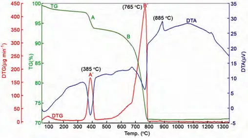
(2) KARACAOGLU & KARASU: LUMINESCENCE PROPERTIES OF ÅKERMANITE TYPE PHOSPHORS. to Ca2MgSi2O7 as luminescent center and were prepared in N2/H2 and/or air (open) atmospheres, respectively. So, there are both similar results with previous researchs and also novel results of this structure with rare-earths. Material and Methods In the present study, the phosphors were synthesized via high temperature solid-state reaction method, also known as ceramic method. Using the base composition of Ca2MgSi2O7: ~0.5 mass% REO (Rare Earth Oxides-REO= Eu2O3, Ce2O3, Pr6O11, Sm2O3, Dy2O3), appropriate amounts of the high purity starting reagents, CaCO3 (Merck, Suprapur, 99.95%), Mg(OH)2 (Aldrich, 95%) and SiO2 (Merck, highly dispersed suitable for use as excipient) and a small amount the fluxing agent, H3BO3 (Eti Maden, Turkiye, 99.9%), were thoroughly mixed and ground in a planetary mill (Fritsch/Pulverisette 5) at 250 rpm for 1.5 h in propanol-2 which was added to facilitate the grinding process and to enhance the particle mixing. Subsequently, the mixed slurry forms were dried in an oven at 100 °C for 5 h. Then, the mixed powders were heated in pure alumina crucibles using a tubular furnace (Protherm PTF 16/50/450) at 1100–1400 °C for 1-4 h under weak reducing atmosphere of 90–97% N2 + 10–3% H2 gas mixture. In addition, the powders were also heated using a open atmosphere furnace and then cooled down to the room temperature slowly. The synthesized phosphors were ground to powder form and screened to pass 170 mesh sieves prior to the characterization.. 1395. Simultaneous differential thermal analysis (DTA) and thermogravimetric (TG) analysis (Seiko Instruments Inc./Exstar TG/DTA 6200) at a heating rate of 10 °C/min from room temperature to 1350 °C were carried out to analyze the decomposition and the oxidation process of the precursor. The particle size analysis was done on a Malvern Instruments Mastersizer Hydro 2000G laser particle size analyzer. The morphology and particle size distribution of the calcined, dry-milled and sieved powders were designated with a Zeiss EVO 50 scanning electron microscope (SEM). A Rigaku Rint 2000 X-ray diffractometer, run at 40 kV and 30 mA (Cu-Kα radiation) in a step-scan mode (2°/2θ), was used to determine phases after calcination. The excitation and emission spectra of the synthesized phosphors were recorded on a fluorometer (Photon Technology International (PTI), QuantaMasterTM 30). Results and Discussion Thermal analysis. To examine the thermal stability of Ca2MgSi2O7 which is composed of CaCO3 and Mg(OH)2 and SiO2, DTA/TG analyses were carried out with the raw material mixture between 50 °C and 1350 °C (Fig. 1). As shown by Fig. 1, the initial weight loss from the former precursor for about 2% from room temperature to 250 °C is due the evaporation of the physically absorbed water from the raw materials mixture. The first major weight loss step is found in the temperature range of 250–440 °C, being due to the transition phase, Mg(OH)2 → MgO + H2O. The first. Fig. 1—TG and DTA results of the oxide/carbonate/hydrate mixtures prepared by ceramic method with identical target lattice composition of Ca2MgSi2O7:0.006RE..
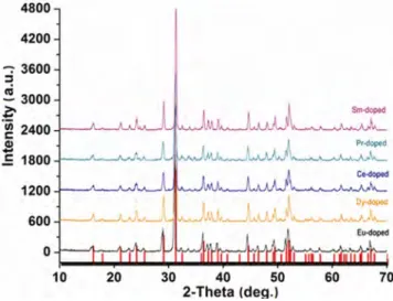
(3) INDIAN J CHEM, SEC A, NOVEMBER 2015. 1396. Fig. 2—(a) SEM image and (b) EDX data of Eu-doped Ca2MgSi2O7 phosphor ground and sieved under 170 mesh.. sharp drop of weight corresponds to the endothermic peak at 385 °C. The second endothermic peak appearing in the range 430–800 °C corresponds to the decomposition of magnesium hydroxide and regulation of the Mg(OH)2 structure to that of pure MgO, accompanied by the loss of structural water. This rapid mass decrease corresponding to the endothermic effect was indicated in the TG results6. The second stage weight loss 510–790 °C is also related to the decomposition of CaCO3 which occurs in a single step in a defined way due to carbon dioxide release, given rise to calcium oxide: CaCO3 (s) → CO2 (g) + CaO(s) corresponding to the endothermic peak at 765 °C in its DTA curve7. The exothermal peak at 884 °C is believed to be the crystallization temperature for the first formation of Ca2MgSi2O7 in the solid state-assisted process; also the precursor continues to crystallize at 1224 °C as indicated by a second clear exothermal peak. The TG curve exhibits a total mass loss of 26.50%, which is almost similar the calculated mass loss (26.48%) attributed to the complete dehydroxylation process of Mg(OH)2 and CaCO3. Particle size and characterization. After calcination at optimum temperature of 1300 °C, for 2 h to get the dominant Ca2MgSi2O7 phase, the samples were dry-ground and sieved under 170 mesh for characterization, e.g. SEM, XRD and PL. The comparative particle size distribution of Ca2MgSi2O7 based phosphor samples, into which the rare earths ions were separately doped, were analyzed by laser particle size analysis. The particle size distribution of the Ca2MgSi2O7 based phosphor samples was found to be d(0.1): 2.535 mm, d(0.5): 15.390 mm and d(0.9): 63.256 mm.. Fig. 3—XRD patterns of Ca2MgSi2O7 phosphors activated with different rare earth ions and heated at 1300 °C for 2 h.. SEM and particle size analysis results show that the grains are irregular in shape with a average particle size of ~15 µm. SEM and EDX of Eu-doped Ca2MgSi2O7 based phosphor samples are shown in Fig. 2 as representative. For the Dy, Ce, Pr and Sm-doped Ca2MgSi2O7 phosphors, see Figs S1-S4 (Supplementary Data). EDX analysis showed that all the Ca2MgSi2O7 based phosphors have mainly Ca, Mg and Si and small amounts of the rare earth elements Eu, Dy, Ce, Pr and Sm (Fig. 2(b) and Supplementary Data, Figs S1 - S4 (b)). X-ray diffraction (XRD) analysis. Figure 3 depicts that the XRD patterns of the samples indicating the major peaks which can be indexed to Ca2MgSi2O7 in a tetragonal structure (JCPDS 87-0052). This single-phase sample is well crystallized at 1300 °C for 2 h. Since no impurity phase was.
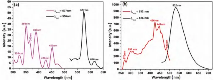
(4) KARACAOGLU & KARASU: LUMINESCENCE PROPERTIES OF ÅKERMANITE TYPE PHOSPHORS. 1397. Fig. 4—Excitation and emission spectra of Ca2MgSi2O7:0.006Eu3+/Eu2+ synthesized in (a) open atmosphere and (b) weak reducing atmosphere.. observed in any of the samples, it clearly implies that the obtained samples are single phase and doping does not cause any significant change in the host structure or lattice distortions. Photoluminescence properties. The excitation and emission spectra of Eu2O3 added Ca2MgSi2O7 phosphors, calcined in open atmosphere as well as weak reducing atmosphere are presented in Fig. 4. Eu2O3 added Ca2MgSi2O7 phosphor calcined in weak reducing atmosphere has an emission band being at 532 nm in yellowish-green color region (Fig. 4(b)). The relative strong intensity of Eu2+ observed in its emission spectrum is due to the 4f 65d 1→4f 7 transition of the Eu2+ ion because of the reducing atmosphere calcination conditions as reported in recent studies2,3. Divalent europium ion (Eu2+) is one of a well known activator in phosphors for application in modern lighting, displays, and optical communications fields such as light-emitting diodes (LEDs), long-lasting phosphors (LLPs), fluorescent lamps and plasma display panels (PDPs)9-12. According to the structural studies, there is only one eightcoordinated Ca2+ site in the tetragonal Ca2MgSi2O7 lattice13. It was predicted that Eu2+ and Mn2+ tend to substitute the Ca2+ and Mg2+ sites, respectively, because the ionic radii of Eu2+ (1.25 Å) and Mn2+ (0.66 Å) are compatible with those of Ca2+ (1.12 Å) and Mg2+ (0.57 Å). Also, the Si4+ site (0.26 Å) is too small for the Eu2+ and Mn2+ occupation to take place. When Eu replaces Ca in the Ca2MgSi2O7 phosphor, it will occupy two different kinds of lattice sites (Eu1 and Eu2) with coordination numbers of six or eight14.. In the emission and excitation spectra of Eu3+doped, or in other words, Eu2O3-activated Ca2MgSi2O7 calcined in oxidized atmosphere measured at room temperature (Fig. 5(b)), the following emission transitions were observed: 5D0→7F1 at 577 nm and 5D0→7F2 at 616 nm (611–620 nm), of which the 5D0→7F1 transition is stronger. Eu3+ ions emit a characteristic red light with several narrow lines due to the 4f→4f (5D0→7Fi = 0,1,2,3,4) transitions. The luminescence spectrum of Eu3+ ion is slightly influenced by the surrounding ligands of the host material because the transitions of Eu3+ involve only a redistribution of electrons within the inner 4f sub-shell14,15. The excitation spectra of these phosphor samples show that they can be well excited by near UV-light which is exactly as required by ultra-violet chip pumped multi-phosphor converted white LEDs. Dy3+ ions have been often used as co-dopant in long afterglow phosphorescent pigments. When divalent alkaline earth ions, such as Ca2+ or Sr2+, are substituted by trivalent Dy3+ in the alkaline earth silicates and aluminates, various defects can be induced due to the charge compensation mechanism, among which alkaline earth ion vacancy (VM), is the principal one. VM is considered to be the hole trap, while Dy3+ occupying alkaline earth ion sites is a very probable source of electron trap. In the Eu2+ activated and the Dy3+ co-doped long afterglow phosphors, such as SrAl2O4: Eu2+, Dy3+, Y3+ or Sr4Al14O25: Eu2+, Dy3+, most of the excitation energy will be transferred from the host or the Dy3+ to the Eu2+. Thus, only 5d-4f emissions of Eu2+ can be observed9-12. However, in the current study it is thought that in Dy3+ singly.
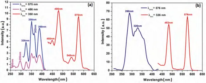
(5) 1398. INDIAN J CHEM, SEC A, NOVEMBER 2015. Fig. 5—Excitation and emission spectra of (a) Ca2MgSi2O7:Dy3+ and (b) Ca2MgSi2O7:Dy3+, Ce3+ (right) synthesized in weak reducing atmosphere.. doped hosts, Dy3+ may be not only the supplier of traps but also an activator itself, as mentioned previously in the literature5. The excitation and the emission spectra of the only Dy3+ doped and also Dy3+-doped with Ce3+-co-doped Ca2MgSi2O7 samples are given in Fig. 5. Dy3+ ions, which show luminescence lines in the 470–500 nm region due to 4F9/2-6H15/2 transition and in the 570–600 nm region due to the 4F9/2-6H13/2 transition, have attracted much attention because of the white emission in the visible region of electromagnetic spectra. For the Dy3+ single-doped Ca2MgSi2O7:Dy3+ phosphor (Fig.5(a)), the excitation spectra for the emission at both 575 nm and 486 nm comprise a series of line spectra in the 250–430 nm region with the strongest line at 350 nm and some lines at 266, 296, 326, 362 and 389 nm, which are ascribed to the transitions from the ground state to excited states in the 4f 9 configuration of Dy3+; however these are not clearly assigned due to the dense and somewhat overlapping levels of 4f configuration of Dy3+ in the high energy region. The blue emission peaking at 486 and yellow emission peaking at 575 nm were observed and may be assigned to the 4F9/2-6H15/2 and 4 F9/2-6H13/2 transitions of Dy3+, respectively16. Both the excitation and the emission spectra of Dy3+-activated and Ce3+ co-doped Ca2MgSi2O7 samples comprise the characteristic lines of Dy3+ within 4f 9 configuration (Fig. 5(b)). The emission spectra composed of two groups of emissions peaking at 483 and 575 nm, may be assigned to the Dy3+:4f 4 4 transitions of F9/2-6H15/2 and F9/2-6H13/2. respectively5,14. The excitation spectra monitoring the 575 and 576 nm emissions consist of a series of lines in the 250–350 nm range with the strongest one at 350 and 326 nm. Also, it can be clearly seen that Ce2O3 co-doping increases the luminescence intensity of the Dy3+-doped host lattice, and requires to be investigated in detail. The after glow process in the CaxMgSi2O5+ x:Dy3+ (x: 1, 2, 3) system has been described earlier by Chen et al.5 According to this study, after irradiation with the ultraviolet light, most of the excitation energy associated with the excited carriers (electrons or holes) will be transferred via the host directly to the luminescence centers, Dy3+, followed by the Dy3+ 4f emissions as the immediate luminescence. However, part of the excitation energy will be stored when some of the excited carriers drop into the traps, instead of returning to the ground state. Later, with thermal excitation at proper temperature, these carriers will be released from the traps and transferred via the host to the Dy3+ ions, followed by the characteristic and persistent Dy3+ emissions. In practise, the electron traps and the hole traps may not be both equally abundant or important in terms of their contribution to the long afterglow emission5. The excitation spectra (λexc: 341 nm and 347 nm) of Ce3+-doped Ca2MgSi2O7 host, calcined in open and weak reducing atmosphere has been observed as one broad band centered at 395 and 398 nm, respectively (Fig. 6). The notable point is that main excitation and emission wavelengths are similar for synthesis in.
(6) KARACAOGLU & KARASU: LUMINESCENCE PROPERTIES OF ÅKERMANITE TYPE PHOSPHORS. 1399. Fig. 6—Excitation and emission spectra of Ca2MgSi2O7:0.006Ce3+ synthesized in (a) open atmosphere and (b) weak reducing atmosphere.. both open and weak reducing atmospheres, but the Ce-activated phosphor synthesized in weak reducing atmosphere shows a broad blue asymmetric emission band at higher intensity, in addition to white light emission under UV-A excitation. The asymmetric band could be excited by the spin orbit coupled into two levels of ground state. The asymmetry peak can be deconvolved into two Gaussian profiles centering at ~390 and ~430 nm, which may be assigned to the transition from the lowest 5d level to 2F5/2 and 2F7/2, respectively. The energy difference between the spin-orbit splitting of the 4f ground state (ΔE = 1/λ1 − 1/λ2) in Ca2MgSi2O7:Ce3+ is about 2254 cm−1, which agrees with the theoretical value (2000 cm−1)17. Ce3+ doping in some hosts results in emission spectra near the ultraviolet region. The luminescent colours or wavelengths of these ions change widely from near UV to red region depending on the nature of the host lattices. Alkaline earth silicates, such as Ce3+ activated Ca3MgSi2O8 phosphor, gave rise to an efficient purple-blue emission under UV excitation and generated two kinds of emission centers in the host14. Also, the long lasting phosphorescence was observed in Ce3+ doped Ca2Al2SiO7 at room temperature18. Ce3+ ion in åkermanite calcined in reducing atmosphere, which is in the 4f 1 configuration, showed efficient white luminescence owing to the 4f–5d transition. The 4f–5d transition of Ce3+ ions in solids is the parity allowed electric dipole transition having large oscillator strengths; such transitions produce efficient broad band luminescence. It has a larger Stokes shift than that of other rare earth ions due to the extended. radial wave functions of the 5d state. Because of the favourable spectroscopic properties of Ce3+ and the ability to incorporate Ce3+ into different host materials, cerium activated materials have received renewed interest18. Generally, lanthanides exhibit the 3+ oxidation state, since they lose two 6s electron and one 5d(4f ) electron to attain the lowest ionization energy. However, due to the presence of either half filled or completely filled or empty 4f sub shell, Ce, Pr, Nd, Tb, and Dy can also present the 4+ oxidation state, i.e. Ce4+ (4f 0), Pr4+ (4f 1), Nd4+ (4f 2), Tb4+ (4f 7), and Dy4+ (4f 8)19. Praseodymium oxides have attracted in the recent years because of their optical, electronic, and chemical properties. The PrOx system has a similar series having the general formula, PrnO2n−2 with n= 4, 5, 6, 7, 9, 10, 11, 12, ∞. The Pr3+ oxide phase, i.e., Pr2O3, has also a hexagonal structure. This structure is characteristic for stoichiometric Pr4+ oxide (α-PrO2). The phases intermediate between Pr3+ and Pr4+ oxides (β-Pr6O11, δ-Pr11O20, ε-Pr5O9, ξ -Pr9O16, ι-Pr7O12, Pr6O10 and Pr5O8) are classified into the oxygendeficient modifications of the fluorite structure. Since the most stable phase of the neighboring rare earth oxide (REO) praseodymia is Pr6O11, exhibiting a mixed valence state (Pr4+/Pr3+) and the highest oxygen exchange activity among all rare earth oxides at atmospheric pressure and room temperature20, Pr6O11 has been used as the source of Pr3+ or Pr4+-ions in the present study. The excitation and emission spectra of Pr6O11doped Ca2MgSi2O7 phosphor which is calcined under open atmosphere is presented in Fig. 7(a). There are.
(7) INDIAN J CHEM, SEC A, NOVEMBER 2015. 1400. Fig. 7—Excitation and emission spectra of Ca2MgSi2O7:0.006Pr3+/Pr4+ synthesized in (a) open atmosphere and (b) weak reducing atmosphere.. three excitation peaks at 447 nm (3H4-3P2), 468 nm (3H4-3P1) and 483 nm (3H4-3P0) and the strongest peak is at 447 nm. Significantly, the sharp emission peak at 600 nm and the half peak which is joined to the main peak can be seen in Fig. 7 (b). The main emission peak corresponds to 1D2-3H4 transition of Pr3+. In the Ca2MgSi2O7 crystal lattice, the praseodymium ions are substituted for the Ca2+ ions, both as trivalent (Pr3+) and tetravalent (Pr4+) states and the red color is exclusively caused by the Pr4+ ions since the calcination atmosphere is appropriate to maintain Pr4+ ions in Pr6O11. Pr6O11-added, or more clearly Pr3+-doped phosphors with full color luminescence have received substantial interest because the Pr3+ ions exhibit different emissions according to the host lattice in which they are incorporated, e.g., red (from the 1D2 level), green (from the 3P0 level), blue (from the 1S0 level) and ultraviolet (from the 4f–5d state)21. The phosphorescence of 640 nm (red) and 553 nm (green) emission in the sample synthesized in weak reducing atmosphere is derived from 3P0→3H6 and 3P0→3H5 transitions of Pr3+ under 299 nm UV excitation (Fig. 7(b)). Pr6O11 (i.e. Pr3+/Pr4+) source for the present study is composed of Pr2O3 and PrO2, and the Pr6O11 doping process of can be defined by Eqs (1-3). Ca MgSi O 2 7 2 Pr V " O . Pr O. Ca. 2. Ca. Ca MgSi O. 3. 2 7 2 2 Pr V " O . Pr O 3. ... (1). 2. Ca. Ca. 2. 2. ... (2). Pr O 3. 3 Ca Mg Si O " 22 7 2 PrCa 2e' VCa O 2 2 . PrCa. ... (3). represents vacancy with two positive charge 4+. when Pr ion takes up the Ca2+ site. PrCa represents a 3+ vacancy with a positive charge when Pr ion takes up. " a Ca2+ site. VCa is the vacant site of a Ca2+ site and e′ is a quasi-free electron. Previous works have shown that RE3+ ions are randomly distributed over the same cationic sites of Ca2+ in the Ca2MgSi2O7 host6. According to KrögerVink notation, Pr4+ occupies the position of Ca2+ on PrO2 doping. Cation vacancy is generated to compensate charge for the non-equivalence susbstitution as in Eq. (1). When Pr2O3 is added to the host, the Pr3+ ions occupy the Ca2+ sites with the cation vacancies created to compensate charge as seen in Eq. (2). The PrCa vacancy may also be compensated by the quasi-free electron, which is generated from the 5d band of Pr3+ (Eq. (3))18. The presence of Pr in Ca2MgSi2O7 requires to be be investigated in detail by X-ray absorption nearedge spectroscopy (XANES) to determine valences of both 3+ and 4+ or one of them. The rare earth Sm3+ with a 4f 5 electron configuration is often regarded as an efficient activator or coactivator to enhance luminescence intensity. It has complex energy levels and various possible transitions between f levels exhibit a strong orangered fluorescence. The transitions obtained between these f levels are highly marked and the spectral lines are sharp23..
(8) KARACAOGLU & KARASU: LUMINESCENCE PROPERTIES OF ÅKERMANITE TYPE PHOSPHORS. 1401. Acknowledgement The authors thank the Small and Medium Enterprises Development Organization Projects (KOSGEB), 2011/7, Republic of Turkiye, for the financial support. References 1. 2 3 4 3+. Fig. 8—Excitation and emission spectra of Ca2MgSi2O7:0.006Sm synthesized in open atmosphere.. The excitation and emission spectra of Sm2O3 added as Sm3+ source for Ca2MgSi2O7 host synthesized in open atmosphere is shown in Fig. 8. The excitation spectra of Sm3+-doped phosphor shows eight excitation bands, i.e., 6H5/2 → 3H7/2, 6H5/2 → 4 F9/2, 6H5/2 → 4K13/2, 6H5/2 →4F7/2, 6H5/2 → (6P5/2,4P5/2), 6 H5/2 → 4G9/2, 6H5/2 → 4F5/2+4I13/2, 6H5/2 → 4I9/2 transitions at 344, 362, 372, 404, 422, 440, 467 and 485 nm respectively. The emission transitions of the same sample are positioned at 565 nm (4G5/2 → 6H5/2), 601 nm (4G5/2 → 6H7/2) and 643 nm (4G5/2 → 6H9/2), respectively15,24,25. The major orange color region emission among three emission transitions is due to the 4G5/2 → 6H7/2 transition at 601 nm. Conclusions Novel phosphors, Eu2+/3+, Dy3+, Ce3+, Pr3+/4+, Sm3+ separately-doped single-phased Ca2MgSi2O7 were synthesized by conventional solid-state reaction method under open (air) atmosphere and/or weak reducing atmosphere comprising nitrogen and hydrogen and its luminescence properties have been investigated. The Ca2MgSi2O7 host structure exhibited emission of different colors due to the transitions of each doped rare earth ions. The difference in the measured emission spectra is either due to different emission centers or same emission center with different ionic states occupying in the same host lattice. The results show that Ce3+-doping generated visible white light emission from the åkermanite type phosphor. The study shows that the rare earth ions (REI3+/4+) change the emission properties of the samples.. 5 6 7 8 9 10 11 12 13. 14 15 16 17 18 19 20 21 22 23 24 25. Birkel A, Darago L E, Morrison A, Lory A, George N C, Mikhailovsky A A, Birkel C S & Seshadri R, Solid State Sci, 14 (2012) 739. Jiang L, Chang C, Mao D & Feng C, Mater Sci Eng: B103 (2003) 271. Kyeong Y J, Kook H H, Kang Y C & Ha-Kyun J, Mater Chem Phy, 98 (2006) 330. Yi T & Dongying J, Adv Mater Sci Tech, 675-677 (2011) 1089. Chen Y, Cheng X, Liu M, Qi Z & Shi C, J Lumin, 129 (2009) 531. Lin L, Zhonghua Z, Weiping Z, Zhiqiang Z & Min Y, J R Earths, 27(5) (2009) 749. Dhaouadi H, Chaabane H & Touati F, Nano-Micro Lett, 3 (3) (2011) 153. Murakami F S, Rodrigues P O, Teixeira de Campos C M & Silva M A S, Ciênc Tecnol Aliment, 27 (2007) 3. El Kazazz H, Karacaoglu E, Karasu B & Agatekin M, J Am Sci, 7 (2012) 12. Kaya Y S, Karacaoglu E & Karasu B, Ceramic Int, 38 (2012) 3701. Kaya Y S, Karacaoglu E & Karasu B, Adv Appl Ceramic: Struct Funct Bio, 111 (2012). Karacaoglu E & Karasu B, Mater Res Bull, 48 (2013) 3702. Hölsä J, Kirm M, Laamanen T, Lastusaari M, Niittykoski J & Raud J, http://hasyweb.desy.de, Annual Reports 2006; Part 1. Jiang L, Chang C & Mao D, J Al Comp, 360 (2003) 193. Öztürk E & Karacaoglu E, J Therm Anal Cal, 119 (2015) 1063. Su Q, Liang H, Li C, He H, Lu Y, Li J & Tao Y, J Lumin, 122 (123) (2007) 927. Gong Y, Wang Y, Xu X, Li Y & Jiang Z, J Electrochem Soc, 156 (10) (2009) 295. Bhatkar V B, Omanwar S K & Moharil S V, Op Mater, 29 (2007) 1066. Zhu F, Xiao Z, Yan L, Zhang F & Huang A, Appl Phys A, 101 (2010) 689. Wang X, Yang C, Wang T M & Liu P, Electrochim Acta, 58 (2011) 193. Shih H-R, Tsai Y-Y, Liu K-T, Liao Y-Z & Chang Y-S, Opt Mater, 35(12) (2013) 2654. Yan X, PhD Thesis, (Wolfson Centre for Materials Processing Brunel Univ), 2012. Han B, Zhang J, Li P & Shi H, Mater Lett, 126 (2014) 113. Rudramadevi B H & Buddhudu D, Indian J Pure Appl Phy, 46 (2008) 825. Li Y-C, Chang Y-H, Lin Y-F, Chang Y-S & Lin Y-J, J Alloys Comp, 439 (2007) 367..
(9)
Şekil
Benzer Belgeler
In the thin section laboratory of the Çukurova University Geological Engineering Department, thin sections were prepared from the corundum and side rock samples
Araştırmada klinik önemi olan Klebsiella bakterilerinde GSBL enzim varlığı Kombine disk yöntemi (Klavulonik asit içeren kombinasyon disklerinin
Ders verirken, bizzat kendisi esa tiri bir yaratık gibi hareketlerle, sanki ders anlatmaz da, bir vol kan gibi adeta indifa ederdi.. İlk dersini hiç unutmam: Ortalıkta
Krom bileşiklerinin toksik etkilerine maruz kalmamak için çeşitli içme suyu standartlarınca içme sularına bazı sınırlamalar getirilmiş ve ağır metal ihtiva
The study describes the evaluation of the angularity characteristics of the aggregates crushed with different types of crushers, and their impact on the surface properties of
Rum, Ermeni, Çerkez ve Sefarad yemekleriyle farklı bir mönüye sahip olan Zarifi'de müzikler de müşterinin ilgisine göre her telden çalıyor.. Zarifi ortamıyla olduğu
Although stability characterizes the Turkish-Iranian relationship, especially from 2011 to 2015 the Syrian crisis had revealed the clashing strategic interests of Turkey and Iran
Ramazan ERDEM, Özlem ERDEM (2017): Elektrik Alan ile Lif Çekimi Yöntemi ile Elde Edilen Ligand Katkılı Poliüretan Nanoliflerin Morfolojik ve Lüminesans Özelliklerinin
