Recurrent renal giant leiomyosarcoma
Primary renal leiomyosarcomas are rare, aggressive tumors. They constitute 1-2% of adult malignant renal tumors. Although leiomyosarcomas are the most common histological type (50-60%) of renal sarcomas, information on renal leiomyosarcoma is limited. Local or systemic recurrences are common. The radiological appearance of renal leiomyosarcomas is not specific, therefore renal leiomyosarcoma cannot be distinguished from renal cell carcinoma by imaging methods in all patients. A 74-year-old female patient presented to our clinic complaining of a palpable mass on the right side of her abdomen in November 2012. The abdominal magnetic resonance imaging revealed a mass, 25 x 24 x 23 cm in size. Her past medical history revealed that she has undergone right radical nephrectomy in 2007, due to a 11 x 12 x 13 cm renal mass that was then reported as renal cell carcinoma on abdominal magnetic resonance imaging, but the pathological diagnosis was low-grade renal leiomyosarcoma. The most recent follow-up of the patient was in 2011, with no signs of local recurrence or distant metastases within this four-year period. The patient underwent laparotomy on November 2012, and a 35 cm retroperitoneal mass was excised. The pathological examination of the mass was reported as high-grade leiomyosarcoma. The formation of this giant retroperitoneal mass in 1 year can be explained by the transformation of the lesion’s pathology from low-grade to a high-grade tumor.
Keywords: Leiomyosarcoma, kidney, recurrence
INTRODUCTION
The incidence of primary renal sarcoma in adults is between 0.8-2.7% (1). This entity includes leiomyosar-coma (60%), liposarleiomyosar-coma (15%), hemangiopericytoma (9%), fibrosarleiomyosar-coma (7%), malignant fibrous histio-cytoma (6%), and rhabdomyosarcoma (5%) (2). Renal leiomyosarcomas are very rare and can be confused with primary renal cell carcinoma. They have a dismal prognosis, and represent 1% of all renal malignan-cies. Renal leiomyosarcoma may present with symptoms like blunt abdominal pain, abdominal distension, hematuria and oliguria that may be present in a variety of diseases. However, the tumor usually does not cause symptoms until it is too large. Therefore, the tumor is generally large at diagnosis. In this article we wanted to discuss a patient who underwent surgery twice for renal leiomyosarcoma, and who showed pathologic transformation from low-grade leiomyosarcoma to high-grade leiomyosarcoma.
CASE PRESENTATION
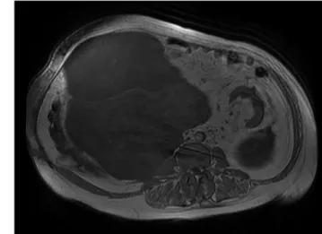
The patient to be presented is a 74-year-old female. She had undergone total abdominal hysterectomy and bilateral salpingo-oophorectomy surgery 35 years ago for myoma and right radical nephrectomy for renal leiomyosarcoma five years ago. Our clinic was called for consultation from the nephrology clinic due to nausea, vomiting, fatigue, abdominal pain and a mass palpable on the right side of her abdomen. She had diabetes mellitus, hypertension and chronic renal failure. On physical examination, she had tender-ness especially in the right quadrants, and a mass completely filling the right abdomen. The biochemical tests showed blood urea nitrogen: 77 mg/dL, creatinine: 3.8 mg/dL, white blood cell count: 11600/mm3, hemoglobin: 8.2 g/dl, sedimentation rate: 109 mm/h, and C-reactive protein: 146 mg/L. Tumor markers were within normal limits (carcinoembryonic antigen: 1.42, alpha fetoprotein: 3.2, CA19-9: 0.16, CA125: 15.7, CA15-3: 16.7, and CA72-4: 1.4 U/ml). The abdominal ultrasound revealed a 25x20x20 cm in size, heter-ogenous, partially echogenic solid mass lesion with septa, which occupied the right quadrant and the mid-line. The abdominal magnetic resonance imaging showed a mass approximately 25x24x23 cm in size that was limited by the abdominal wall in the anterior and lateral borders, by the psoas muscle and vertebrae in the posterior and medial borders, extending to the left of the midline medially, compressing the inferior vena cava, distorting the cecum and ascending colon, that begins from the pelvis and extends superiorly to the gallbladder (Figure 1, 2). The thoracic computed tomography and whole body bone scintigraphy that were performed to screen for metastases did not reveal any metastatic foci.
When the patient’s evaluations prior to the right radical nephrectomy in 2007 were reviewed, it was seen that a 10 x 12 x 13 cm in size solid mass was detected in abdominal magnetic resonance imaging at the right kidney lower pole that extended to the vena cava and compressed the right ureter, which was then 1Department of General Surgery,
Ufuk University School of Medicine, Ankara, Turkey 2Department of Pathology, Private Mikro-Pat Laboratory, Ankara, Turkey
Address for Correspondence
Salih Erpulat Öziş e-mail: [email protected] Received: 30.11.2013 Accepted: 06.04.2014 Available Online Date: 25.12.2014 ©Copyright 2016
by Turkish Surgical Association Available online at www.ulusalcerrahidergisi.org
Salih Erpulat Öziş
1, Kamil Gülpınar
1, Zafer Şahlı
1, Baha Burak Konak
1, Mete Keskin
1, Süleyman Özdemir
1, Ömür Ataoğlu
2145
ABSTRACT
Ulus Cerrahi Derg 2016; 32: 145-148
interpreted as renal cell carcinoma. The pathologic examina-tion of the right radical nephrectomy material for a prelimi-nary diagnosis of renal cell carcinoma has been reported as low-grade renal leiomyosarcoma (Figure 3). It was identified that the patient did not receive chemotherapy or radiotherapy during that period. The abdominal computed tomography in 2011 did not show an abdominal mass.
According to this information, the patient was transferred to our clinic from the nephrology clinic to be operated with a preliminary diagnosis of recurrent renal leiomyosarcoma. The patient’s pathology slides of the first operation were obtained, the pathological examination was performed again and was reported as low-grade renal leiomyosarcoma. After obtaining informed consent and completing preoperative preparations, the patient underwent laparotomy in November 2012. On exploration, a mass that filled the right half of the abdominal cavity, adhering to the ascending colon and transverse colon was identified. The mass was separated from surrounding tis-sues and the retroperitoneal mass excision was completed en-bloc with sharp dissection over the regular-bordered cap-sule. On macroscopic appearance, the mass appeared yellow-creamy colored, hard in consistency, and contained necrotic areas (Figure 4, 5).
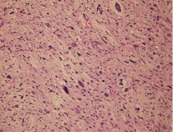
Pathologic findings revealed a high-grade leiomyosarcoma with atypical fusiform nuclei within scattered areas of necrosis, abundant eosinophilic cytoplasm, prominent mitotic activity, originating from smooth muscle (Figure 6). The patient was re-ferred to the medical oncology clinic. The patient was not con-sidered appropriate for chemotherapy due to her age. She was planned for radiotherapy after discharge. However, she died on the 38th postoperative day due to cardiopulmonary failure. DISCUSSION
Leiomyosarcoma is a sarcoma derived from the smooth mus-cle and is usually detected in the elderly. It constitutes 11% of all soft tissue sarcomas and 30% of all uterine sarcomas. Nearly half of all leiomyosarcomas are seen in the uterus. It can also be seen in the retroperitoneum, bones, blood vessels and the skin. In the literature, leiomyosarcoma of the thyroid gland, gallbladder, tongue, liver, bronchus, pancreas, and kidney has been reported as case reports (3).
Leiomyosarcomas are generally associated with unbalanced translocations and activating mutations in the tyrosine kinase receptor. Similarly, mutations in the p53 gene, phosphatase and tensin homolog gene (PTEN) inactivation, or activation of mTOR were detected in some subtypes (4).
146
Öziş et al.
Renal leiomyosarcoma
Figure 1. Magnetic resonance imaging view of renal leiomyosarcoma-1 Figure 3. Microscopic view of the radical nephrectomy material in 2007
Despite the histological resemblance to each other, these tumors vary clinically according to anatomic area. These dif-ferences also make diagnosis difficult. Ultrasound, computed tomography, magnetic resonance imaging and positron emis-sion tomography are important to determine the location of the tumor, preoperative staging and reveal any metastasis if present, while a definitive diagnosis is established by patho-logic examination. There are no specific immunochemical markers for leiomyosarcoma. The presence of smooth muscle cell specific actin, desmin and caldesmon in the surgical ma-terial is diagnostic. Similarly, keratin, epithelial membrane an-tigen and S100 may be determined as positive in pathologic examination. The estrogen and progesterone receptors can be detected as positive in uterine leiomyosarcomas. Cellular atypia, mitotic activity, and necrosis determine the degree of differentiation (5).
Currently, the only treatment modality that could provide a cure for leiomyosarcoma is surgical resection. Adjuvant che-motherapy and radiotherapy can be applied in selected cases. The main drug in conventional chemotherapy is doxorubicin. Gemcitabine and docetaxel can be combined with that drug (6). Conventional chemotherapy is usually suggested for pal-liation. Gene therapy has been used in selected patients in a limited number of studies (7).
Anatomic location is important for prognosis. Retroperitoneal tumors or those of vascular origin have a poor prognosis while cutaneous leiomyosarcoma have the best prognosis. Also, one of the most important prognostic factors is tumor differentia-tion (8).
Renal leiomyosarcomas constitute 1% of malignant renal cancers in adults. There is no difference in the incidence be-tween men and women, and it is usually detected in the 6th de-cade. Renal leiomyosarcoma most commonly originates from smooth muscle cells of the renal veins. More rarely, they are derived from the renal parenchyma, renal pelvis, renal hilum and renal capsule smooth muscle fibers (2, 9). A few reports of renal leiomyosarcoma originating from the renal hilus and renal capsule have been reported in the literature. In our case, the 13 cm mass that has been removed in 2007 involved the renal parenchyma and thus avoided determination of the par-ticular renal origin.
It causes non-specific symptoms such as abdominal pain, a palpable mass in the abdomen, weight loss, hematuria and oliguria that can mimic renal cell carcinoma. Symptoms usu-ally do not occur until the tumor reaches an advanced size. Preoperative imaging methods are important to determine the location of the tumor, its relation with surrounding tis-sues, and reveal any metastasis if present, while a definitive diagnosis is only established by pathologic examination of the surgical specimen. However, for a definitive preoperative diagnosis, ultrasound-guided fine needle aspiration cytology and ureteroscopic biopsy can be used. On computed tomog-raphy and magnetic resonance imaging, they appear as het-erogenous lesions with irregular borders, with seldom cystic components without calcification or fat tissue. They can result in compression or shift in surrounding organs according to their size. However, imaging methods can not distinguish leio-myosarcoma from renal cell carcinoma. In our case, the lesion has been reported as renal cell carcinoma in the abdominal magnetic resonance imaging performed in 2007. The diag-nosis, however, has been made by postoperative pathologic examination.
On pathology, presence of classic spindle cells with hyper-chromatic nuclei that show varying degrees of mitotic activity is diagnostic for renal leiomyosarcoma. The curative treatment for renal leiomyosarcoma is surgery. The role of chemotherapy and radiotherapy is limited. Although complete surgical resec-tion with at least 3 cm margin of safety is the treatment opresec-tion, it is rarely applied due to local invasion (10). This is usually not possible due to invasion of surrounding organs since the le-sion is very large by the time of diagnosis. The most important prognostic factor is a tumor-free surgical field. The large size of the lesion and invasion to surrounding organs worsen the outcome.
Miyajama et al. (11) reported that age, sex, tumor size (stage), mitotic activity, necrosis intensity (grade) are important for prognosis. While the five-year survival is 77% in tumors smaller than 5cm, this rate decreases to 41% when the tumor is larger than 5 cm. In the presented case, the approximately five-year survival can be explained by low mitotic activity, absence of areas of necrosis, and the low pathologic grade of the leiomyo-sarcoma. The development of a 35-cm mass in about a year
147
Ulus Cerrahi Derg 2016; 32: 145-148
Figure 5. Postoperative view of the 35 cm lesion
Figure 6. Microscopic view of the retroperitoneal mass excision material in 2012
after no intraabdominal lesion was detected in the abdominal computed tomography obtained in 2011 can be explained by the transformation of the pathologically low grade leiomyo-sarcoma to a high grade leiomyoleiomyo-sarcoma.
The leiomyosarcoma presented herein is not the largest one detected by radiologic methods in the literature, but when the ones surgically removed are taken into consideration, our case is one of the largest. The average tumor size within 13 leiomyosarcoma in an article published in 2012 from China was reported as 11.1 cm, with a maximum tumor size of 34 cm (12). In another article published in 2010 that compiled 27 cases the average tumor size was reported as 13.4 cm and the maximum tumor size as 26 cm (13).
CONCLUSION
Renal leimyosarcoma is a rare tumor that can be mistaken for renal cell carcinoma. The treatment of this rare disease is surgery. Chemotherapy and radiotherapy can be used for pal-liative purposes. The main treatment strategy should be early diagnosis of the recurrence by periodic follow-up and timely surgical excision.
Informed Consent: Written informed consent was obtained from pati-ent who participated in this case.
Peer-review: Externally peer-reviewed.
Author Contributions: Data Collection and/or Processing - S.E.Ö., K.G., Ö.A.; Literature Review - B.B.K., M.K.; Writer - S.E.Ö., Z.Ş.; Critical Review - S.E.Ö., S.Ö.
Conflict of Interest: No conflict of interest was declared by the authors. Financial Disclosure: The authors declared that this study has recei-ved no financial support.
REFERENCES
1. Vogelzang NJ, Fremgen AM, Guinan PD, Chmiel JS, Sylvester JL, Sener SF. Primary renal sarcomas in adults. A natural history and management study by the American Cancer Society, Illinois Divi-sion. Cancer 1993; 71: 804-810. [CrossRef]
2. Kavantzas N, Pavlopoulos PM, Karaitianos I, Agapitos E. Renal lei-omyosarcoma: report of three cases and rewiev of literature. Arch Ital Urol Androl 1999; 71: 307-311.
3. Ducimetiere F, Lurkin A, Ranchere-Vince D, Decouvelaere AV, Péoc’h M, Istier L, et al. Incidence of sarcoma histotypes and molecular subtypes in a prospective epidemiological study with central pathology review and molecular testing. PloS One 2011; 6: e20294. [CrossRef]
4. Gokaslan H, Turkeri L, Kavak ZN, Eren F, Sişmanoğlu A, Ilvan S, et al. Differential diagnosis of smooth muscle tumors utilizing p53, pTEN and Ki-67 expression with estrogen and progesterone re-ceptors. Gynecol Obstet Invest 2005; 59: 36-40. [CrossRef] 5. Gadducci A. Prognostic factors in uterine sarcoma. Best Pract Res
Clin Obstet Gynaecol 2011; 25: 783-795. [CrossRef]
6. Hensley ML. Update on gemcitabine and docetaxel combination therapy for primary and metastatic sarcomas. Curr Opin Oncol 2010; 22: 356-361. [CrossRef]
7. Hernando E, Charytonowicz E, Dudas ME, Menendez S, Matus-hansky I, Mills J, et al. The AKT-mTOR pathway plays a critical role in the development of leiomyosarcomas. Nat Med 2007; 13: 748-753. [CrossRef]
8. Kieffer E, Alaoui M, Piette JC, Cacoub P, Chiche L. Leiomyosarcoma of the inferior vena cava experience in 22 cases. Ann Surg 2006; 244: 289-295. [CrossRef]
9. Singh I, Joshi M, Mishra K. Capsular renal leiomyosarcoma with encasement of the inferior vena cava-Diagnosed by immunostai-ning and review of literature. Urol Ann 2010; 2: 36-38. [CrossRef] 10. Chung YG, Kang SC, Yoon SM, Han JY, Seong DH. Leiomyosarco-ma arising from the blind end of a bifid renal pelvis. Yonsei Med J 2007; 48: 557-560. [CrossRef]
11. Miyajima K, Oda Y, Oshiro Y, Tamiya S, Kinukkawa N, Masuda K. Clini-copathological prognostic factors in soft tissue leiomyosarcoma: a multivariate analysis. Histopatology 2002; 40: 353-359. [CrossRef] 12. Xiong ZB, Shi M, Kunwar A, Chen N, Zhang P, Zeng H, et al.
Pri-mary leiomyosarcoma of the kidney: a clinicopathological study of 13 cases at a high-volume institution. Sichuan Da Xue Xue Bao Yi Xue Ban 2012; 43: 86-90.
13. Miller JS, Zhou M, Brimo F, Guo CC, Epstein JI. Primary leiomyo-sarcoma of the kidney: a clinicopathologic study of 27 cases. Am J Surg Pathol 2010; 34: 238-242. [CrossRef]
Öziş et al.
Renal leiomyosarcoma