Case Report
The removal of shrapnel from deep tissue with a magnet:
a novel approach
Abstract
Penetrating injuries due to the presence of suspected foreign bodies are a common problem for the emergency department (ED). Foreign bodies should be extracted from wounds as soon as possible during the primary wound management stage. Foreign body injuries may be located in superficial and deep tissue. In addition to determining the correct location, correct diagnosis may be quite difficult. Superficial foreign bodies are easily removed, but the management of those in deep tissue can be extremely difficult and therefore requires surgical removal.
The case is here presented of the novel approach of shrapnel re-moved from deep tissue with a magnet in the ED. The correct localiza-tion of the foreign bodies was determined, and the patient was successfully treated with removal by magnet.
To the best of our knowledge, there have been no previous studies or case reports in literature related to treatment in the ED of shrapnel re-moval from deep tissue.
Penetrating injuries due to the presence of suspected foreign bodies are a common problem for the emergency department (ED). Approxi-mately one-third of all foreign bodies are missed in thefirst primary sur-vey[1]. With such a high incidence, retained foreign bodies constitute 14% of medical lawsuits and 5% of legal settlements[2]. Foreign bodies come in many different shapes and sizes but are typically splinters or fragments of metal, wood, and glass[3]. Metallic foreign bodies can be determined and localized by radiography, computed tomography (CT), sonography, or electromagnetic metal detector[4].
The magnet used in the case presented in this article was a gravity-powered, long, thin device, which is a tool used in industry to prevent screws or other loose metal parts falling into a machine.
A 30-year-old man was admitted to the ED after having been injured in a barrel bomb explosion in the Syrian war. The patient had com-plaints of pain in the left lower extremity. In the ED examination, vital signs were normal, and there was no medical history of disease, opera-tion, medical allergies, or trauma. The patient was fully conscious, alert, and answering questions appropriately. The physical examination re-vealed only a wound in the left lateral 1/3 of the lower femoral area, which had been sutured and stapled by a paramedic in the ambulance. The patient had a strong femoral and left dorsalis pedis arterial pulse.
Intravenous antibiotics, saline, and intramuscular tetanus toxoid were administered for empirical coverage. The wound on the distal left femur was 5 × 1.5 cm in size, and the metal staple sutures which had been applied were opened before x-rays were taken. The wound extended to a depth of 7.5 cm subcutaneously. The left lower extremity
neurovascular examination result was normal. Complete blood count and serum blood markers showed only leukocytosis (18 × 103/L) with
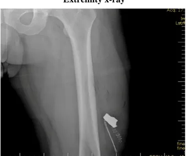
a left shift. To visualize the potential complications, radiography was ap-plied to the extremity. The x-ray examination showed a foreign body with intact femoral bone (Fig. 1). The vascular Doppler ultrasound ex-amination result was normal. The shrapnel fragment could not be re-moved with standard surgical instruments. The use of magnets is known to be useful in the localization of a foreign body. An industrial magnet with reinforced magnetic property was used for the procedure. The magnet was wrapped in a sterile glove and pushed 7.5 cm into the wound. The shrapnel (4 × 2 × 0.5 cm) stuck to the magnet, and the mag-net together with the attached shrapnel was immediately withdrawn from the wound (Fig. 2). This process was completed in only 1 minute. When the procedure was completed and the vascular Doppler ultrasound and neurovascular examination results were normal, the wound was closed. The patient was discharged after 2 hours with a prescription.
The presence of subcutaneous foreign bodies presents many challenges. Clinical diagnosis of the presence of foreign bodies may not be easy at the initial examination. Foreign bodies are usually dirty and carry multiple microorganisms, and a retained foreign body can cause high levels of pain[5]. Previous reports in literature have focused on infection associated with retained foreign bodies in the extremities[1]. Therefore, rapid removal and antibiotic therapy are essential.
Determining the location of the foreign object is the most important part of the treatment. Plain radiography does not provide enough information about the right localization. Deeply embedded foreign bodies are best visualized with CT imaging. If a CT scan does not reveal a suspected fragment, magnetic resonance imaging should be applied[6,7]. Although radiation exposure for the patient is much higher in a CT scan, magnetic resonance imaging is not used for metallic foreign bodies. In the current case, the presence of the metallic body was determined on x-ray, but accurate localization was made with the magnet.
The long, reinforced, industrial magnet, wrapped in sterile gloves, was inserted into the wound without the need for extensive dissection. With this approach, the metallic object was removed quickly from the deep tissue. This could be a beneficial treatment option as the use of a magnet is not only rapid and cost-effective but also the need for anes-thetic agents, surgical requirements, the size of incision, pain, risk of in-fection, and development of scar tissue are all reduced. In the light of this information, the use of a magnet might be a good selection for me-tallic foreign body removal from deep tissue in the ED.
American Journal of Emergency Medicine 35 (2017) 194.e5–194.e6
0735-6757/© 2016 Elsevier Inc. All rights reserved.
Contents lists available atScienceDirect
American Journal of Emergency Medicine
j o u r n a l h o m e p a g e :w w w . e l s e v i e r . c o m / l o c a t e / a j e mIn conclusion, this case demonstrates that the use of a magnet could be an effective choice for both diagnosis and treatment of patients with metallic foreign bodies embedded in deep tissue. As the magnet is easy to use and provides rapid diagnosis, thereby allowing early treat-ment, it should be considered in the ED before preparations are made for surgery.
Mustafa Bolatkale, MD Department of Emergency Medicine Medipol University Hospital, Istanbul, Turkey Corresponding author. Tel.: +90 5336111123 E-mail address:[email protected] Çağdaş Can, MD Department of Emergency Medicine Merkezefendi State Hospital, Manisa, Turkey E-mail address:[email protected] Aydın Sarıhan, MD Department of Emergency Medicine Manisa State Hospital, Manisa, Turkey E-mail address:[email protected]
Ahmet Cagdas Acara, MD Department of Emergency Medicine Gaziemir State Hospital, Izmir, Turkey E-mail address:[email protected] http://dx.doi.org/10.1016/j.ajem.2016.06.100
References
[1]Anderson M, Newmeyer WL, Kilgore Jr ES. Diagnosis and treatment of retained for-eign bodies in the hand. Am J Surg 1982;144:63–5.
[2]Gui H, Yang H, Shen SG, Xu B, Zhang S, Bautista JS. Image-guided surgical navigation for removal of foreign bodies in the deep maxillofacial region. J Oral Maxillofac Surg 2013;71:1563–71.
[3]Kadish HA, Corneli HM. Removal of nasal foreign bodies in the pediatric population. Am J Emerg Med 1997;15:54–6.
[4]Veselco M, Trobec R. Intraoperative localization of retained metallic fragments in mis-sile wounds. J Trauma 2000;49:1052.
[5]Dort JC, Robertson D. Nonmetallic foreign bodies of the skull base: a diagnostic chal-lenge. J Otolaryngol 1994;24(1):69–72.
[6]Ng SY, Songra AK, Bradely PF. A new approach using intraoperative ultrasound imag-ing for the localization and removal of multiple foreign bodies in the neck. Int J Oral Maxillofac Surg 2003;32:433–6.
[7]Bradely M, Kadzombe E, Simms P, Eyes B. Percutaneous ultrasound guided extraction of non palpable soft tissue foreign bodies. Arch Emerg Med 1992;9:181–4.
Magnet-shrapnel duo
Fig. 2. Magnet-shrapnel duo.
Extremity x-ray
Fig. 1. Extremity x-ray.