Copyright c 2012 by UniversityPublications.net
THE EFFECTS OF SPRINT INTERVAL TRAINING ON
PERFORMANCE AND IONIC REGULATION DURING EXHAUSTIVE
EXERCISE IN YOUNG MEN
Aslankeser Zübeyde
Selcuk University,Turkey
Korkmaz Selcen
Erciyes University, Turkey
Özdemir Çi÷dem and Kurdak S.Sadi
Çukurova University, Turkey
Sprint Interval Training (SIT) includes high intensity intermittent forms of cycling or running repeated sprints loads. Fatigue can be defined as impairment in muscle performance. Neuro-muscular and-or ionic perturbation during fatigue have investigated before. The aim of this study was to investigate the effects of SIT on sprint performance, ionic regulation and levels of creatine kinase (CK) and lactate dehydrogenase (LDH) during exhaustive exercise in young men. Fourteen male (22.3± 3.1 year) participated the study voluntarily. Before the training program, maximal 30 m sprint time and maximum voluntary contraction force (MVC) were measured. The subjects perform the exhaustion cycle test before and after the SIT. Earlobe capillary blood lactate concentration was measured just before and after the exhaustion test. The blood serum potassium (K+), inorganic phosphate (Pi), CK and LDH were measured just
before and immediately after the exhaustion test from the antecubital vein. After the exhaustion test serum K+, Pi and lactate concentrations increased significantly (p<0.05). MVC,
sprint performance, exhaustion time and exhaustion bicycle load and lactate concentration improved significantly after the SIT (p<0.05). It was concluded that SIT improved performance in very short training time. SIT improves against the exhaustion by endurance the fatigue; ion homeostasis and lactate transport capacity.
Keywords: Sprint interval training, Exhaustive exercise, Lactate, Potassium, Inorganic
phosphate.
Introduction
Sportive performance can be modulated by the structure of a training program. Repeated Sprint Interval Training (SIT) involves repeated short to long bouts of high intensity exercise (equal or superior to maximal velocity) and interspersed with recovery periods, which contain light exercise or rest (Billat, 2001). The recovery periods are inadequate for the fully resynthesis of phosphocreatine that a major energy substrate in brief high intensity exercise (Dawson et al., 1997). The possible keys variable determining the adaptations caused by SIT are the sprint
duration, sprint intensity, recovery duration, recovery type (passive or active), the ratio of sprint to recovery time and training volume (Billat, 2001; Balsom et al., 1992).
Low volume sprint training represents a time efficient strategy to induce performance adaptations similar to high volume endurance training (Coyle, 2005). SIT causes performance increasing due to several metabolic milieu and ion homeostasis (Iaia et al., 2009; Mohr et al., 2007; McKenna et al.,1997, Cadefau, 1990).
SIT increases sprint performance and fatigue resistance, through a variety of changes in muscle metabolites, proteins and morphology (Dawson et al., 1998.). The potency of SIT to induce rapid improvement in aerobic and anaerobic metabolism has been examined in many studies before (Babak et al., 2011; Burgomaster et al., 2007; MacDougall et al., 1998). Far less is known about the effects of sprint training on acute fatigue on the already sprint trained athletes. Fatigue can be defined as impairment in muscle performance, which manifests as a reduction in force and power generation (Ditroilo et al., 2011). Fatigue may be due to peripheral muscular changes and central origin (Ditroilo et al., 2011). During fatigue, it is indicated that changes serum K+, Pi, lactate and hydrogen (H+) ion concentrations. The marked cellular K+ efflux may cause muscle membrane depolarization and reduced excitability (McKenna et al, 1997). Lactic acidosis and [H+] increasing are thought to be a major factor in such metabolic fatigue (Posterino&Fryer, 2000). Pi, the end product of the keratin phosphate breakdown, may depress contraction function (Westerblad et al., 2002).
Plasma LDH and CK activity show different behavior before and after physical exercise, depending on different protocols, intensity and levels of training (Brancaccio et al., 2008). When the exercise loading exceeds a certain limit of muscle ability such as exhausting loads, CK leaks into the interstitial fluid, is taken up by the lymphatic system, and returned into the circulation. Efflux of intramuscular enzyme CK and LDH into the blood is assessed as an indicator of the muscle fatigue and damage (Brancaccio et al., 2008; Raimondi et al., 1975). After prolonged exercise such as a marathon, the levels of LDH double and remain elevated for days (Kobayashi et al., 2005).
Training loads purpose to endurance against fatigue or keep performance at high level. Training at high intensity work rates such as sprint training will provide a large adaptive stimulus to regulatory mechanisms controlling ionic homeostasis. Such training- might potentially decrease the rate of fatigue during exercise (McKenna et al., 1997).
In summary the purpose of the present study was to examine the changes in maximal performance during fatigue test and how would change performance after sprint interval training. This study investigated the effects of sprint interval training on performance and metabolic and ionic perturbations during exhaustive test.
Materials and Methods
Experimental design and participants
Fourteen healthy male volunteers ranging 22.3± 3.1 yr, with an average height of 177.2± 7.1 cm and a mean weight of 71.5± 6.2 kg, participated in the study. The subjects were all university level athletes from various team sport backgrounds competing and training at least once per week. The subjects were informed about the experimental procedures and any risks and discomfort associated with the experiments before they gave their informed consent. The Cukurova University Ethics Committee approved the study. The subjects were physically
active without engaging in any training and they did not regular physical activity or exercise during the experimental period.
Preliminary measurements Sprint test
Subjects were familiarized with sprint running until fully confident of producing an all-out effort from a stationary start. The sprint test was conducted on an indoor track. Infrared light sensors connected to a timing device were placed at 30 m distance. The subjects completed two maximally sprint over 30 m, which were separated by 60 seconds of recovery and were recorded the best measurement.
Maximum voluntary contraction
Maximum voluntary contraction force (MVC) was measured in the right knee extensor muscles using an isokinetic dynamometer (Cybex, Norm 6000). The subjects sat with a hip angle of 900 in an experimental chair. The knee joint was aligned with the dynamometer’s axis of rotation and all subject’s knee ROM were 900(complete knee extension was 00). The MVC test was measured during right knee extension, and was maintained 30 sec. Standardized verbal instructions and encouragements were provided throughout the protocol. The best peak MVC value was used in the data analysis.
Lactate measurement
Blood samples taken from the ear lobe were analyzed enzimatically to determine blood lactate concentration (YSI 1500 lactate analyzer). Measurements were performed at rest, after the exhaustion test within five minutes.
Exhaustion exercise
Subjects were requested to refrain from heavy exercise or traveling across several time zones a couple of days prior to testing. It was also instructed to avoid alcohol at least 24 h and heavy meal at least 2 h before the test. Acute infection (cold, sore throat etc.) postponed the test. Before the test, rest level of blood lactate was measured by earlobe capillary samples. Blood ion and enzyme levels measured from antecubital vein. After rest blood measures, subjects warmed up 5 min on electronic bicycle (Monark 839 Electronic Ergometer; Monark Exercise Sweden) with 60 W- 60 rpm.
Then, the subjects started the exhaustive exercise which was a step incremental exercise protocol begun with 60 W / 80 rpm. The test consisted of stages included 3 minutes loading. After every stage, 3 minutes passive recovery was applied on the cycle. The cycle load was increased every minute and start of the every stage (80 rpm invariable). The subjects continued exercising until exhaustion (e.g. a drop in the cycling cadence under 60 rpm despite verbal encouragement, volitional fatigue). During the test, heart rate (HR) was monitored continuously using a wireless HR monitor (S610i, Polar, Finland).
Blood analyses
Blood samples (10 ml) were collected from the antecubital vein before exhaustion test and within 5 minutes after exhaustion for determining the levels of serum K+, Pø, CK and LDH. Serum CK and LDH were measured with colorimetric assay procedure measured with electrochemiluminescence immunoassay (Elecsys E-170, Roche, Mannheim, Germany). All blood sampling process were performed at the same time of the day. Serum K+ and Pi ion concentrations were analyzed with the ion selective method (DPP).
Training
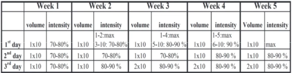
The subjects were trained 3 days per week for 5 weeks. In each week, there was at least 24 h resting period between 1st and 2nd sessions and at least 48 h between 2nd and 3rd training sessions. One set was composed of 10 repetitions.
All training was performed on a 60-m indoor track. Before the training sessions, the subjects were obligated to complete a standardized 15 min warm-up period, involving light jogging, stretching, faster strides and change of direction activities. Training sessions were consisted of repeated 30-m sprints that were interjected by low-intensity jogs (30 ± 5 seconds), in which after the sprinting subject was ought to jog around a cone and return to the starting point. One set consists of repeated 10×30-m sprints and each sets interspersed with 5-minute active recovery. After a standing start, the subjects’ 30 meters sprint times were recorded with infrared light sensors connected to a timing device. The subjects’ jog time were controlled by chronometer. A cone was placed in the 25th meter and in this way, the subjects had chance to decelerate their speed. In each training the maximal 30 m sprint performance was measeured for monitoring the improvement.
Statistical Analysis
The values are reported as means ± standard error mean (SEM). Statistical significance was accepted at p<0.05. Within group, the pre-training and post-training values were compared using paired t-test for MVC, sprint time and lactate. A two-way repeated-measure ANOVA was used to compare the blood ion and enzyme changes, with time serving as the within-group (exhaustion X training). Post hoc analyses were carried out using the Bonferroni test.
Results
After training period the mean MVC force increased significantly (p<0.001). Training also caused significant improving the sprint performance (p<0.001).
• There was no significant change in rest lactate values between pre and post training levels (p>0.05). After the exhaustion exercise blood lactate values were higher than rest values both before and after training (p<0.001). Moreover, after the training, exhaustion blood lactate values were higher than before training exhaustion lactate concentration (p<0.005).
SIT caused the improvement in exhaustion time significantly (p<0.005). Training also caused an increase in the exhaustion load significantly (p<0.01). Pre and post training exhaustion HR values were not significant statistically (p>0.05).
Serum [LDH] was shown in figure 1. There was no significant effect of training on LDH levels after the exhaustion exercise (F=2.59, P>0.05). Acute exhaustion exercise increased serum [LDH] before (F=49.89, p<0.005) and after (F=49.89, p<0.001) training. Changes in serum [CK] are shown in figure 2. There was no significant change of the training on rest or exhaustion values (F=0.08, p>0.05). Acute exhaustion exercise increased serum [CK] before and after training. These values were significant statistically compared to rest values before and after training (F=27.74, p<0.05). There was no significant change of the training on rest or exhaustion serum K+ levels (F=0.1, p>0.05). Exhaustion exercise increased blood [K+] concentration significantly before and after training (F= 13.77, p<0.05). There was no significant effect of training on Pi levels after the exhaustion exercise (F=1.9, p>0.05). Acute exhaustion exercise increased serum Pi levels before and after training (F=9.22, p<0.05).Exhaustion exercise increased serum K+ and Pi concentration significantly (p<0.05).
Discussion
• The main finding of this study indicates that the SIT had a marked improvement 30 m sprint performance, maximal voluntary contraction, endurance to exhaustion test despite very short total training duration. In one training day, active sprint load time nearly three minutes maximum. This training duration is remarkable for enhancing of sportive performance. In this study after the SIT program, sprint performance increased 6 %, exhaustion maximal lactate increased 20.3 %, exhaustion time increased 12.7 %, exhaustion load increased 8.1 %, MVC increased 11.1 %. The increase performance against to exhaustion test may be due to reduced local fatigue in the contracting muscles. There has been much investigation on low volume sprint training to induce performance adaptations similar to high volume endurance training (TØnnessen et al., 2011; Coyle., 2005; Iaia et al., 2009; Mohr et al., 2007; McKenna et al., 1997, Cadefau., 1990).
Lactate
• After the SIT, exhaustion time and exhaustion load increased, accompanied by greater blood lactate. Higher blood lactate concentration with exhaustive exercise suggests that sprint training may enhance blood flow and/ or lactate transport capacity (Pilegaard et al., 1999). Some previous studies have investigated unchanged (Stathis et al., 1994) or lower (Harmer et al., 2000) muscle lactate accumulation during exhaustive exercise after sprint training. Training volume, intensity, rest time and exercise are differential factors. The exhaustion exercise in this study is high intensity (80 rpm, 93 % HRmax) and more anaerobic.
• The process of performance increasing may have associated with anaerobic enzymes activity increasing. In fact, high intensity training increases lactate and H+ bearing capacity (McKenna et al., 1997). In numerous studies, it has been stated that this type of overloading training increases anaerobic metabolism (Harmer et al., 2000; Iaia et al., 2009; Farzad et al.,2011) and lactate tolerance (McKenna et al., 1997; Pilegaard et al., 1999,). The adaptive response to sprint training is the increasing of the muscle buffer capacity (Gibala et al., 2006; Edge et al., 2006)
After anaerobic training, higher concentrations of blood lactate may occur during maximal exercise and likely is due to higher stores of intramuscular glycogen and glycolytic enzymes (Sharp et al., 1986). An improvement in buffer capacity should have resulted in higher lactate concentrations and more work during the fatigue test and longer exhaustion time (Mohr et al., 2007). In a study, where sprint training has been performed for 7 weeks (McDougall et al., 1998), aerobic and anaerobic enzyme activities have significantly increased.
Serum lactate levels were higher as exhaustion after the training. However, the respondents have ensured this by performing more. This situation makes us think that more anaerobic end products and improved lactate tolerance (McKenna et al.1997; Pilegaard et al., 1999). As a matter of fact, high intensity training increases lactate and H+ bearing capacity (Juel, 2004; McKenna et al., 1997).
Serum ions and enzymes
We demonstrated that serum [K+] concentration increases during intense exercise. Serum [K+] increasing and Na+-K+ ATPase activity reduction during fatiguing exercise have been reported
before (Fraser et al., 2002; McKenna et al., 2008). Serum [K+] increases as a result of a shift in water from the extracellular to the intracellular compartment; due to lactate increase influencing the extrusion of K+ out of the exercising muscle; increasing the action potentials during exercise and incomplete reuptake by K+ by Na+/K+ ATPase pump after each repolarization (Dolezal and Fedde,1988). In this investigation, increases in extracellular [K+] during high-intensity activity have been suggested to be an important contributor of muscle fatigue. Serum [K+] may increase due to the above factors and depression of the Na+/K+ ATPase pumps due to the peripheral changes during fatigue.
Serum [K+] increased after the exhausting test before and after SIT. However, exhaustion serum [K+] has unchanged after training in this study, despite greater work output like as demonstrated before (McKenna et al., 1997; Harmer et al., 2000). The lower ǻ [K+]/work ratio has been indicated during intermittent exercise after sprint training (McKenna et al., 1993). Broch-Lips et al (2011) have indicated that the depressing effects of elevated extracellular [K+] on the excitation-induced force production were smaller in rats that had performed 8 wk of running than in sedentary rats. SIT may induce K+ regulation include reduced K+ release from muscle, and/or enhanced K+ clearance due to the increased Na+/K+ ATPase content and/or activation (Harmer et al., 2000; Fraser et al., 2002).
Moreover, it was recently demonstrated that intermittent intensive exercise is even more efficient than high-volume exercise at lower intensities in increasing the muscle content of Na+/K+ pumps (Bangsbo et al., 2009; Nielsen et al., 2004). A high Na+-K+ pump activity protects against the depressive effect that contraction-induced disturbances in ion homeostasis and thereby may be of importance for preserving muscle force production during intense exercise (Iaia et al., 2011; Fraser et al., 2002).
In exhausting, serum [Pi] increased before and after the training. An increase in [Pi] is one of the significant findings of fatigue and may depress contraction function (Westerblad et al., 2002; Allen&, Westerblad, 2001). Pi accumulation may affect cross bridge function in fatigue (Coupland et al., 2001) especially relatively early in the contraction. In this study serum Pi levels increased before and after SIT during exhaustion test. However, exhaustion Pi values between before and after SIT are not significantly different. This may show that phosphocreatine (CrP) catabolism and re-synthesis have not changed during fatigue test against more power generation during the fatigue test.
During fatiguing exercise serum LDH and CK levels increase (Brancaccio et al., 2008). An eccentric bout of exercise induces increase of serum enzyme activity much more than concentric bout and serum enzyme values are high after 24-48 hours depending on damages (Magal et al., 2010). In our fatigue protocol, which contains bicycle test against increasing load, the LDH and CK increase in blood makes us think that there are slight muscle damages.
The study has a number of limitations. Firstly, muscle samples were not studied, muscle biopsy may elicit about muscle enzyme, ion and lactate levels, secondly Na+/ K+ pumps concentration was not studied by muscle samples.
Conclusion
High intensity sprint training can prefer performance increasing in shorter time than long duration low volume training. Many coaches can apply like this training load in a sportive season for rapid improves such as after injures, illness, or when it is necessary to make sudden changes in the training schedule. SIT increased the muscular capacity and improved muscle ion regulation during exercise. Increased muscle K+ uptake during exercise after SIT are consistent
with a greater activation of the N+-K+ ATPase pump activation and/or amount. Increasing exhaustion lactate concentrations after the training suggests that improved lactate transport capacity. In summary, in the present investigation high intensity sprint intervals running training improved sprint performance. Furthermore, during exercise continued until exhaustion, greater work and more lactate was performed after SIT, and muscle ionic disturbances were similar compared before training.
References
1. Allen DG &Westerblad H (2001). Role of phosphate and calcium stores in muscle fatigue. J Physiol 536, 657–665.
2. Babak F, Gharakhanlou, R, Agha-Alinejad, H, Curby, DG, Bayati, M, Bahraminejad, M, and Mäestu, J (2011). Physiological and performance changes from the addition of a sprint-interval program to wrestling training. J Strength Cond Res 25(9): 2392-2399.
3. Balsom PD, Seger YJ, Sjödin B, Ekblom B (1992). Maximal-Intensity intermittent exercise: Effect of recovery duration. International Journal of sports medicine, 13: 528-533.
4. Bangsbo J, Gunnarsson TP, Wendell J, Nybo L, Thomassen M (2009). Reduced volume and increased training intensity elevate muscle Na+-K+ pump alpha2-subunit expression as well as short- and long-term work capacity in humans. J Appl Physiol 107: 1771–1780.
5. Billat LV (2001). Interval training for performance: a scientific and empirical practice. Sports Med ;31(1):13-31.
6. Brancaccio P, Maffuli N, Buonauro R, Limongelli FM (2008). Serum Enzyme Monitoring in Sports
Medicine. Clin Sports Med 27:1-18.
7. Broch-Lips MB, Paoli F, Pedersen TH, Kristian Overgaard, Nielsen OB (2011). Effects of 8 wk of voluntary
unloaded wheel running on K+ tolerance and excitability of soleus muscles in rat. J Appl Physiol 111: 212– 220.
8. Burgomaster KA, Cermak NM, Phillips SM, Benton CR, Bonen A, Gibala MJ (2007). Divergent response of
metabolite transport proteins in human skeletal muscle after sprint interval training and detraining. Am J Physiol Regul Integr Comp Physiol 292: R1970–R1976.
9. Cadefau J, Casademont J, Grau JM, Fernandez J, Balaguer A, Vernet M, Cusso R, Urbano-Marquez A (1990). Biochemical and histochemical adaptation to sprint training in young athletes. Acta Physiologica Scandinavia 140(3): 341-351.
10. Coupland ME, Puchert E, Ranatunga KW (2001). Temperature dependence of active tension in mammalian
(rabbit psoas) muscle fibres:effect of inorganic phosphate. J Physiol 536:879-891.
11. Coyle EF (2005). Very intense exercise –training is extremely potent and time efficient: a reminder. J Appl Physiol 98: 1983-1984.
12. Dawson B, Fitzsimons M, Green S, Goodman C, Carey M, Cole K (1998). Changes in performance, muscle
metabolites, enzymes and fiber types after short sprint training. Europen Journal of Applied Physiology, 78: 163-169.
13. Dawson B, Goodman C, Lawrence S, Preen D, Polglaze T, Fitzsimons M et al (1997). Muscle
phosphocreatine repletion following single and repeated short sprint efforts. Scandinavian Journal of Medicine and Science in Sports, 7,206-213.
14. Ditroilo M, Watsford M, Fernández-Peña E, D'Amen G, Lucertini F, De Vito G (2011). Effects of fatigue on
muscle stiffness and intermittent sprinting during cycling. Med Sci Sports Exerc. May;43(5):837-45.
15. Doleozal-Fosha SR and Fedde MR (1988). Serum potassium during exercise in Hereford calves: influence of
physical conditioning. J Appl Physiol.65(3):1360-1366.
16. Edge J, Bishop D, Goodman C (2006). The effects of training intensity on muscle buffer capacity in fremales, Eur J Appl Physiol 96:97-105.
17. Farzad B, Gharakhanlou R, Alinejad HA, Curby D, Bayatõ M, Bahraminejad M, Maestu J (2011). Physiological and performance changes from the addition of a sprint interval program to wrestling training. J Strength Cond Res 25(9): 2392-2399.
18. Fraser SF, Li JL, Carey MF, Wang XN, Sangkabutra T, Sostaric S, Selig SE, Kjeldsen K, McKenna MJ (2002). Fatigue depresses maximal in vitro skeletal muscle Na+-K+-ATPase activity in untrained individuals, J Appl Physiol 93: 1650-1659.
19. Gibala MJ, Little PJ, van Essan M, Wilkin GP, Burgomaster KA, Safdar A, Raha S, Tarnopolsky MA (2006).
Short- term sprint interval versus traditional endurance training:similar initial adaptations in human skeletal muscle an exercise performance, J Physiol , 575: 901-911.
20. Harmer AR, McKenna MJ, Sutton JR, Snow RJ, Ruell PA, Booth J, Thompson MW, Mackay NA, Stathis CG, Crameri RM, Carey MF, Eager DM (2000). Skeletal muscle metabolic and ionic adaptations during intense exercise following sprint training in humans. J.Appl Physiol 89:1793-1803.
21. Iaia FM, Gomez JP, Thomassen M, Nordsborg NB, Hellsten Y, Bangsbo J (2011). Relationship between performance at different exercise intensities and skeletal muscle characteristics. J Appl Physiol 110: 1555– 1563.
22. Iaia FM, Hellsten Y, Nielsen JJ, Fernström M, Sahlin K, Bangsbo J (2009). Four weeks of speed endurance
training reduces energy expenditure during exercise and maintains muscle oxidative capacity despite a reduction in training volume. J Appl Physiol.106: 73-80.
23. Juel C, Klarskov C, Nielsen JJ, Krustrup P, Mohr M, Bangsbo J. Effect of high-intensity intermittent training
on Lactate and H+ release from human skeletal muscle.Am J Physiol Endocrinol Metab 2004:286:E245-E251.
24. Kobayashi Y, Takeuchi T, Hosoi T, Yoshizaki H, Loeppky Jack A (2005). Effect of a marathon run on serum
lipoproteins, creatine kinase, and lactate dehydrogenase in recreational runners. Research Quarterly for Exercise and Sport, 76(4): 450-455.
25. MacDougall JD, Hicks AL, MacDonald JR, McKelvie RS,Green HJ, Smith KM. Muscle performance and enzymatic adaptations to sprint interval training. J.Appl.Physiol.1998,84(6):2138-2142.
26. Magal M, Dumke CL, Urbiztondo ZG et al (2010) “Relationship between serum creatine kinase activity following exercise induced muscle damage and muscle fibre composition,” Journal of Sports Sciences, 28(3): 257–266.
27. McKenna MJ, Bangsbo J, Renaud JM (2008). Muscle K+, Na+, and Cl- disturbances and Na+ -K+ pump inactivation: implications for fatigue. J Appl Physiol 104: 288– 295.
28. McKenna MJ, Heigenhauser GJF, McKelvie RS, MacDougall JM, Jones NL (1997). Sprint training enhances
ionic regulation during intense exercise in men. Journal of Physiology ,501.3:687-702.
29. McKenna MJ, Schmidt TA, Hargreaves M, Cameron L, Skinner SL, Kjeldsen K (1993). Sprint training õncreases human skeletal muscle Na+-K+ ATPase concentration and improves K+ regulation, J Appl Physiol, 75(1):173-180.
30. Mohr M, Krustrup P, Nielsen JJ, Nybo L, Rasmussen MK, Juel C et al (2007). Effect of two different intense training regimens on skeletal muscle ion transport proteins and fatigue development. American Journal of Physiology: Regulatory, Integrative and Comparative Physiology, 292:R1594-R1602.
31. Nielsen JJ, Mohr M, Klarskov C, Kristensen M, Krustrup P, Juel C, Bangsbo J (2004). Effects of high-intensity intermittent training on potassium kinetics and performance in human skeletal muscle. J Physiol 554: 857–870.
32. Pilegaard H, Terzis G, Halestrap A, Huel C (1999). Distribution of the Lactate/H+ ion transporter isoforms MCT1 and MCT2 in human skeletal muscle. Am J Physiol Endocrinol Metab 276: 843-848.
33. Posterõno, GS. & Fryer, M W. (2000). Effects of high myoplasmic L-lactate concentration on E-C coupling in mammalian skeletal muscle. Journal of Applied Physiology 89, 517–528.
34. Raimondi GA., Puy RJM., Raimondi AC., Schwarz ER., Rosenberg M (1975). Effects of physical training on
enzymatic activity of human skeletal muscle. Biomedicine. 22: 496-501.
35. Sharp RL, Costill DL, Fink WJ, and King DS (1986). Effects of eight weeks of bicycle ergometer sprint training on human muscle buffer capacity. Int JSports Med 7: 13-17.
36. Stathis CG, Febbraio MA, Carey MF, Snow RJ (1994). Influence of sprint training on human skeletal muscle purine nucleotide metabolism. J Appl Physiol 76: 1802-1809.
37. TØnnessen E, Shalfawi SA, Haugen T, Enoksen E (2011). The effect of 40-m sprint training on maximum sprinting speed, repeated sprint speed endurance, vertical jump, and aerobic capacity in young elite male soccer players. J Strength Cond Res 25(9): 2364-2370.
38. Westerblad H, Allen DG, Lannergren J (2002). Muscle Fatigue: Lactic Acid or Inorganic Phosphate the Major Cause?, News Physiol Sci, 17: 21.