European Journal of Orthodontics 36 (2014) 86–92 doi:10.1093/ejo/cjt006
Advance Access publication 4 March 2013
© The Author 2013. Published by Oxford University Press on behalf of the European Orthodontic Society. All rights reserved. For permissions, please email: [email protected]
Effects of maxillary protraction for early correction of class III
malocclusion
Mevlut Celikoglu
*and Hüsamettin Oktay
***Department of Orthodontics, Faculty of Dentistry, Karadeniz Technical University, Trabzon and **Istanbul Medipol
University, Istanbul, Turkey
Correspondence to: Mevlut Celikoglu, Department of Orthodontics, Faculty of Dentistry, Karadeniz Technical University, Trabzon 61080, Turkey.E-mail: [email protected]
SUMMary This prospective study investigated the skeletal, dental, and soft-tissue effects of a mini
maxil-lary protractor appliance in class III subjects with maxilmaxil-lary retrusion and mandibular protrusion and compared these changes with those of untreated, well matched control sample with normal occlusions.
Twenty patients with class III malocclusion (mean age 11.1 ± 0.8 years) and 20 subjects with normal occlusion (mean age 10.9 ± 0.4 years) were included to this study. The class III subjects were treated with the mini maxillary protractor appliance, and the others were used as control subjects. Paired t-test and Student’s t-test were used to determine the within- and between-group differences, respectively.
In the study group, the maxilla moved forward (SNa, 2.0 degrees and a–y, 2.4 mm) (P < 0.001) with a slight rotation of palatal and occlusal planes (SN–PP, −0.8 degree and SN–OP, −0.7 degree) (P > 0.05). The mandible displaced backwards and downwards (SNB, −1.1 degrees; SND, −0.9 degree; B–y, −0.9 mm and Pog–y, −0.3 mm; P < 0.001). These movements in the maxilla and mandible caused a significant improve-ment in intermaxillary sagittal relationship (aNB, 3.0 degrees; Convexity, 6.3 degrees; Wits, 4.6 mm;
P < 0.001). The maxillary incisors moved forward (2.2 degrees) while the mandibular ones backward (−1.9
degrees). The improvement in overjet was 5.0 mm, and 66.1 per cent of this change (3.3 mm) was skeletal (a–y; 2.4 mm and B–y; −0.9 mm), and the remaining (1.7 mm) dentoalveolar (U1–Na; 0.9 mm and L1-NB; −0.8 mm). The change in Ls–E measurement was more in the study group (2.1 mm), and the difference between the groups was statistically significant (P < 0.001).
Introduction
Class III skeletal pattern is one of the most difficult maloc-clusions to diagnose and treat in orthodontics and is charac-terized by mandibular prognathism, maxillary retrognathism, retrusive mandibular dentition, protrusive maxillary denti-tion, and a combination of these components (Sanborn, 1955;
Nanda, 1980; Guyer et al., 1986; Pangrazio-Kulbersh et al., 1998; Kilic et al., 2010b). It has been reported that two-thirds of skeletal class III malocclusions are due to either maxillary retrognathism or a combination of maxillary retrognathism and mandibular prognathism (Sanborn, 1955; Nanda, 1980;
Guyer et al., 1986). The prevalence of this anomaly was found to be approximately 1–5 per cent in white populations; (Emrich et al., 1965; Thilander and Myrberg, 1973), while this prevalence was as high as 14 per cent for Asian popula-tions (Iwagaki, 1938; Irie and Nakamura, 1975).
Class III skeletal patterns often exhibit a high incidence of deficient transverse maxillary growth (Hata et al., 1987), and maxillary expansion is often the first treatment procedure. According to Proffit and Fields (1993), maxillary expansion should be applied to mobilize the maxillary sutures before maxillary protraction. Furthermore, McNamara (1987) and
Turley (1988) have recommended the use of bonded rapid maxillary expansion appliance for several days before
beginning the protraction in order to facilitate maxillary movement. Optimal treatment timing is one of the most important factors to be taken in consideration while treating skeletal class III patients with orthopedic forces (Melsen and Melsen, 1982; Proffit, 1992). Orthopedic treatment applied during prepubertal and pubertal periods can shorten the treatment time, and favourable anterior occlusion can be obtained if mandibular growth is properly controlled during and after retention period (Campbell, 1983; Chong et al., 1996). Some researchers (Proffit, 1992; Sung and Baik, 1998; Yavuz et al. 2009) reported that greater skeletal changes with the use of maxillary protraction appliances are possible in young patients. Nonetheless, some researchers (Baik, 1995; Sung and Baik, 1998; Yüksel et al., 2001) com-pared the treatment changes obtained at different ages and found no statistically significant difference.
Protraction of maxilla with a facemask is the most common treatment procedure for class III malocclusions with maxillary retrusion and mandibular protrusion. The effects of maxillary protraction have been investigated by many authors and following changes were reported: acceleration of forward growth of maxilla with a counterclockwise rotation, forward movement of maxillary dentition, retardation of mandibular growth, and backward
prospective clinical study was to investigate skeletal, den-tal, and soft tissue effects of the mini maxillary protractor in class III subjects with maxillary retrusion and mandibu-lar protrusion, and to compare these changes with those of untreated control sample with normal occlusion.
Subjects and Methods
Ethical approval for the present prospective study was obtained from the Ethical Committee of Ataturk University, Faculty of Dentistry. An informed consent was signed by parents of the subjects included to this study.
Sample size for the groups was calculated based on a sig-nificance level of 0.05 and a power of 80 per cent to detect a clinically meaningful difference of 1 mm (±0.98 mm) for the distance from point A to PM Vertical (Y) passing through ethmoid registration point and pterygo-maxillary fissure inferior. Power analysis showed that 16 subjects in each group were required. To compensate for possible dropouts during the trial, we decided to enroll more patients.
The study group was selected according to the follow-ing criteria: (1) Skeletal class III malocclusion character-ized by maxillary deficiency and/or mandibular protrusion (ANB less than 0 degrees, Wits less than −1 mm, overjet less than 0 mm); (2) Vertically normal growth pattern (SN– GoMe = 32 degrees ± 6); (3) Minimum or moderate crowd-ing (less than 5 mm) in both dental arches; (4) Late mixed or early permanent dentition. Exclusion criteria were craniofa-cial anomalies, lingual nonocclusion of posterior dentition, and previous orthodontic treatment. The patients and their parents were informed about the mini maxillary protractor appliance and study protocol. Those accepted to enroll the study were included to the study group (10 males and 10 females; mean age 11.1 ± 0.8 years).
Age and gender matched control subjects were cho-sen from the longitudinal archive of the Department of Orthodontics at Ataturk University (10 males and 10 females; mean age: 10.9 ± 0.4 years). The selection was based on presence of normal growth and development (skeletal class I; 0 degrees<ANB<4 degrees), a balanced facial appearance, class I molar relationship, minimal den-tal crowding (less than 2 mm), absence of anterior and/or posterior crossbites, and no previous orthodontic treatment.
The patients were treated by one author (MC) by means of the mini maxillary protractor appliance until 2 mm positive overjet was obtained (Figure 1). The appliance consisted of intra- and extra-oral parts. Maxillary part was a full-cover-age acrylic splint type maxillary expander with two hooks
extended to the vestibular fornix of the canines. Mandibular part composed of an acrylic plate covering all posterior teeth and a chin cup, which were connected each other by a special bow. The bow was bent from 1.2 mm stainless steel round arch wire, and a horizontal bar was soldered on it to apply protraction forces from the hooks on the maxillary expander. Extra-oral part of the appliance was a cervical headgear.
Palatal screw on the maxillary part of the appliance was activated twice a day for 5 days (total of 2 mm expansion) even if a posterior crossbite was absent. At the end of the fifth day, the protraction headgear was applied. A protrac-tion force of 450–500 g was applied bilaterally between the hooks and horizontal bar. Direction of the force was adjusted to be approximately 30 degrees downward from occlusal plane. The patients were instructed to wear their appliances for at least 20 hours per day until a 2 mm positive overjet was achieved. Edgewise fixed appliances were used for alignment of the teeth if needed.
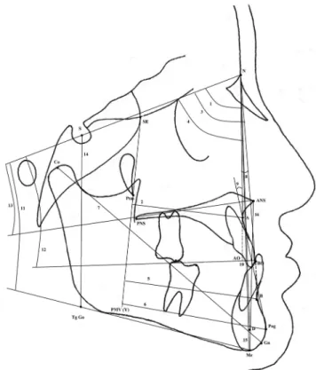
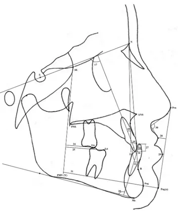
Cephalometric radiographs were obtained before (T1) and after (T2) protraction therapy in the study group. The films were taken in a standard position by one operator using the same X-ray machine and cephalostat (Siemens Nanodor 2; Siemens AG, Munich, Germany). Films of the control sub-jects were obtained from files of the longitudinal archive, which were obtained previously by the same X-ray machine and cephalostat. In order to determine skeletal, dental, and soft tissue changes, 17 linear and 14 angular measurements were used. Landmarks and measurements used in this study are shown in Figures 2–4. All measurements were made by one author (MC).
Statistical analysis
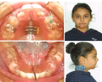
To determine the errors associated with the radiographic measurements, 15 radiographs were selected randomly. Figure 1 Intraoral and extraoral photographs of mini maxillary
protrac-tor used in the study.
Landmark identification, tracing, and measurements were repeated after a period of 1 month by the same author (MC). Intra-class correlation coefficients were performed to assess the reliability of measurements as described by Houston (1983). The coefficients of reliability for all measurements were above 0.92, confirming the measurement reliability.
Descriptive statistics of all variables were computed for T1 and T2 periods in each group. Shapiro–Wilks normality test showed a normal distribution, and thus parametric tests were used in statistical evaluations. Since there was no gender difference in both groups, the data for males and females were pooled. The initial measurements and treatment/control changes were compared between the groups by means of Student’s t-test. All statistical analyses were performed by the Statistical Package for Social Sciences (Windows 7, version 15.0, SPSS Inc., Chicago, IL, USA).
Results
Mean ages of the study and control groups were 11.1 ± 0.8 and 10.9 ± 0.4 years, respectively (P = 0.891). Mean
treatment and observation periods were 8.10 ± 2.53 and 11.95 ± 0.22 months in the study and control groups, respec-tively (P < 0.001).
Table 1 shows the results of Student’s t-test compar-ing the initial measurements of both groups. SNA, ANB, convexity, SN/OP, IMPA, and L1-NB angles and Wits appraisal, A–Y distance, overjet, overbite, and Ls–E meas-urements were significantly smaller in the study group, whereas SNB, U1–SN, and U1–NA angles and Co–Gn, B–Y, Pog–Y, U1–NA, and Pog (s)–Y measurements were significantly larger in the study group as compared with the control group.
Table 2 shows the comparison of the changes in the study and control groups. Notable changes were observed in most of the variables in the study group. Maxilla moved forward (SNA, 2.0 degrees and A–Y, 2.4 mm) (P < 0.001) in combi-nation with an insignificant rotations of palatal and occlusal planes (SN–PP, −0.8 degrees and SN–OP, −0.7 degrees) (P > 0.05). The mandible was displaced backward and downward (SNB, −1.1 degrees; SND, −0.9 degrees; B–Y, −0.9 mm and Pog–Y, −0.3 mm) (P < 0.001). Mandibular length (Co–Gn) increased 2.5 mm in the control group and 1.6 mm in the study group (P < 0.01). Combined movements of maxilla and mandible caused a signifi-cant improvement in intermaxillary sagittal relationship (ANB, 3.0 degrees; Convexity, 6.3 degrees; Wits, 4.6 mm) Figure 2 Landmarks used in the study: S, sella; N, nasion; Or,
orbit-ale; Ls, labiale superior; Li, labiale inferior; Pog(s), soft tissue pogonion; A, Point A; B, Point B; D, Point D; Pog, pogonion; Gn, Gnathion; Co, Condylon; ANS, Spina nasalis anterior; PNS, Spina nasalis posterior; Me, menton; Tg Go, Tangent gonion; Se, intersection of the greater wing of sphenoid bone with the floor of anterior cranial fossa; ptm, the most infe-rior and posteinfe-rior point on the anteinfe-rior outline of pterygomaxillary fissure; Prn, pronasale.
Figure 3 Skeletal measurements used in the study: (1) SNA; (2) A–PMV
(Y line); (3) SNB; (4) SND; (5) B–Y; (6) Pog–Y; (7) Co-Gn; (8) ANB; (9) Convexity angle; (10) Wits; (11) SN–GoMe; (12) SN–OP; (13) SN–PP; (14) S–Go; (15) N–Me; (16) ANS–Me.
(P < 0.001). In addition, significant increases were observed in SN–GoMe angle (1.5 degrees; P < 0.001) and lower face height (Study Group, 2.6 mm and Control Group, 1.5 mm) (P < 0.05).
At least 2 mm positive overjet was obtained in all of the subjects with class III malocclusion. Mean overjet change was 5.0 mm in the study group and 0.1 mm in the control group (P < 0.001). Class III molar occlusion was overcor-rected to class I or class II molar relationship by the aid of sagittal movements of maxillary (4.1 mm) and mandibu-lar (−1.4 mm) first momandibu-lars. The changes in maxilmandibu-lary and mandibular first molar positions were statistically signifi-cant when compared with those of control group (P < 0.05 and P < 0.001, respectively). Overbite increased 0.1 mm in the study group and 0.7 mm in the control, and the differ-ence between the groups was not statistically significant (P > 0.05).
In the study group, maxillary incisors proclined (2.2 degrees, P < 0.001) and mandibular incisors retroclined (−1.9°, P < 0.01) significantly. The change in Ls–E measurement was more in the study group (2.1 mm), and the difference between the groups was statistically significant (P < 0.001). Pog (s)–Y distance decreased (−0.6 mm) in the study group, whereas it increased (2.5 mm) in the control group with growth (P < 0.001).
Discussion
In the present study, treatment effects of the maxillary pro-tractor appliance were evaluated by means of untreated and age and gender matched control group. Since ethi-cal considerations did not allow postponing the treatment of class III subjects for scientific purposes, the control group was formed by the subjects with dental and skele-tal class I relationship. Similarly, many authors (Battagel, 1993; Takada et al., 1993; Kapust et al., 1998; Sung and Baik, 1998; Kiliçoglu and Kirliç, 1998; Mouakeh, 2001;
Arman et al., 2004; Altug and Arslan, 2006; Kilic et al., 2010a; Kilic et al., 2010b) have used class I untreated sub-jects as control like in this study. Comparison of the first records showed that the study group had a retrusive maxilla and upper lip, protrusive mandible and lower lip, proclined maxillary incisors, retroclined mandibular incisors, and reduced overjet and overbite, which were the main charac-teristics of a class III malocclusion.
Figure 4 Dental and soft tissue measurements used in the study: (17)
U1–SN (18) U1–NA degree; (19) U1–NA mm; (20) IMPA; (21) L1– NB degree; (22) L1–NB mm; (23) U1–L1; (24) U6–Y; (25) L6–Y; (26) Overjet; (27) Overbite; (28) Ls–E; (29) Li–E; (30) Nasolabial angle; (31) Pog (s)–Y. Skeletal measurements SNA (°) 76.8 ± 2.0 80.3 ± 3.1 *** A-Y (mm) 47.8 ± 2.6 50.6 ± 1.4 *** SNB (°) 79.2 ± 2.3 77.0 ± 2.8 * SND (°) 76.6 ± 2.3 74.5 ± 2.6 NS B–Y (mm) 52.7 ± 3.0 50.7 ± 3.3 * Pog–Y (mm) 56.5 ± 3.9 52.8 ± 6.0 * Co–Gn (mm) 117.4 ± 5.4 113.4 ± 4.7 * ANB (°) −2.4 ± 1.0 3.3 ± 1.2 *** Convexity (°) −7.0 ± 2.6 5.1 ± 3.7 *** Wits (mm) −6.4 ± 2.3 −0.2 ± 1.9 *** SN/GoMe (°) 32.7 ± 3.6 32.3 ± 3.8 NS SN/OP (°) 17.3 ± 3.6 19.5 ± 3.0 ** SN/PP (°) 11.5 ± 3.6 9.5 ± 3.6 NS S–Go (mm) 72.8 ± 4.6 74.7 ± 4.8 NS N–Me (mm) 112.5 ± 6.6 112.7 ± 6.0 NS ANS–Me (mm) 52.1 ± 3.5 50.9 ± 3.1 NS Dental measurements U1–SN (°) 104.9 ± 5.5 100.3 ± 4.5 * U1–NA (°) 26.4 ± 4.3 20.4 ± 4.9 ** U1–NA (mm) 5.0 ± 1.5 3.7 ± 1.2 ** IMPA (°) 87.1 ± 4.6 94.4 ± 4.6 *** L1–NB (°) 20.2 ± 4.5 24.0 ± 4.1 ** L1–NB (mm) 4.1 ± 1.6 4.6 ± 1.4 NS U1–L1 (°) 136.1 ± 8.2 132.9 ± 5.8 NS U6–Y (mm) 25.8 ± 3.5 25.8 ± 3.0 NS L6–Y (mm) 28.8 ± 2.8 27.2 ± 3.5 NS Overjet (mm) −1.8 ± 1.1 2.9 ± 0.8 *** Overbite (mm) 1.3 ± 1.7 3.0 ± 1.3 ** Soft tissue measurements
Ls–E (mm) −5.7 ± 1.8 −2.0 ± 2.4 *** Li–E (mm) −2.0 ± 1.3 −0.8 ± 2.6 NS Nasolabial (°) 110.2 ± 7.9 108.3 ± 8.3 NS Pog (s)–Y (mm) 67.7 ± 4.5 63.2 ± 7.3 * NS: Not significant; *P < 0.05; **P < 0.01; ***P < 0.001.
The decision to start early orthopedic treatment or to wait until growth is completed is not easy. The advantages of early treatment include minimizing dental compensation and overclosure of mandible, which can lead to a better facial esthetics and self-esteem during this important growth period. Many researchers (Takada et al., 1993;
Chong et al., 1996; Kapust et al. 1998; Baccetti et al., 2000) recommended early orthopedic treatment in order to obtain more skeletal effect and they showed that late treatment, alternatively, resulted in more mandibular rotation and dentoalveolar changes. Maxillary protraction has been applied during late-mixed and/or early permanent dentition stages in order to obtain maximum growth advantage (Irie and Nakamura, 1975; Ishii et al., 1987; Takada et al., 1993). On the other hand, it has been known that these patients might still need some surgical approaches after early orthopedic and orthodontic treatment and that treatment time with mandibular surgery alone would be
shorter (Cha et al., 2011). Facial growth pattern and pre-treatment overbite measurement are the factors to be considered for treatment prognosis and also for prevention of relapse (Battagel, 1993; Uner et al., 1995). The patients included to the present study had normal vertical growth patterns (SN–GoMe: 32.7 ± 3.6) and nearly normal overbite relationships (1.3 ± 1.7), and they were in late mixed and/or early permanent dentition stages.
Different types of maxillary protraction appliances have been used successfully for treatment of class III malocclu-sions (Haas, 1965; Dellinger, 1973; Nanda, 1980; Turley, 1988; Tanne and Sakuda, 1991; Ngan et al., 1992; Takada et al., 1993; Alcan et al., 2000, Altug and Arslan, 2006) and orthopedic maxillary expansion, which was reported to facilitate the maxillary protraction (Haas 1965; Turley, 1988; Ngan et al., 1992; Altug and Arslan, 2006). Turley (1988) suggested that maxillary expansion ‘disarticulates’ the maxilla and initiates cellular response in the circum-maxillary sutures, allowing a more positive reaction to protraction forces. Baik (1995) treated 60 patients with protraction facemask, 47 patients with maxillary expansion and 13 patients without expansion, and found significantly greater forward movement of maxilla in the maxillary expansion group. In the present study, maxillary expansion was applied for disarticulating of maxillary sutures rather than correction of posterior crossbite, since the improve-ment of maxillomandibular relationship into a class I occlu-sion was sufficient to eliminate the posterior crossbite.
The treatment changes showed maxillary anterior placement. SNA angle increased 2.0 degrees and the dis-tance from point A to Y axes increased 2.4 mm. The same increases were 0.3 and 1.8 mm in the control group, respec-tively. The difference between the groups regarding these parameters was statistically significant (P < 0.001). Forward displacement of maxilla with maxillary protraction has been demonstrated both clinically and experimentally (Dellinger, 1973; Kambara, 1977; Jackson et al., 1979; Tanne and Sakuda, 1991; Baik, 1995; Sung and Baik, 1998; Alcan et al., 2000; Yüksel et al., 2001; Cha et al., 2011). Sung and Baik (1998) found 1.7 mm anterior movement of point A in 12-year-old age group treated with facemask and expansion. In a study accomplished by Cha et al. (2011), anterior move-ment of point A was 1.0 mm in an older age group (mean age, 13.07 years) treated with similar treatment protocol. In the present study, mean age of the study group was approxi-mately 11 years, and we are in agreement with the literature that showed greater skeletal changes at younger ages.
The present study also showed that positional changes of mandible contributed to class III correction. Downward and backward rotation of mandible (SNB, −1.1 degrees; SND, −0.9 degrees; B–Y, −0.9 mm; and Pog–Y, −0.3 mm) improved the maxillomandibular skeletal relationship and facial convexity but resulted in an increase in lower ante-rior face height (2.6 mm) and mandibular plane angle (1.4 degrees). These findings were consistent with the results of Table 2 The results of Student’s t-test comparing the changes in
the study and control groups.
Measurements Study Group Control Group Mean and SD Mean and SD P
Skeletal measurements SNA (°) 2.0 ± 0.8 0.3 ± 0.7 *** A–Y (mm) 2.4 ± 1.0 1.2 ± 0.7 *** SNB (°) −1.1 ± 0.8 0.5 ± 0.6 *** SND (°) −0.9 ± 0.8 0.5 ± 0.6 *** B–Y (mm) −0.9 ± 1.7 1.8 ± 1.0 *** Pog–Y (mm) −0.3 ± 2.1 2.2 ± 1.4 *** Co–Gn (mm) 1.6 ± 2.7 2.5 ± 2.0 ** ANB (°) 3.0 ± 1.2 −0.2 ± 0.5 *** Convexity (°) 6.3 ± 2.9 −0.1 ± 1.1 *** Wits (mm) 4.6 ± 1.9 0.2 ± 1.4 *** SN/GoMe (°) 1.4 ± 0.7 −0.3 ± 1.1 *** SN/OP (°) −0.7 ± 2.6 0.1 ± 1.3 NS SN/PP (°) −0.6 ± 1.6 −0.1 ± 1.2 NS S–Go (mm) 0.6 ± 0.9 1.9 ± 1.0 NS N–Me (mm) 4.1 ± 1.9 2.4 ± 1.5 NS ANS–Me (mm) 2.6 ± 1.3 1.5 ± 1.1 * Dental measurements U1–SN (°) 2.2 ± 2.3 −1.1 ± 2.3 *** U1–NA (°) 0.1 ± 4.2 −1.3 ± 3.1 NS U1–NA (mm) 0.9 ± 1.5 0.2 ± 0.9 ** IMPA (°) −1.9 ± 2.6 1.3 ± 3.1 ** L1–NB (°) −2.5 ± 3.3 0.9 ± 3.9 ** L1–NB (mm) −0.8 ± 0.7 0.1 ± 0.8 *** U1–L1 (°) −0.1 ± 5.7 0.1 ± 4.5 NS U6–Y (mm) 4.1 ± 2.1 2.7 ± 2.4 * L6–Y (mm) −1.4 ± 2.2 1.6 ± 0.9 *** Overjet (mm) 5.0 ± 1.7 −0.1 ± 0.7 *** Overbite (mm) 0.1 ± 2.1 0.7 ± 1.5 NS Soft tissue measurements
Ls–E (mm) 2.1 ± 1.6 −0.3 ± 0.9 *** Li–E (mm) −0.6 ± 1.8 −0.4 ± 1.0 NS Nasolabial (°) −1.7 ± 7.9 −1.1 ± 6.4 NS Pog(s)–Y (mm) −0.6 ± 1.7 2.5 ± 1.2 *** NS: Not significant; *P < 0.05; **P < 0.01; ***P < 0.001.
angle. In agreement with our finding, Yavuz et al. (2009)
and Kapust et al. (1998) found that SNA angle showed an increase more than the decrease in SNB angle.
Counterclockwise rotation of palatal plane found by
Takada et al. (1993), Kapust et al. (1998), and Yavuz et al. (2009) was not observed in the present study (−0.8 degrees). Westwood et al. (2003) and Altug and Arslan (2006) reported an insignificant increase in the palatal plane angle. Possible causes of palatal rotation may be the varia-tions in the application point and/or direction of protraction force and facial pattern of patient. To minimize the rotation of palatal plane, in the present study, protraction force was applied from the hooks near maxillary canines on each side, and its direction was adjusted to be downward 30 degrees from the occlusal plane.
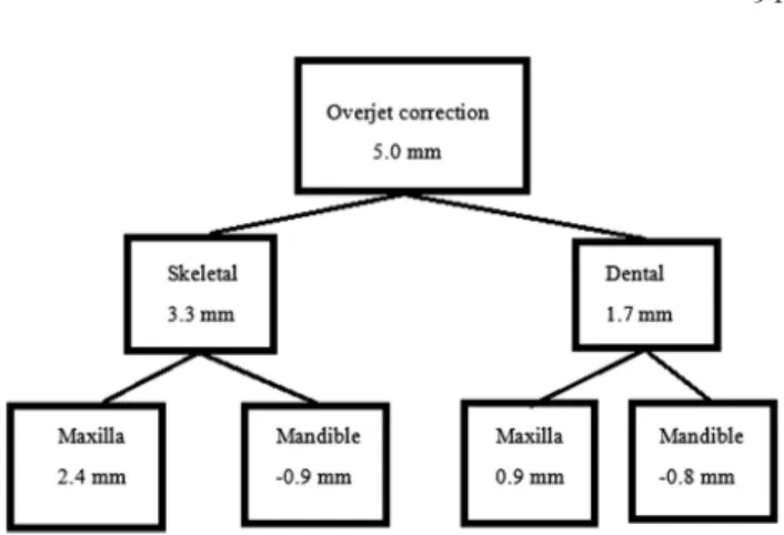
Upper incisors protruded and lower incisors retruded at a statistically significant level, resulting in an increase in overjet measurement. The change in overbite was found to be insignificant. Similar dentoalveolar changes have been reported in the previous studies (Arman et al. 2004; Altug and Arslan, 2006; Yavuz et al., 2009; Kilic et al. 2010a). Possible explanations of these dentoalveolar changes may be mesial drifting of maxillary dentition (U6–Y, 4.1 mm) resulting from the protraction forces and retrusive effect of chin cup component of the appliance on lower inci-sors. Merwin et al. (1997) reported that skeletal and den-tal contributions to overjet correction were 63 and 37 per cent, respectively. According to Cha et al. (2011), skeletal contribution to overjet correction was 63.1 per cent. In the present study, overjet correction (5.0 mm) was achieved by 66.1 per cent skeletal (A–Y; 2.4 mm and B–Y; −0.9 mm, a total of 3.3 mm) and 33.9 per cent dental (U1–NA; 0.9 mm and L1–NB; −0.8 mm, a total of 1.7 mm) changes. In addi-tion, maxillary skeletal contribution (A–Y; 2.4 mm; 74.2 per cent) to overjet correction was higher than mandibu-lar skeletal contribution (B–Y; −0.9 mm; 25.8 per cent) (Figure 5).
Soft tissue changes contributed to improve class III pro-files of the subjects. Upper lip showed significant forward movement (2.1 mm), while the retrusion in lower lip was not significant (−0.6 mm). This movement of upper lip in conjunction with the downward and backward movement of soft tissue chin (−0.6 mm) caused the facial profile to become more convex. These findings regarding soft tissue profile are consistent with the literature (Arman et al. 2004;
Kilic et al. 2010a).
The results from the present study are limited to a short-term observation period of maxillary protraction.
Long-term studies are needed to evaluate the stability of the results of mini maxillary protractor appliance and to com-pare its effects with those of similar appliances and treat-ment protocols. Another limitation of the present study is that observation periods of the subjects in the control group were 12.0 ± 0.2 months, while it was 8.1 ± 2.5 months for the patients in the study group. This longer observation period of the control subjects might have masked some effects of mini maxillary protractor appliance. On the other hand, this new appliance is smaller than the conventional face mask, and thus it is more esthetic and acceptable. An additional advantage is also that it is cheap. Since mini maxillary pro-tractor appliance is as effective as the conventional face masks in class III malocclusion treatment, it might be used in class III subjects who do not wish to use conventional face mask due to its appearance.
Conclusions
1. The maxillary protraction appliance caused significant changes in skeletal, dental, and soft tissue structures. 2. The skeletal contribution (66.1 per cent) to overjet
cor-rection of 5.0 mm was more than the dentoalveolar con-tribution (33.9 per cent).
3. The maxillary skeletal contribution (74.2 per cent) to overjet correction was more than the mandibular skeletal contribution (25.8 per cent).
References
Alcan T, Keles A, Erverdi N 2000 The effects of a modified protraction headgear on maxilla. American Journal of Orthodontics and Dentofacial Orthopedics 117: 27–38
Altug Z, Arslan A D 2005 A mini-maxillary protractor for class III correc-tion. Journal of Clinical Orthodontics 39: 522–5; quiz 531
Altug Z, Arslan A D 2006 Skeletal and dental effects of a mini maxillary protraction appliance. The Angle Orthodontist 76: 360–368
Arman A, Toygar T U, Abuhijleh E 2004 Profile changes associated with different orthopedic treatment approaches in class III malocclusions. The Angle Orthodontist 74: 733–740
Figure 5 Maxillary and mandibular skeletal and dentoalveolar changes
(mm) contributing to overjet correction in class III subjects.
Baccetti T, Franchi L, McNamara J A Jr 2000 Treatment and posttreat-ment craniofacial changes after rapid maxillary expansion and facemask therapy. American Journal of Orthodontics and Dentofacial Orthopedics 118: 404–413
Baik H S 1995 Clinical results of the maxillary protraction in Korean chil-dren. American Journal of Orthodontics and Dentofacial Orthopedics 108: 583–592
Battagel J M 1993 The aetiological factors in class III malocclusion. European Journal of Orthodontics 15: 347–370
Campbell P M 1983 The dilemma of class III treatment. Early or late? The Angle Orthodontist 53: 175–191
Cha B K, Choi D S, Ngan P, Jost-Brinkmann P G, Kim S M, Jang I S. 2011 Maxillary protraction with miniplates providing skeletal anchor-age in a growing class III patient. American Journal of Orthodontics and Dentofacial Orthopedics 139: 99–112
Chong Y H, Ive J C, Artun J 1996 Changes following the use of protrac-tion headgear for early correcprotrac-tion of class III malocclusion. The Angle Orthodontist 66: 351–362
Dellinger E L 1973 A preliminary study of anterior maxillary displace-ment. American Journal of Orthodontics and Dentofacial Orthopedics 63: 509–516
Emrich R E, Brodie A G, Blayney J R 1965 Prevalence of Class 1, Class 2, and Class 3 malocclusions (Angle) in an urban population. An epide-miological study. Journal of Dental Research 44: 947–953
Guyer E C, Ellis E E III, McNamara J A Jr, Behrents R G 1986 Components of class III malocclusion in juveniles and adolescents. The Angle Orthodontist 56: 7–30
Haas A J 1965 The treatment of maxillary deficiency by opening the mid-palatal suture. The Angle Orthodontist 35: 200–217
Hata S et al. 1987 Biomechanical effects of maxillary protraction on the craniofacial complex. American Journal of Orthodontics and Dentofacial Orthopedics 91: 305–311
Houston W J 1983 The analysis of errors in orthodontic measurements. American Journal of Orthodontics 83: 382–390
Irie M, Nakamura S 1975 Orthopedic approach to severe skeletal class III malocclusion. American Journal of Orthodontics 67: 377–392
Ishii H, Morita S, Takeuchi Y, Nakamura S 1987 Treatment effect of combined maxillary protraction and chincap appliance in severe skel-etal class III cases. American Journal of Orthodontics and Dentofacial Orthopedics 92: 304–312
Iwagaki H 1938 Hereditary influence of malocclusion. American Journal of Orthodontics and Oral Surgery 24: 328–338
Jackson G W, Kokich V G, Shapiro P A 1979 Experimental and postexperi-mental response to anteriorly directed extraoral force in young Macaca nemestrina. American Journal of Orthodontics 75: 318–333
Kambara T 1977 Dentofacial changes produced by extraoral forward force in the Macaca irus. American Journal of Orthodontics 71: 249–277 Kapust A J, Sinclair P M, Turley P K 1998 Cephalometric effects of face
mask/expansion therapy in class III children: a comparison of three age groups. American Journal of Orthodontics and Dentofacial Orthopedics 113: 204–212
Kilic N, Catal G, Kiki A, Oktay H 2010a Soft tissue profile changes fol-lowing maxillary protraction in class III subjects. European Journal of Orthodontics 32: 419–424
Kilic N, Celikoglu M, Oktay H 2010b Effects of the functional regulator III on profile changes in subjects with maxillary deficiency. European Journal of Orthodontics 32: 729–734
Kiliçoglu H, Kirliç Y 1998 Profile changes in patients with class III maloc-clusions after Delaire mask therapy. American Journal of Orthodontics and Dentofacial Orthopedics 113: 453–462
McNamara J A Jr 1987 An orthopedic approach to the treatment of class III malocclusion in young patients. Journal of Clinical Orthodontics 21: 598–608
Melsen B, Melsen F 1982 The postnatal development of the palatomax-illary region studied on human autopsy material. American Journal of Orthodontics and Dentofacial Orthopedics 82: 329–342
Merwin D, Ngan P, Hagg U, Yiu C, Wei S H 1997 Timing for effective application of anteriorly directed orthopedic force to the maxilla. American Journal of Orthodontics and Dentofacial Orthopedics 112: 292–299
Mouakeh M 2001 Cephalometric evaluation of craniofacial pattern of Syrian children with class III malocclusion. American Journal of Orthodontics and Dentofacial Orthopedics 119: 640–649
Nanda R 1980 Biomechanical and clinical considerations of a modified protraction headgear. American Journal of Orthodontics 78: 125–139 Ngan P, Wei S H, Hagg U, Yiu C K, Merwin D, Stickel B 1992 Effect of
pro-traction headgear on class III malocclusion. Quintessence International 23: 197–207
Pangrazio-Kulbersh V, Berger J, Kersten G 1998 Effects of protraction mechanics on the midface. American Journal of Orthodontics and Dentofacial Orthopedics 114: 484–491
Proffit W R 1992 Contemporary Orthodontics. CV Mosby, St Louis, MO, pp. 229–244
Proffit W R, Fields H W J 1993 Contemporary Orthodontics. CV Mosby, St Louis, MO
Sanborn R T 1955 Differences between the facial skeletal patterns of class III malocclusion and normal occlusion. Angle Orthodontist 25: 208–222
Sung S J, Baik H S 1998 Assessment of skeletal and dental changes by max-illary protraction. American Journal of Orthodontics and Dentofacial Orthopedics 114: 492–502
Takada K, Petdachai S, Sakuda M 1993 Changes in dentofacial morphol-ogy in skeletal class III children treated by a modified maxillary protrac-tion headgear and a chin cup: a longitudinal cephalometric appraisal. European Journal of Orthodontics 15: 211–221
Tanne K, Sakuda M 1991 Biomechanical and clinical changes of the craniofacial complex from orthopedic maxillary protraction. The Angle Orthodontist 61: 145–152
Thilander B, Myrberg N 1973 The prevalence of malocclusion in Swedish schoolchildren. Scandinavian Journal of Dental Research 81: 12–21
Turley P K 1988 Orthopedic correction of class III malocclusion with palatal expansion and custom protraction headgear. Journal of Clinical Orthodontics 22: 314–325
Uçüncü N, Uçem T T, Yüksel S 2000 A comparison of chincap and maxil-lary protraction appliances in the treatment of skeletal class III maloc-clusions. European Journal of Orthodontics 22: 43–51
Uner O, Yüksel S, Uçüncü N 1995 Long-term evaluation after chincap treatment. European Journal of Orthodontics 17: 135–141
Westwood P V, McNamara J A Jr, Baccetti T, Franchi L, Sarver D M 2003 Long-term effects of class III treatment with rapid maxillary expansion and facemask therapy followed by fixed appliances. American Journal of Orthodontics and Dentofacial Orthopedics 123: 306–320
Yavuz I, Halicioğlu K, Ceylan I 2009 Face mask therapy effects in two skel-etal maturation groups of female subjects with skelskel-etal class III maloc-clusions. The Angle Orthodontist 79: 842–848
Yüksel S, Uçem T T, Keykubat A 2001 Early and late facemask therapy. European Journal of Orthodontics 23: 559–568