ORIGINAL ARTICLE
Transcriptional analyses of the effects of
Catharanthus roseus L.
medicinal plant extracts on some markers related to obesity
and inflammation in 3T3-L1 mouse cell lines
Gülben Uytan1
&Hilal Büşra Tokgöz1&Reşat Ünal1&Filiz Altan1 Received: 8 March 2020 / Accepted: 9 July 2020
# Institute of Molecular Biology, Slovak Academy of Sciences 2020 Abstract
Catharanthus roseus L. (C. roseus) is one of the medicinal plants used to treat diabetes. In this study, 3T3-L1 preadipocyte cell lines which are fully differentiated into adipocytes were utilized and these were treated with extracts derived from above ground part ofC. roseus. The effect of these extracts on obesity and inflammation markers in cells was examined at the transcriptional level. Adipocyte lipid contents were measured by Oil Red O staining. Analyses, including changes related to adiposity and inflammation, were evaluated by measuring the relative mRNA expression levels of the genes of interest by the Real Time Reverse Transcription-Polymerase Chain Reaction (RT-PCR) method. The appropriate dose and durations forC. roseus were determined to be 12.5μg/mL and 24- and 48 h respectively. The expression of the inflammation marker Interleukin-6 (Il-6) was decreased whenC. roseus extract was administered to fully differentiated 3T3-L1 cells according to the determined dose and durations. Lipoprotein lipase (Lpl) and Fatty acid synthase (Fasn) gene expressions in fully differentiated cells decreased compared to the control after 24 h however, this effect was not observed after 48 h. Collagen V, has also been shown to be affected by treatment of fully differentiated 3T3-L1 cells with plant extracts in both 24- and 48-h periods.
Keywords Catharanthus roseus-diabetes-inflammation-medicinal plants-obesity
Abbreviations
C.roseus Catharanthus roseus L
MCP Monocyte chemoattractant protein CLS Crown like structures
IL-1β Interleukin-1β Il-6/IL-6 Interleukin-6
Tnf-α/TNF-α Tumor necrosis factor α Lpl Lipoprotein lipase
LPS Lipopolysaccharide
Fasn Fatty acid synthase TIAs Terpenoid indole alkaloids WHO World Health Organization ATCC American type culture collection IBMX 3-isobutyl-1-methylxanthine DMEM Dulbecco’s modified eagle medium SGBS Simpson Golabi Behmel Syndrome
ColV Collagen V
18S 18S ribosomal RNA
Pln1 Perilipin
NCBI National Center for Biotechnology Information
Fabp 4 Fatty acid-binding protein 4
SD Standard deviation
Atgl Adipose triglyceride lipase
ns Not significant
Glut 4 Glucose transporter type 4 ECM Extracellular matrix
VLDL Very low-density lipoprotein RT-PCR Reverse transcription-polymerase
chain reaction
Introduction
Obesity is an overexpansion of adipose tissue stores resulting from chronic calorie consumption that exceeds an individual’s energy needs (Hardy et al. 2012). Obesity has reached epi-demic proportions all over the world today because of its * Filiz Altan
1 Department of Molecular Biology and Genetics, Faculty of Science,
Muğla Sıtkı Koçman University, 48000, Kötekli, Muğla, Turkey
association with many diseases (Goossens et al.2011). In 2014, more than 600 million adults worldwide (13% of the total adult population) were classified as obese and it has been reported that the global obesity prevalence increased by about 2% every 10 years (Abarca-Gómez et al.2017; Pineda et al.
2018). According to World Health Organization (WHO), con-trary to what is known, obesity is not only limited to industri-alized societies, over 115 million people in developing coun-tries are estimated to suffer from obesity related problems. The prevalence of obesity has increased threefold in the last two decades in Europe, and the obesity prevalence is estimated to be 23% (Uerlich et al.2016; Pineda et al.2018). In the United States, 68.5% of the adult population has overweight or obe-sity problems (Bray et al.2018). Although there are not any publications about the distribution of obesity mediated inflam-mation worldwide to our knowledge, it is well established that there is a tight relation between obesity and inflammation. There are reports showing that obesity is associated with chronic systemic inflammation and that there are components of signaling pathways involved in inflammatory responses which is conditioned by the innate immune system activation in adipose tissue and promotes an increase in the production and release of pro-inflammatory cytokines in response that contribute to the triggering of the systemic acute-phase re-sponse which is characterized by elevation of acute-phase protein levels (Olefsky 2009; Rodríguez-Hernández et al.
2013). So, it therefore is probable that distribution of obesity mediated inflammation correlates positively with that of obesity.
Increased obesity rates have emerged as a major health prob-lem and cause economic burden in all countries (Engin2017). Although there are behavioral approaches and pharmacological treatments for obesity, it is not possible to say that they are very effective in healing. Although it is effective in reducing body weight, associated comorbidities and improving quality of life, bariatric surgery is associated with many complications (Qasim et al.2018). It has been reported by previous studies that obe-sity is characterized as a state of systemic chronic low inflam-mation (Park et al.2005; Goossens et al.2011; Liu et al.2016). In response to excessive intake of nutrients and energy, meta-bolic signals and inflammatory responses damaging homeosta-sis are initiated by the cells (Gregor and Hotamisligil2011). The secretion of pro-inflammatory cytokines by both adipo-cytes and adipose tissue macrophages contributes to the devel-opment of insulin resistance and atherosclerosis (Zieger et al.
2018). 3T3-L1 preadipocyte cell line that is derived from mouse cells and has the capability to differentiate into an adipocyte-like phenotype under appropriate conditions is orig-inally of fibroblast cell type and has therefore a fibroblast-like morphology (Green and Kehinde1975). The number of articles published on adipogenesis and the biochemistry of adipocytes where these cell lines were utilized reached a significant value. Although 3T3-L1 cells have the potential to convert from
fibroblastic phenotype to adipocytes, this alteration does not occur under normal circumstances, if necessary stimulating agents are required. Only in the presence of the agents insulin, dexamethasone and 3-isobutyl-1-methylxanthine (IBMX) at certain concentrations, fibroblast-like morphology having 3T3-L1 preadipocyte cells convert to fully differentiated adipo-cyte cells. There are reports about the protocols of how to effectively differentiate 3T3-L1 cells to adipocytes which indi-cates that the differentiantion of 3T3-L1 cells to adipocytes requires an optimal conversion condition (Zebisch et al.
2012). Once 3T3-L1 preadipocyte cells are stimulated to dif-ferentiate into adipocytes, adipogenesis is triggered and this in turn leads to the secretion of adipocytokines. Adipocytokine secretion plays a crucial role in the generation of an inflamma-tory microenvironment. In a study where human preadipocyte Simpson Golabi Behmel Syndrome (SGBS) cells were con-verted to differentiated SGBS cells, it was shown that the in-crease in adipogenesis correlated positively with an accumula-t i o n o f i n f l a m m aaccumula-t o r y m a r k er s su c h a s Mo n o c yaccumula-t e chemoattractant protein (MCP), (IL-1β) and IL-6 in the super-natant of differentiated SGBS cells. There are also a significant number of reports showing that adipocytes are secreting pro-inflammatory components such as IL-6 and TNF-α (Smitka and Marešová2015; Nickel et al.2018; Fuggetta et al.2019). Based on these information we hypothesised that the creation of an inflammatory microenvironment without making use of additional treatments with agents such as (LPS) and TNF-α by solely due to the conversion of preadipocyte 3T3-L1 cells to adipocytes would be sufficient to result in an enrichment of pro-inflammatory markers and this would mimic an in vitro chronic inflammation state to a mild chronic inflammation of an adipose tissue in vivo.
C. roseus is a perennial plant belonging to the family of Apocynaceae (Espejel-Nava et al.2018). It is widely distrib-uted throughout the world due to its high ability to survive in various habitats and its use as ornamental plant (Al-Shaqha et al. 2015). It produces more than 130 different terpenoid indole alkaloids (TIAs), some of which exhibit strong and important pharmacological activities (Almagro et al.2015). We in one of our previous studies have investigated the alka-loids of this plant in callus and multiple shoots obtained from C. roseus (Altan and Duru 2017). It is used in traditional practice for the treatment of diabetes in many countries such as Malaysia, India, China, South Africa and Mexico. Studies to investigate oral hypoglycemic effects have led to the dis-covery of the first clinically used natural anticancer agents vinblastine and vincristine (Tiong et al. 2015). Extract of C. roseus flower dissolved in ethanol has been reported to have wound healing activity (Nayak and Pereira 2006). There are also studies showing the hypolipidemic and anti-inflammatory activity ofC. roseus (Patel et al.2011; Rasineni et al.2013; Wahjuni et al.2015; Rajabi et al.2018). Based on these observations, the ability of C. roseus to inhibit
inflammation and adipogenesis in 3T3-L1 cells was investi-gated. The results support the view that the plant may be a potential alternative treatment for obesity and associated inflammation.
Materials and methods
Plant material
TheC. roseus plant to be used for the study was obtained commercially from a wholesaler in Köyceğiz/Muğla. The plants were dried for 5 weeks in a cool and moisture-free environment. The above-ground parts of the dried plants were ground to a powder. The plant powder was incubated in ab-solute ethanol for 48 h at room temperature (Shanmugaraju and Bhakyaraj2016). After the extract was filtered, the sol-vent was removed by evaporation. The stock solutions were dissolved in ethanol to a final concentration of 20 mg/mL and stored at−20 °C until use.
Cell culture and differentiation
3T3-L1 mouse cell lines were purchased from the American Type Culture Collection (ATCC). Cells were maintained in Dulbecco’s modified eagle medium (DMEM) (Capricorn, Cat. no. DMEM-HA) supplemented to 10% with newborn calf serum (Capricorn, Cat. no. DMEM-HA), 1% penicillin/ streptomycin (Multicell, Cat. no. 450–201-EL). For experi-ments the cells were grown to a confluence of 70% and stim-ulated to differentiate in predifferentiation medium containing 1 μM dexamethasone (Sigma, BCBV5460), 0.5 μM 3-isobutyl-1-methylxanthine (IBMX) (Sigma,STBF6061V), 1.0 μg/mL insulin (Sigma, SLBV1793) (Zebisch et al.
2012). The predifferentiation medium then was replaced after 48 h with differentiation medium in DMEM containing 10% fetal bovine serum and 1% penicillin/streptomycin, 1.0μg/ mL insulin (Sigma, SLBV1793) for 7–10 days.
Evaluation of changes in morphology and viability of
cells
The cells were placed onto petri dishes and incubated within a media containing 12.5 μg/mL, 25 μg/mL and 50 μg/mL C. roseus extracts for 24-, 48- and 72 h (van de Venter et al.
2008). The adherence and morphology of the cells on petri dishes were evaluated using light microscopy.
Staining of the 3T3-L1 cells with oil red O dye
The differentiation of 3T3-L1 adipocytes was done as de-scribed previously. Oil Red O staining was done according to manufacturer’s instructions using the Biovision Oil Red O
Staining kit. The procedures that adipocytes went through can be summarized so that they were initially fixed at room tem-perature for 20 min in 3.7% formaldehyde followed by rinsing them with distilled water and staining them for 2 h at room temperature. The dye was extracted for 30 min in 0.5 mL of dye extraction solution. The procedure ended by the aspiration of the dye and rinsing of the plates that after with distilled water. Removal of the dye was performed using 0.5 mL of dye extraction buffer for 30 min.
Total RNA isolation and real time reverse
transcription-polymerase chain reaction (RT-PCR)
Total RNA from adipocyte cells was extracted using a RiboEx™ (Cat. No.301–001) total RNA isolation solution from GeneAll (Cat. No.301–902). Ultraviolet light spectro-photometry followed by formaldehyde- agarose gel electro-phoresis was used to determine the quality and quantity of the isolated RNA. Five hundred nanograms of total RNA was reverse transcribed using oligo-dT primers with EasyScript Plus cDNA synthesis kit from ABM Alfagen (Cat. No. G236). The amplification of reverse transcribed RNA was achieved with the use of Ampliqon RealQ Plus 2 × Master Mix Green in the presence of 0.3 mM gene-specific forward and reverse primers by a thermocycling on a Roche Light Cycler 96 Real Time PCR machine for 45 cycles. The tem-peratures and durations for denaturation, annealing and exten-sion were 95 °C for 30 s; 55–58 °C for 30 s and 72 °C for 30 s, respectively. The normalization of the differences in individ-ual samples was done by the use of amplified 18S expression as a standard control. The description of the mouse Lipoprotein lipase (Lpl), Fatty acid synthase (Fasn), Collagen V (ColV), Interleukin-6 (Il-6) and 18S ribosomal RNA (18S) primer sequences are given below in Table 1. All expression data for mouse Lpl, Fasn, ColV and Il-6 were interpreted by relating them to 18S RNA. Pooled cDNA of the samples being assayed were used to generate standard curves. This enables the accurate comparison of samples within each assay but do not necessarily enable accurate comparison of samples between different assays represented as arbitrary units. The analyses of all samples were done in duplicates; DNA contamination was eliminated by designing primers spanning an intron. The primers were designed based on the information of the sequences of the genes of interest using The National Center for Biotechnology Information (NCBI2018) as a source. The information about the primer sequences used for Real Time PCR and the accession numbers of the genes analyzed are shown in Table1.
Statistical analysis
The comparison of groups with respect to continuous vari-ables and comparison of baseline and posttreatment
measurements were done by using Student’s two-sample t-test and a paired t-test respectively. Differentially expressed genes with significance were identified using aP value <0.03.
Results
The effect of
Catharanthus roseus extract on cell
viability
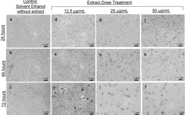
To observe the effect ofC. roseus extracts on undifferentiated 3T3-L1 cells, cell morphology and cell survival circumstances were examined. 3T3-L1 cells are adherent cells and they need to be attached to the surface to maintain their viability and the tendency of them to detach will be indicative of a deteriorated cell morphology. Therefore, it was investigated whether the doses of 12.5μg/mL, 25 μg/mL and 50 μg/mL of ethanol-dissolved extracts obtained from the above-ground parts of the plant along with the solvent itself alone had unfavorable ef-fects on the cells. Cells were exposed to these determined doses for 24-, 48- and 72 h. At the end of the treatment of cells with the solvent itself alone in all time durations, it was observed that the solvent used had no toxic effect on the cells and was suitable for the experiment since there was neither a detachment of the cells from surface nor a change in cell morphology (Fig.1a, b, c). The treatment of the cells with the extract for 72 h, regardless of concentration and time, both affected the cells’ adherence to the surface to which they were attached and caused deterioration in their morphology. Based on this, it was concluded that the application of extracts for 72 h had a negative effect on cell viability (Fig.1f, i, l). It was also demonstrated in this study that the treatment of the cells with extracts at doses of 25μg/mL and 50 μg/mL for 48 h, resulted in cell detachment and morphology deterioration. Thus, it was concluded that the treatment of cells with an extract dose of 25μg/mL and above for 48 h and more had no favorable effect on cell viability (Fig.1g–l). It was ob-served that treatment of the cells with 12.5μg/mL extract for 24 h and 48 h did not cause a significant decrease in cell
attachment to the surface and did not result in a negative effect on cell morphology. Based on all these evaluations, it was concluded that the best condition for the cells to be treated with C. roseus extract is 12.5 μg/mL dose application for 24- and 48 h (Fig.1d, e).
The information about the size of the cells both in Fig.1
and Fig.2were estimated using the bar graphs shown in each figure introduced by using the ImageJ fromhttp://rsb.info.nih. gov/ij/download.html (National Institutes of Health, (NIH)
2020). It is well established that obesity is a low grade chronic inflammation, and obese individuals undergo an increase in lipid accumulation in their adipocytes which in turn leads to the enlargement of their adipose tissue. Extraordinary lipid accumulation of adipocytes causes the transformation of monocytes in heterogeneous adipose tissue to macrophages and these macrophages infiltrate adipocytes and form crown like structures (CLS) in which the case obesity has now be-come an inflammatory syndrome. In this study, cells were used as an in vitro model and preadipocyte 3T3-L1 cells were first transformed into fully differentiated adipocytes to mimic the fat accumulation state of adipocytes in obese individuals and thus to evaluate the markers of inflammation. The results show that the differentiation conditions used to treat cells with extract were appropriate and that undifferen-tiated preadipocyte cells were converted to fully differ-entiated adipocyte cells as expected (Fig. 2).
Alterations of lipogenic Lpl and Fasn enzyme
expressions due to the treatment with
Catharanthus
roseus extract
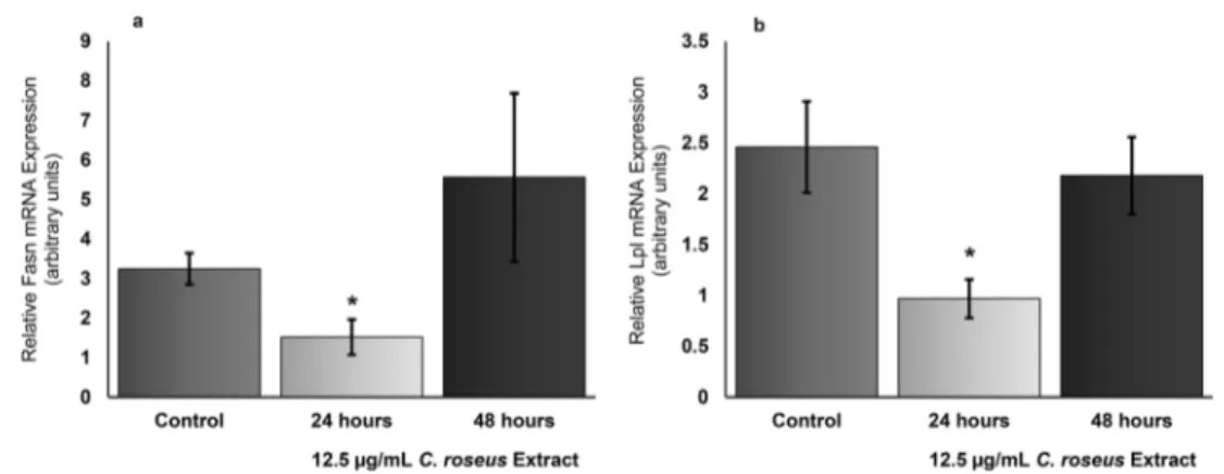
Gene expression measurements of lipogenic Lp1 and Fasn genes were performed to understand the molecular mecha-nism underlying the hypolipidemic effect ofC. roseus extract. Treatment of fully differentiated 3T3-L1 adipocyte cells with the extract at a dose of 12.5 μg/mL for 24 h resulted in a significant decrease in Fasn mRNA level. Exposure of adipo-cyte cells to extracts at a dose of 12.5μg/mL for 24 h resulted in a reduction of Fasn gene expression levels in these cells by Table 1 Sequences, lengths, melting temperatures and NCBI accession numbers for primers designed for real time PCR reactions
Gene Primer sequence (5′ 3′) Primer length (bases) Tm (°C) GenBank accession number
18S Forward: TTCGAACGTCTGCCCTATCAA 21 63 NR_003278.3
Reverse: ATGGTAGGCACGGCGACTA 19 63
Lpl Forward: ACTCGCTCTCAGATGCCCTA 20 65 NM_008509.2
Reverse: TTGTGTTGCTTGCCATTCTC 20 60
Fasn Forward: CTGAGATCCCAGCACTTCTTGA 22 64 NM_007988.3
Reverse: GCCTCCGAAGCCAAATGAG 19 63
Il-6 Forward: ACCAAACTGGATATAATCAGGAAA 24 56 NM_031168.2
Reverse: GAGAGCATTGGAAATTGGGG 20 57
ColV Forward:CTCAGGGGTAACGAAAACCA 20 60 NM_015734.2
53% compared to their control counterparts. Although the administration of the extract at this dose for 48 h appears to show an increase in the Fasn mRNA level of the cells, this increase is statistically not significant. Thus, it was concluded that treatment of adipocyte cells with extract at a dose of 12.5μg/mL had an effect on Fasn mRNA expression in the first 24 h, which reduced gene expression, and then this effect disappeared (Fig.3a).
Following the same experimental setup, the gene expres-sion pattern obtained by looking at the mRNA levels of an-other lipogenic enzyme Lpl revealed a great parallelism with the expression pattern of the Fasn gene. In this case, treatment of fully differentiated 3T3-L1 cells with extract at a dose of 12.5μg/mL for 24 h significantly reduced the Lpl mRNA level too. After 24 h, Lpl gene expression decreased by
61%. Although the treatment of cells with extract for 48 h seemed to reduce Lpl gene expression, this decrease was not significant when statistical analyzes were considered. These results concluded a significant reduction in the lipogenic en-zyme Lpl gene expression when the extract used was admin-istered to fully differentiated adipocytes at a dose of 12.5μg/ mL for 24 h, but this reduction effect disappeared after 48 h at the same dose (Fig.3b).
The data obtained regarding the alterations of 3T3-L1 Fasn and Lpl expression profiles in response to 12.5 μg/mL C. roseus extract treatments for 24 h and 48 h in arbitrary units is summarized in Table2. Values shown in bold indicate that the percent changes analyzed are of statistical significance. The values having the descriptions“(ns)” indicate that these values are statistically not significant.
Fig. 1 Microscopic visualization and evaluation of changes in morphology and viability of 3T3-L1 cells treated with plain solvent or C. roseus extracts with a dose of 12.5 μg/mL, 25 μg/mL and 50 μg/mL
for 24 h (a, d, g and j), 48 h (b, e, h and k) and 72 h (c, f, i and l) by light microscopy. (Scale bars in each micrograph represent a length of 1μm)
Fig. 2 Oil Red O Staining to determine the lipid accumulation in fully differentiated 3T3-L1 cells. Cells were visualized before staining and after staining in a 10 × magnitude (a, b) respectively and after staining
at a 40 × magnitude (c) by light microscopy. (Scale bars in each micro-graph represent a length of 1μm)
Effects on expression of extracellular matrix
component collagen V and pro-inflammatory marker
Il-6 after treatment with
Catharanthus roseus extract
In addition to the metabolic complications, obesity appears as a condition that manifests itself with some changes in the remodeling of the extracellular matrix (ECM) components in adipose tissue. The extracellular matrix components of adi-pose tissue undergo a constant remodeling so that the shapes and functions of adipocytes and their precursor cells respond to nutritional cues. It is well established in the literature that obesity causes accumulation of ECM components and their modifiers in adipose tissue (Lin et al.2016). Collagens are the most abundant scaffolding in ECM. Collagen V has been chosen to investigate the potential changes in ECM remodel-ing as a consequence of an increase in adiposity since it is essential for fibrillation of types I and III collagen, and conse-quently for optimal fibrillary formation and tissue quality. Changes in Collagen V mRNA levels were evaluated to get an opinion about the role ofC. roseus extract on extracellular matrix remodeling. Exposing fully differentiated 3T3-L1 adi-pocyte cells to the extract at a dose of 12.5μg/mL for both 24 h and 48 h resulted in a significant decrease in Collagen V mRNA expression by 97% and 91% respectively (Fig.4a).
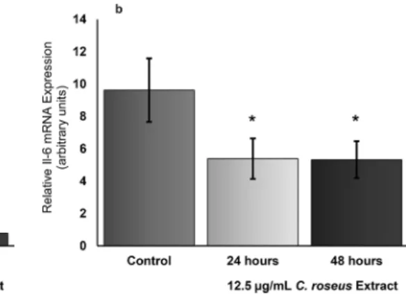
The molecular mechanisms underlying the relation-ship between obesity and inflammation has been uncov-ered to a significant extent. The change in the interac-tion of adipocytes and monocytes of heterogeneous ad-ipose tissue due to obesity, causes the secretion of some inflammatory cytokines which in turn results in the con-version of native monocytes to pro-inflammatory macro-phages infiltrating adipocytes and consequently form “crown like structures” (CLS). An adipose tissue under such stress results in a state of mild chronic inflamma-tion. When this is the case, knowing what effect C. roseus has in terms of reducing the inflammatory condition that will arise due to the increase of adiposity has been one of the questions sought for this study. For this purpose, C. roseus extracts were applied to fully differentiated 3T3-L1 cells at a dose of 12.5 μg/mL for 24 h and 48 h periods. At this dose, pro-inflammatory Il-6 mRNA level used as a marker in both time periods decreased by approximately 40% (Fig. 4b). The data obtained regarding the alterations of 3T3-L1 ColV and Il-6 expression profiles in response to 12.5μg/mL C. roseus extract treatments for 24 h and 48 h in arbitrary units is summarized in Table3. Values shown in bold indicate that the percent changes analyzed are statistically significant. Fig. 3 Quantitative measurement of Fasn and Lpl mRNA in 3T3-L1
cells. Fasn and Lpl quantitation of mRNA in 3T3- L1 adipocytes differ-entiated with IBMX, Dexamethasone and insulin were done by Real Time RT-PCR using duplicate measurements in three separate
experiments. The changes in Fasn mRNA and Lpl mRNA expression in fully differentiated adipocytes indicated with asterisks were significant (P < 0.03). (Error bars show standard deviation (SD))
Table 2 Fully differentiated 3T3-L1 gene expressions and percent changes of Fasn and Lpl in response to 12.5μg/mL C. roseus treatment for 24- and 48 h in comparison to control. Plus (+) indicates an increase
and minus (−) a decrease in gene expression. Bold labeling mean that the alterations are statistically significant whereas“(ns)” indicates that the change is not of significance
Samples genes of interest Relative mRNA expression (arbitrary units)
Control 12.5μg/ml C. roseus extract
24 h % change to control 48 h % change to control
Fasn 3.25 + 0.40 1.52 + 0.45 −53% 5.56 + 2.13 +71% (ns)
Discussion
Today, obesity is a global disease that affects individuals from each age group in developing and developed countries (Ofei
2005). Studies have been able to relate obesity to many health problems such as insulin resistance, cancer, heart and respira-tory diseases and hypertension. The risk of morbidity and premature death is directly related to excessive accumulation and distribution of fat in the body (Greco et al.2002). When health expenditures are taken into consideration, it has been demonstrated that expenditures for obese individuals are 1.5– 1.8 times higher than for non-obese individuals (Cox et al.
2015). Many pharmacological drugs used to combat obesity have also been reported to have significant side effects (Kang and Park2012; Huang et al. 2019). Considering all these, medicinal plants stand out as potential useful alternatives to pharmacological drugs due to their cheapness, reliability and low side effects. Preadipocyte 3T3-L1 cells acquire adipocyte morphology after differentiation induction, accumulate large amounts of triacylglycerol and become sensitive to lipolytic and lipogenic hormones, thereby providing an excellent mod-el system to study the biochemical and molecular mechanisms involved in adipose tissue development and lipid metabolism (Moustaid and Sul1991). In adipogenesis, the interaction of proteins such as fatty acid-binding protein with lipid
metabolizing enzymes such as lipoprotein lipase and fatty acid synthase induces various transcription factors to mediate adi-pocyte differentiation (Kim and Spiegelman1996; Tsai et al.
2017). Therefore, as an important approach for the prevention and/or treatment of obesity, it is important to suppress both adipocyte cell count (hyperplasia) and cell size (hypertrophy) induced adipose tissue expansion.C. roseus is an important plant in this sense because there are previous studies showing the effect of it decreasing lipid parameters. Rasineni et al. showed in their study where they used a high-fat diet fed mice model thatC. roseus leaf extract solution given to the mice at a certain dose on a daily basis (100 mg/kg body weight/day) reduced the amount of free fatty acid in the tissue by providing a decrease in the activity of Fasn (Rasineni et al.2013). In our study, we observe a positive correlation between this decrease in Fasn activity and our finding of the decrease of mRNA expression obtained by applyingC. roseus plant extracts to adipocytes.
It is well established that adipogenesis plays a prominent role in the expansion of a healthy adipose tissue which in turn leads to obesity (Kershaw and Flier 2004; Vishvanath and Gupta 2019). It is also very well known that diabetes and obesity are two diseases that are significantly interdependent (Golay and Ybarra2005).C. roseus is a medicinal plant that is commonly used to treat diseases in a wide spectrum including Fig. 4 Quantitative measurement of ColV and Il-6 mRNA in 3T3-L1
cells. ColV and Il-6 quantitation of mRNA in 3T3- L1 adipocytes differ-entiated with IBMX, Dexamethasone and insulin were done by Real Time RT-PCR using duplicate measurements in three separate
experiments. The changes in ColV mRNA and Il-6 mRNA expression in fully differentiated adipocytes indicated with asterisks were significant (P < 0.03). (Error bars show standard deviation (SD))
Table 3 Fully differentiated 3T3-L1 gene expressions and percent changes of ColV and Il-6 in response to 12.5μg/mL C. roseus treatment for 24- and 48 h in comparison to control. Plus (+) indicates an increase
and minus (−) a decrease in gene expression. Bold labeling mean that the alterations are statistically significant
Samples genes of interest Relative mRNA expression (arbitrary units)
Control 12.5μg/ml C. roseus extract
24 h % change to control 48 h % change to control
CoIV 3.68 + 1.10 0.12 + 0.03 −97% 0.33 + 0.12 −91%
diabetes all over the world. Although there are quite a number of reports regarding antihyperglycemic effects ofC. roseus, the influence of this plant on adipogenesis and obesity has not been studied extensively. It is important to know whether metabolic improvements mediated throughC. roseus might be linked to its effects on adipogenesis. In a recent study it has been reported that 1α, 25-dihydroxy Vitamin D3 contain-ing fractions ofC. roseus leaf aqueous extract inhibited preadipocyte differentiation and induced lipolysis in 3T3-L1 cells. The authors showed in their study in detail that the differentiation of 3T3-L1 preadipocytes was inhibited and the lipid accumulation in differentiating 3T3-L1 cells was re-duced uponC. roseus extract administration (Borah et al.
2019). This is the first report to our knowledge that provides a direct answer to the question whetherC. roseus extracts have an effect on adipogenesis. They also showed in their study using RT-PCR analysis that the genes such as Perilipin 1 (Pln1), Fatty acid-binding protein 4 (Fabp 4), Adipose triglyc-eride lipase (Atgl), Lipoprotein lipase (Lpl), Glucose trans-porter type 4 (Glut4), and Adiponectin which they called “ad-ipocyte factors” were downregulated upon C. roseus extract treatment. The downregulation of Lpl afterC. roseus treat-ment that we obtained in our current study is consistent with this report.
Adipocytes have the property of storing fatty acids that are generated during the process starting with the metabolism of circulating exogenous fat as triglycerides and also fatty acids as the result of endogenous fatty acid biosynthesis. Lipoprotein lipase hydrolyzes triacylglycerol-rich portions of both very low-density lipoprotein (VLDL) and of chylomi-crons, resulting in fatty acid release. These nonesterified fatty acids are then imported by fat cells and mainly serve as a precursor for fat synthesis (Kim and Spiegelman 1996; Mead et al.2002). Studies in diabetic mice and pigs fed on a high-fat diet treated withC. roseus leafs dissolved in ethanol have been reported to reduce the levels of triglyceride and cholesterol levels in blood (Ara et al.2009; Patel et al.2011; Muralidharan2014). This decrease in lipid levels may be as-sociated with the plant down-regulating Lpl mRNA expres-sion, which is increased in adipose tissue.
120 alkaloids were shown to be produced inC. roseus of which 70 were pharmacologically active (Barrales-Cureño et al.2019). Pharmacokinetics of alkaloids are changing from one to another, which in turn may influence their effect on the biological question of interest. Aconitum is one of the plants, which is shown to contain alkaloids and in a study it was reported that the concentration of all of the six analyzed Aconitum alkaloids were decreased within a time frame of 24 h in rat plasma (Zhang et al.2019). There are also similar studies that characterized the pharmacokinetics ofC. roseus alkaloids such as vinblastine (van Tellingen et al. 1993), vindoline and catharanthine (Lin et al.2015). In all these re-ports it was consistently shown that the plasma levels of all of
the alkaloids decreased within 24 h. For some alkaloids pres-ent inC. roseus this effect was even observed between 8- and 24 h, We, in this study, observed that the downregulatory effect ofC. roseus extract on 3T3-L1 cells after 24 h was no longer the case after 48 h of administration. In line with pre-vious reports, we believe that the reason of the attenuation of the inhibitory effects ofC. roseus on the expression of Lpl and Fasn is likely to be the consequence of the much reduced amounts ofC. roseus taken into cells between 24- and 48 h, hence decreasing its effect further. In addition, as a result of the increase in the incubation period due to the mixed multiple componential nature of plant extracts, the structures of some substances in the extract may deteriorate or change and lose their effectiveness, so the expected effect may not be seen. Apart from this, our plant extract may also have blocked gene expression via a cytoplasmic signaling pathway.
Adipose tissue also secrets cytokines that can affect energy metabolism. Tumor necrosis factor (Tnf-α) and interleukin-6 (Il-6) are prominent pro-inflammatory cytokines (Zieger et al.
2018). Dysregulated and continuous synthesis of Il-6 is asso-ciated with chronic inflammation and autoimmune disorders (Tanaka et al. 2014). Increased cytokine levels such as of Tnf-α and Il-6 were observed in the blood of mice fed with a high-fat diet for 6 weeks. C. roseus leaf extract led to a decrease in the levels of these cytokines. In our study, plant extract provided a decrease in the mRNA levels of the inflam-matory agent Il-6, consistent with the observed drop in blood serum levels mentioned in the study above.
An increase in inflammatory macrophages, fibrosis, many E C M c o m p o n e n t s i n c l u d i n g C o l l a g e n V I a n d thrombospondin has been reported to be the characteristics of adipose tissues obtained from obese, insulin resistant ro-dents and humans (Spencer et al. 2011). In an experiment conducted by Spencer et al. (2011), it was found that there was a significant increase in both mRNA and protein levels of Collagen V in fat tissue of obese individuals compared to lean individuals. In our study, C. roseus plant rearranged ECM components and reduced Collagen V mRNA expression in 3T3-L1 cells that have been dysregulated due to obesity.
Conclusion
This study focused on molecular changes in gene regulation at the transcriptional level in order to reveal the underlying mechanisms of the results obtained in studies in the literature, which were rather in vivo studies in which the effects of C. roseus plant extracts were studied. Although we observed that the results obtained by in vivo studies are mostly in pos-itive correlation with the results we received at the transcrip-tional level, there is a need for more detailed studies to under-stand the molecular mechanisms underlying these changes. In this sense, we believe that our study is important at the
molecular level and also constitutes a basis for conducting more detailed studies.
Acknowledgments This work was supported by a Scientific Research Project (BAP) Grant (19/084/05/1/2) from the Muğla Sıtkı Koçman University to Dr. Filiz ALTAN. We also would like to thank Dr. Mehmet VAROL for his expertise and assistance in language and tech-nical editing.
Compliance with ethical standards
Competing interests The authors declare that they have no competing interest.
References
Abarca-Gómez L, Abdeen ZA, Hamid ZA, Abu-Rmeileh NM, Acosta-Cazares B, Acuin C, Adams RJ, Aekplakorn W, Afsana K, Aguilar-Salinas CA (2017) Worldwide trends in body-mass index, under-weight, overunder-weight, and obesity from 1975 to 2016: a pooled anal-ysis of 2416 population-based measurement studies in 128· 9 mil-lion children, adolescents, and adults. Lancet 390(10113):2627– 2642.https://doi.org/10.1016/S0140-6736(17)32129-3
Almagro L, Fernández-Pérez F, Pedreño MA (2015) Indole alkaloids from Catharanthus roseus: bioproduction and their effect on human health. Molecules 20(2):2973–3000.https://doi.org/10.3390/ molecules20022973
Al-Shaqha WM, Khan M, Salam N, Azzi A, Chaudhary AA (2015) Anti-diabetic potential of Catharanthus roseus Linn. And its effect on the glucose transport gene (GLUT-2 and GLUT-4) in streptozotocin induced diabetic wistar rats. BMC Complement Altern Med 15(1): 1–8.https://doi.org/10.1186/s12906-015-0899-6
Altan F, Duru ME (2017) Catharantus roseus L.’den Elde Edilen Kallus ve Çoklu Sürgünlerde Alkaloidlerin Araştırılması. Yüzüncü Yıl Üniversitesi Tarım Bilimleri Dergisi 27(3):337–346.https://doi. org/10.29133/yyutbd.290146
Ara N, Rashid M, Amran S (2009) Comparison of hypotensive and hy-polipidemic effects ofCatharanthus roseus leaves extract with aten-olol on adrenaline induced hypertensive rats. Pak J Pharm Sci 22(3).
https://doi.org/10.3923/jbs.2008.1082.1086
Barrales-Cureño HJ, Reyes CR, García IV, Valdez LGL, De Jesús AG, Ruíz JAC, Herrera LMS, Caballero MCC, Magallón JAS, Perez JE (2019) Alkaloids of pharmacological importance in Catharanthus roseus. Alkaloids-their importance in nature and human life. IntechOpen.https://doi.org/10.5772/intechopen.82006
Borah AK, Singh A, Yasmin R, Doley R, Mattaparthi VSK, Saha S (2019) 1α, 25-dihydroxy vitamin D3 containing fractions of Catharanthus roseus leaf aqueous extract inhibit preadipocyte differ-entiation and induce lipolysis in 3T3-L1 cells. BMC Complement Altern Med 19(1):338.https://doi.org/10.1186/s12906-019-2754-7
Bray GA, Heisel WE, Afshin A, Jensen MD, Dietz WH, Long M, Kushner RF, Daniels SR, Wadden TA, Tsai AG (2018) The science of obesity management: an endocrine society scientific statement. Endocr Rev 39(2):79–132.https://doi.org/10.1210/er.2017-00253
Cox AJ, West NP, Cripps AW (2015) Obesity, inflammation, and the gut microbiota. The Lancet Diabetes Endocrinol 3(3):207–215.https:// doi.org/10.1016/S2213-8587(14)70134-2
Engin A (2017) The definition and prevalence of obesity and metabolic syndrome. In: Engin AB, Engin A (eds) Obesity and Lipotoxicity, Springer, pp 1–17.https://doi.org/10.1007/978-3-319-48382-5_1
Espejel-Nava JA, Vega-Avila E, Alarcon-Aguilar F, Contreras-Ramos A, Diaz-Rosas G, Trejo-Aguilar G, Ortega-Camarillo C (2018) A
phenolic fraction fromCatharanthus roseus L. stems decreases gly-cemia and stimulates insulin secretion. Evid-Based Complement Altern Med 2018.https://doi.org/10.1155/2018/7191035
Fuggetta MP, Zonfrillo M, Villivà C, Bonmassar E, Ravagnan G (2019) Inflammatory microenvironment and adipogenic differentiation in obesity: the inhibitory effect of theobromine in a model of human obesity in vitro. Mediat Inflamm 2019.https://doi.org/10.1155/ 2019/1515621
Golay A, Ybarra J (2005) Link between obesity and type 2 diabetes. Best Pract Res Clin Endocrinol Metab 19(4):649–663.https://doi.org/10. 1016/j.beem.2005.07.010
Goossens GH, Bizzarri A, Venteclef N, Essers Y, Cleutjens JP, Konings E, Jocken JW,Čajlaković M, Ribitsch V, Clément K (2011) Increased adipose tissue oxygen tension in obese compared with lean men is accompanied by insulin resistance, impaired adipose tissue capillarization, and inflammation. Circulation 124(1):67–76.
https://doi.org/10.1161/CIRCULATIONAHA.111.027813
Greco L, Romino R, Coto I, Di Cosmo N, Percopo S, Maglio M, Paparo F, Gasperi V, Limongelli M, Cotichini R (2002) The first large population based twin study of coeliac disease. Gut 50(5):624– 628.https://doi.org/10.1136/gut.50.5.624
Green H, Kehinde O (1975) An established preadipose cell line and its differentiation in culture II. Factors affecting the adipose conversion. Cell 5(1):19–27.https://doi.org/10.1016/0092-8674(75)90087-2
Gregor MF, Hotamisligil GS (2011) Inflammatory mechanisms in obesi-ty. Annu Rev Immunol 29:415–445.https://doi.org/10.1146/ annurev-immunol-031210-101322
Hardy OT, Czech MP, Corvera S (2012) What causes the insulin resis-tance underlying obesity. Curr Opin Endocrinol Diabetes Obes 19(2):81.https://doi.org/10.1097/MED.0b013e3283514e13
Huang D, Deng M, Kuang S (2019) Polymeric carriers for controlled drug delivery in obesity treatment. Trends Endocrinol Metab.
https://doi.org/10.1016/j.tem.2019.09.004
Kang JG, Park C-Y (2012) Anti-obesity drugs: a review about their ef-fects and safety. Diabetes Metab J 36(1):13–25.https://doi.org/10. 4093/dmj.2012.36.1.13
Kershaw EE, Flier JS (2004) Adipose tissue as an endocrine organ. J Clin Endocrinol Metab 89(6):2548–2556. https://doi.org/10.1016/ S1043-2760(00)00301-5
Kim JB, Spiegelman BM (1996) ADD1/SREBP1 promotes adipocyte differentiation and gene expression linked to fatty acid metabolism. Genes Dev 10(9):1096–1107.https://doi.org/10.1101/gad.10.9. 1096
Lin C, Cai J, Yang X, Hu L, Lin G (2015) Liquid chromatography mass spectrometry simultaneous determination of vindoline and catharanthine in rat plasma and its application to a pharmacokinetic study. Biomed Chromatogr 29(1):97–102.https://doi.org/10.1002/ bmc.324
Lin D, Chun T-H, Kang L (2016) Adipose extracellular matrix remodel-ling in obesity and insulin resistance. Biochem Pharmacol 119:8– 16.https://doi.org/10.1016/j.bcp.2016.05.005
Liu W, Crott JW, Lyu L, Pfalzer AC, Li J, Choi S-W, Yang Y, Mason JB, Liu Z (2016) Diet-and genetically-induced obesity produces alter-ations in the microbiome, inflammation and Wnt pathway in the intestine of Apc+/1638N mice: comparisons and contrasts. J Cancer 7(13):1780.https://doi.org/10.7150/jca.15792
Mead JR, Irvine SA, Ramji DP (2002) Lipoprotein lipase: structure, function, regulation, and role in disease. J Mol Med 80(12):753– 769.https://doi.org/10.1007/s00109-002-0384-9
Moustaid N, Sul H (1991) Regulation of expression of the fatty acid synthase gene in 3T3-L1 cells by differentiation and triiodothyro-nine. J Biol Chem 266(28):18550–18554.https://doi.org/10.1042/ bj2920767
Muralidharan L (2014) Catharanthus roseus leaves as an anti-diabetic and Hypolipidemic agents in Alloxan-induced diabetic rats. Am J Phytomedicine Clin Ther 2(12):1393–1396
National Institutes of Health (NIH) (2020) Image J, Image J download for windows.http://rsb.info.nih.gov/ij/download.html. Accesed 05 May 2020
Nayak B, Pereira LMP (2006)Catharanthus roseus flower extract has wound-healing activity in Sprague Dawley rats. BMC Complement Altern Med 6(1):41.https://doi.org/10.1186/1472-6882-6-41
Nickel A, Blücher C, Al Kadri O, Schwagarus N, Müller S, Schaab M, Thiery J, Burkhardt R, Stadler SC (2018) Adipocytes induce distinct gene expression profiles in mammary tumor cells and enhance in-flammatory signaling in invasive breast cancer cells. Sci Rep 8(1):1– 13.https://doi.org/10.1038/s41598-018-27210-w
Ofei F (2005) Obesity-a preventable disease. Ghana Med J 39(3):98 Olefsky JM (2009) IKKɛ: a bridge between obesity and inflammation.
Cell 138(5):834–836.https://doi.org/10.1016/j.cell.2009.08.018
Park HS, Park JY, Yu R (2005) Relationship of obesity and visceral adiposity with serum concentrations of CRP, TNF-α and IL-6. Diabetes Res Clin Pract 69(1):29–35.https://doi.org/10.1016/j. diabres.2004.11.007
Patel Y, Vadgama V, Baxi S, Tripathi CB (2011) Evaluation of hypolip-idemic activity of leaf juice of Catharanthus roseus (Linn.) G. Donn. In Guinea pigs. Acta Pol Pharm 68(6):927–935
Pineda E, Sanchez-Romero LM, Brown M, Jaccard A, Jewell J, Galea G, Webber L, Breda J (2018) Forecasting future trends in obesity across Europe: the value of improving surveillance. Obes Facts 11(5):360– 371.https://doi.org/10.1159/000492115
Qasim A, Turcotte M, De Souza R, Samaan M, Champredon D, Dushoff J, Speakman J, Meyre D (2018) On the origin of obesity: identifying the biological, environmental and cultural drivers of genetic risk among human populations. Obes Rev 19(2):121–149.https://doi. org/10.1111/obr.12625
Rajabi M, Mohaddes G, Farajdokht F, Nayebi Rad S, Mesgari M, Babri S (2018) Impact of loganin on pro-inflammatory cytokines and depression-and anxiety-like behaviors in male diabetic rats. Physiol Int 105(3):199–209. https://doi.org/10.1556/2060.105. 2018.1.8
Rasineni K, Bellamkonda R, Singareddy SR, Desireddy S (2013) Abnormalities in carbohydrate and lipid metabolisms in high-fructose dietfed insulin-resistant rats: amelioration by Catharanthus roseus treatments. J Physiol Biochem 69(3):459–466.https://doi. org/10.1007/s13105-013-0233-z
Rodríguez-Hernández H, Simental-Mendía LE, Rodríguez-Ramírez G, Reyes-Romero MA (2013) Obesity and Inflammation: Epidemiology, Risk Factors, and Markers of Inflammation. Int J Endocrinol 2013.https://doi.org/10.1155/2013/678159
Shanmugaraju V, Bhakyaraj R (2016) Antimicrobial potential activity of leaf extracts of Catharanthus roseus against human pathogens under laboratory conditions. Int J Curr Res Biol Med 1:35–51
Smitka K, Marešová D (2015) Adipose tissue as an endocrine organ: an update on pro-inflammatory and anti-inflammatory microenviron-ment. Prague Med Rep 116(2):87–111.https://doi.org/10.14712/ 23362936.2015.49
Spencer M, Unal R, Zhu B, Rasouli N, McGehee RE Jr, Peterson CA, Kern PA (2011) Adipose tissue extracellular matrix and vascular
abnormalities in obesity and insulin resistance. J Clin Endocrinol Metab 96(12):E1990–E1998.https://doi.org/10.1210/jc.2011-1567
Tanaka T, Narazaki M, Kishimoto T (2014) IL-6 in inflammation, immu-nity, and disease. Cold Spring Harb Perspect Biol 6(10):a016295.
https://doi.org/10.1101/cshperspect.a016295
van Tellingen O, Beijnen JH, Nooijen WJ, Bult A (1993) Plasma phar-macokinetics of vinblastine and the investigational Vinca alkaloid N-(deacetyl-O-4-vinblastoyl-23)-L-ethyl isoleucinate in mice as de-termined by high-performance liquid chromatography. Cancer Res 53(9):2061–2065
The National Center for Biotechnology Information (2018)https://www. ncbi.nlm.nih.gov/genbank/. Accesed 10 Ocak 2018
Tiong SH, Looi CY, Arya A, Wong WF, Hazni H, Mustafa MR, Awang K (2015) Vindogentianine, a hypoglycemic alkaloid from Catharanthus roseus (L.) G. Don (Apocynaceae). Fitoterapia 102: 182–188.https://doi.org/10.1016/j.fitote.2015.01.019
Tsai Y-C, Yang B-C, Peng W-H, Lee Y-M, Yen M-H, Cheng P-Y (2017) Heme oxygenase-1 mediates anti-adipogenesis effect of raspberry ketone in 3T3-L1 cells. Phytomedicine 31:11–17.https://doi.org/10. 1016/j.phymed.2017.05.005
Uerlich MF, Yumuk V, Finer N, Basdevant A, Visscher TL (2016) Obesity management in Europe: current status and objectives for the future. Obes Facts 9(4):273–283.https://doi.org/10.1159/ 000445192
van de Venter M, Roux S, Bungu LC, Louw J, Crouch NR, Grace OM, Maharaj V, Pillay P, Sewnarian P, Bhagwandin N (2008) Antidiabetic screening and scoring of 11 plants traditionally used in South Africa. J Ethnopharmacol 119(1):81–86.https://doi.org/10. 1016/j.jep.2008.05.031
Vishvanath L, Gupta RK (2019) Contribution of adipogenesis to healthy adipose tissue expansion in obesity. J Clin Investig 129(10):4022– 4031.https://doi.org/10.1172/JCI129191
Wahjuni S, Santi SR, Gunawan AN (2015) Supplementation of Cantharanthus Roseus leaf extract as anti-inflammatory substance to hypercholesterolemic Wistar derived-rats. Pure Appl Chem Sci 3: 1–9.https://doi.org/10.12988/pacs.2015.41011
Zebisch K, Voigt V, Wabitsch M, Brandsch M (2012) Protocol for effec-tive differentiation of 3T3-L1 cells to adipocytes. Anal Biochem 425(1):88–90.https://doi.org/10.1016/j.ab.2012.03.005
Zhang J, Wang L, Zhu X, Bai X, Yin H (2019) Simultaneous quantitation of aconitum alkaloids from you-Gui-yin in rat plasma by UPLC– ESI–MS and its application to a pharmacokinetic study. Acta Chromatogr 31(1):23–27.https://doi.org/10.1556/1326.2017.00316
Zieger K, Weiner J, Krause K, Schwarz M, Kohn M, Stumvoll M, Blüher M, Heiker JT (2018) Vaspin suppresses cytokine-induced inflam-mation in 3T3-L1 adipocytes via inhibition of NFκB pathway. Mol Cell Endocrinol 460:181–188.https://doi.org/10.1016/j.mce.2017. 07.022
Publisher’s note Springer Nature remains neutral with regard to jurisdic-tional claims in published maps and institujurisdic-tional affiliations.