ISSN 0015–5659 journals.viamedica.pl
Greater palatine foramen: assessment with
palatal index, shape, number and gender
A. Ortug
1, M. Uzel
21Department of Anatomy, School of Medicine, Istanbul Medipol University, Istanbul, Turkey
2Departmeny of Anatomy, Cerrahpasa Faculty of Medicine, Istanbul University-Cerrahpaşa, Istanbul, Turkey [Received: 21 May 2018; Accepted: 27 July 2018]
Background: Accurate knowledge of location and relation with different para-meters of the greater palatine foramen (GPF) is a crucial necessity in performing a variety of anaesthesiological, dental and surgical procedures. The main aim of this study was to identify the GPF’s locations, numbers and shapes via associating with gender and palatal indices and compare with literature results.
Materials and methods: This study was held on the cranium collections of the many anatomy departments in Turkey. Various metric assessments were observed on sexed, dry, total of 97 craniums.
Results: Thirty-eight male cranium observed and mean values of palatal indices was 86.28 ± 10.75 and for 48 female craniums mean value for palatal indices was 81.06 ± 10.56. Location of GPF observed bilaterally and mostly found near the third upper molar in either of both males and females. It was 62.7% (right), 60.9% (left) for male craniums and 49.0% (right) and 47.1% (left) for female craniums observed near the lateral border of upper third molar. GPF found oval shape for male craniums 62.8% (right) and 61.0% (left) and for female craniums 66.0% (right) and 66.0% (left). Conclusions: These results were compared with already existing anatomical data in other races and populations. These results would have great clinical influence in localising the palatine foramina toward better palatal area surgical approaches to and anaesthetise this area. (Folia Morphol 2019; 78, 2: 371–377)
Key words: greater palatine foramen, lesser palatine foramen, hard palate, palatal index, palatal anaesthesia
INTRODUCTION
Hard and soft palate innervation is supplied by great-er and lessgreat-er palatine ngreat-erves. Those ngreat-erves approach related area inside the canals known by same as their name and reaches to roof of oral cavity via greater pala-tine foramen (GPF) and lesser palapala-tine foramen (LPF).
Anaesthetic block of greater palatine nerve was firstly described in 1927 [24] and recommended for surgical practices involving upper molars, maxillary sinus and nasal region. However, common problem reported for this application is being not easy to
de-termine exact location of greater and lesser palatine foramens so giving insufficient anaesthesia solution [8]. Previous research indicates that success for palatal anaesthesia only possible with the correct identifica-tion of greater palatine nerves locaidentifica-tion [8].
In addition to the distress associated with the emerging location of greater palatine nerve in palate, possible problems can be experienced where greater palatine arteries emergence is important such as free vascular flap, cleft palate or maxillary sinus surgery [8, 9, 18]. On the other hand; the detailed clinical anat-Address for correspondence: Dr. A. Ortug, Istanbul Medipol University, School of Medicine, Department of Anatomy, Istanbul, Turkey, tel: +90 532 468 46 80, e-mail: [email protected]
omy knowledge of greater palatine nerve could be ben-eficial for research related with craniofacial growth [30]. Yet, the developmental distances of maxillary molars and premolars are obtained by the development process of the transverse palatine suture [14].
Tomaszewska et al. [34] state that, general de-scription resides in classical anatomy lecture books about the location of greater palatine nerve; however, there is not always a consensus on this information. For example, previous researches reported different information as lateral border of posterolateral margin [11], medial to last molar teeth [23] or in front of last molar teeth [27]. In addition, anaesthesia books state the location little more detailed but variable as in front of second maxillary molar teeth [31], third maxillary molar teeth or anywhere close to second and third molars [32].
It is obvious that many researchers evaluated the location of GPF for surgical approaches planned to be performed at palatal area. Besides, it is suggested that location and number of this foramen could differ because of the difference among the ethnic groups. Studies revealed variable results in Mongo-lian [2, 3], Caucasians [1] and African population [15]. In our population, except a few studies about the location of GPF, information and comparison of the location, number, ethnicity and gender is miss-ing [7, 10, 28].
In this respect, the aim of this study is to deter-mine the location of GPF evaluate its shape, number and distribution across genders and observe relation with palatal index. Then, the data obtained will be compared with different populational results.
It is believed obtained results would have great contribution for clinical success in maxillofacial and oral surgeries regional anaesthesia and establish a basic index for antropomorphological studies.
MATERIALS AND METHODS
Skull collections
This study was conducted on sexed, intact, with-out developmental malformations, 97 dry craniums belong to Turkish population. Cranium collections of different medical and dentistry faculties in Turkey were used upon their permission.
Measurements
Digital calliper (Altas 905, 150 mm), were used for measurements. Palates were photographed in multiple views for multiple times (LUMIX Panasonic DMC-T25). Each parameter was measured double
blinded and in case of different value, average re-corded. The parameters below were chosen in light of previous research as clear and exact locations, and direct measurements were done (Fig. 1).
Parameters were:
— calculation of palatal index (PI) and classification of palatal shape;
— distance of GPF to closest maxillary molar (MM); — distance of GPF to sagittal plane (SP);
— distance of GPF to posterior nasal spine (PNS); — distance of GPF to incisive foramen (IF); — shape and number of GPF bilaterally; — number of LPF bilaterally.
Palatal index is the ratio of palatal breadth to palatal length (Fig. 2). Types of palate are classified according to the formula given below:
Palatal index (PI) x = (palatal breadth) / (palatal length) × 100 Palates were classified as leptostaphyline for (x < 80), mesostaphyline for (80 ≤ x < 85), and brachy-staphyline for (85 ≤ x) the obtained rational value.
Statistical analysis
Fitness of our right and left GPF measurements to normal distribution was analysed by the Shapiro-Wilk test. As the measurements were distributed normally, they were expressed as values of average ± standard deviation (AVG ± SD) and minimum–maximum (min– max). Categorical variables such as sex and type of palate were shown numerically (percentage).
An unpaired t test was used analyse the variance of right and left GPF measurements by sex, and a
one-Figure 1. Image indicates the measurement parameters: M1, M2,
M3 — maxillary molar I, II, III; A — measurement of greater pala-tine foramen (GPF) to closest maxillary molar teeth; B — measure-ment GPF to sagittal plane; C — measuremeasure-ment of GPF to incisive foramen (IF); D — measurement of GPF to posterior nasal spine (PNS).
-way ANOVA test was used to analyse their variance by palate type. As regards types of palate, variance of the variables were homogenous and therefore ANOVA F-test statistics were conducted, and paired comparisons of the variables involving statistically sig-nificant variance were made with the Tukey test. A c2
analysis was applied to examine the distribution of types of palate by sex, and the distribution of right and left GPF shapes by types of palate, and the Pearson’s c2 value was given. The distribution of the number of right and left lesser palatine foramina by sex and type of palate, and the distribution of the closest maxillary molar at right and left were expressed. No c2 analysis
was made due to the insufficient number in cells. The values of all the individuals in the study whose GPF and LPF numbers were measurable were found to be 1. These values were not included in statistical analyses as there was no variance in GPF and LPF numbers. The statistical significance level was taken as p < 0.05.
IBM SPSS Statistics 21.0 (IBM Corp. Released 2012. IBM SPSS Statistics for Windows, Version 21.0. Ar-monk, NY: IBM Corp.) software was utilised for the statistical analyses and calculations in this study.
RESULTS
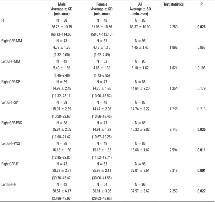
The palatal index for the 43 male and 54 female adults who were included in the study, and the dis-tributions of the values observed in the right and left GPF measurements are given in Table 1. Accordingly, the average PI of the 38 measurable adult males was 86.28 ± 10.75, whereas the PI of the 48 measurable adult females was calculated to be 81.06 ± 10.56. It was found that the PI values of adult male were statistically significantly higher than those of adult female (t = 2.260, p = 0.026).
The individuals in the study were grouped ac-cording to PI as leptostaphyline, mesostaphyline or brachystaphyline. As for the distribution of these types of palate by sex, the brachystaphyline type was observed in 50.0% (n = 19) of males, and the leptostaphyline type palate was observed in 43.8% (n = 21) of females. No difference was observed in the distribution of palate types by sex (c2 = 3.964,
p = 0.138).
The distribution of the closest maxillary molar at right side by sex and type of palate was determined accordingly that the third molar was the closest MM at right in 49.0% (n = 26) and 62.7% (n = 27) of females and males, respectively. It was found that the rate of those for whom third molar was the closest MM at right was the highest among all palate types (leptostaphyline: 53.1%, mesostaphyline: 65.0%, brachystaphyline: 48.4%).
As for the distribution of the closest MM at left side by sex and type of palate, the number of those whose third molar was the closest MM at left was calculated to be 47.1% (n = 25) and 60.9% (n = 25) for females and males, respectively. Among those with the mes-ostaphyline type of palate, no one had the second molar as the closest MM at left.
At right, the distance of GPF to the posterior nasal spine was measured to be 15.84 ± 2.05 mm (n = 38) in males and 14.91 ± 1.93 (n = 47) mm in females, averaging to 15.33 ± 2.02 mm (n = 85). And at the left side, the values obtained were 16.18 ± 1.80 mm (n = 38) for males and 15.16 ± 1.82 mm (n = 48) for females, averaging to 15.66 ± 1.87 mm (n = 86).
Distance to incisive foramen from GPF, at the right side found 38.27 ± 3.61 (n = 43) in males and 35.99 ± 3.11 (n = 53) in females, with an average total of 37.01 ± 3.51 (n = 96). And the left side values were 38.54 ± 4.17 (n = 42) for males and 36.81 ± 3.06 (n = 54), with the average 37.57 ± 3.67 (n = 96).
As for distance to sagittal plane, at the right side found 14.99 ± 2.45 (n = 39) in males and 14.35 ± ± 1.95 (n = 47) in females, with an average total of 14.64 ± 2.20 (n = 86). And the left side values were 15.07 ± 2.39 (n = 38) for males and 14.47 ± 2.06 (n = 47), with the average 14.74 ± 2.22 (n = 86).
Except for one case in our study, the number of GPF was found to be one each at two sides. In the excep-tional case, no GPF was found at the right side (Fig. 3). Our data demonstrated 64.94% oval/round shape and an antero-posterior extension. A compar-ison according to sex and types of palate showed that the shape of GPF was oval in 62.8% (n = 27) of
Figure 2. Calculation of palatal index; A — length of palate; B —
males, and round and 34.0% (n = 18) of females at right. No statistically significant difference was found in the distribution of the right GPF shape by sex. As for the left side, GPF’s shape was oval in 61.0% (n = 25) of males and 66.0% (n = 35) of females. No statistically significant difference was determined in the distribution of the left GPF shape by sex.
Lesser palatine foramen associated with sex and type of palate, and one left-side LPF was found in 40.5% (n = 17) of males and 63.0% (n = 34) of females. In adults with the leptostaphyline type of palate, two left-side LPF was observed at the most. In a great majority of the cases, one LPF was found regardless of sex and type of palate.
Table 1. Palatal index, right-left greater palatine foramen (GPF) distribution according to genders and overall samples Male Average ± SD (min–max) Female Average ± SD (min–max) All Average ± SD (min–max) Test statistics P PI N = 38 N = 48 N = 86 86.28 ± 10.75 (66.13–114.00) 81.06 ± 10.56 (58.67–112.10) 83.37 ± 10.90 2.260 0.026 Right GPF-MM N = 43 N = 53 N = 96 4.77 ± 1.75 (1.32–9.00) 4.18 ± 1.15 (1.92–7.49) 4.45 ± 1.47 1.892 0.063 Left GPF-MM N = 42 N = 53 N = 95 5.40 ± 1.86 (1.46–9.48) 4.86 ± 1.38 (1.73–7.85) 5.10 ± 1.62 1.624 0.108 Right GPF-SP N = 39 N = 47 N = 86 14.99 ± 2.45 (11.32–23.11) 14.35 ± 1.95 (10.96–18.57) 14.64 ± 2.20 1.354 0.179 Left GPF-SP N = 39 N = 48 N = 87 15.07 ± 2.39 (10.28–23.63) 14.47 ± 2.06 (10.56–18.46) 14.74 ± 2.22 1.255 0.213 Right GPF-PNS N = 38 N = 47 N = 85 15.84 ± 2.05 (11.68–21.42) 14.91 ± 1.93 (10.67–19.25) 15.33 ± 2.02 2.143 0.035 Left GPF-PNS N = 38 N = 48 N = 86 16.18 ± 1.80 (12.95–22.85) 15.16 ± 1.82 (11.52–19.74) 15.66 ± 1.87 2.594 0.011 Right GPF-IF N = 43 N = 53 N = 96 38.27 ± 3.61 (30.76–45.47) 35.99 ± 3.11 (30.06–41.55) 37.01 ± 3.51 3.318 0.001 Left GPF-IF N = 42 N = 54 N = 96 38.54 ± 4.17 (30.86–48.92) 36.81 ± 3.06 (29.63–43.63) 37.57 ± 3.67 2.259 0.027
IF — incisive foramen; LPF — lesser palatine foramen; MM — maxillary molar; PI — palatal index; PNS — posterior nasal spine; SD — standard deviation; SP — sagittal plane
Figure 3. One exceptional sample includes just one greater palatine
DISCUSSION
Classical anatomy and surgery books provide some general information about the localisation of GPF. Some problems with surgical interventions at this region were reported due to the lack of detailed information about some parameters [20, 25, 33].
In recent years, studies were carried out attract-ing attention to the fact that GPF’s localisation is a clinically important anatomic point [25, 33]. The possibility of stimulating the pterygopalatine gan-glion through GPF also resulted in getting more at-tention [16, 26, 34]. It was reported upon these developments that GPF could be utilised for alleviat-ing the effects of paralysis in paralytic patients, and also for patient interventions in the cases of cerebral vasospasms or cluster and migraine headaches [26].
In the current study, the subjects were also classi-fied according to their palate types. The participants were grouped as leptostaphyline (narrow), mes-ostaphyline (medium) and brachystaphyline (wide) according to their PI. In previous palatal morphometry studies carried out in Turkey, no distinction was made according to sex. In Turkey, Gözil et al. [13] identified 58.1% leptostaphyline, 17.4% mesostaphyline and 24.4% brachystaphyline with an average value of 77.94 ± 9.54. In Kenya, Hassanali and Mwaniki [15] found 43.2% leptostaphyline, 23.7% mesostaphyline, and 33.1% brachystaphyline with an average value of 82.0 ± 7.84. In India, Dave et al. [6] found the lep-tostaphyline type of palate in 61.5% of females and 63.3% of males, the mesostaphyline type in 30.8% of females and 20.0% of males, and the brachy-staphyline type in 7.7% of females and 16.7% of males (Table 2). Dave’s findings demonstrated 63% leptostaphyline in the evaluation of all crania. In our study, the general PI was 83.37 ± 10.90 (n = 86). The average PI of the 38 measurable adult males was 86.28 ± 10.75, whereas the PI of the 48 measurable adult females was calculated to be 81.06 ± 10.56. As for the distribution of these palate types by sex, the brachystaphyline type was observed in 50.0% (n = 19) of males, and the leptostaphyline type palate was observed in 43.8% (n = 21) of females. In their respective studies, Hassanali and Mwaniki [15], Gözil et al. [13] and Dave et al. [6] found a great majority of their cases to belong to the leptostaphyline pal-ate group. In our study, however, this only applied to female crania. A majority of male crania were in the brachystaphyline palate group. As a result, the palatal indexes of males were found to be significantly higher than those of females. We are of the opinion
that the difference of measurement method might account for the index difference with other research-ers. Hassanali and Mwaniki [15] and Gözil et al. [13] measured palatal length from the oral point, which corresponds to the centre of the line that connects the posterior margins of the alveoli of maxillary central incisors, from the staphylon point, which corresponds to the centre of the line that connects the foremost points of the posterior margins of both sides of the hard palate. In the current study, which used Dave’s method, the length from the oral point to the pos-terior nasal spine was measured.
The literature on the subject shows no consensus on whether the location of GPF is influenced by ethnic dif-ferences. Wang et al. [35] supported the idea of ethnic effects, while Jaffar and Hamadah rejected this theory. Although homogeneous results were obtained in the studies carried out in Europe, significant variations were found by Indian researchers in their studies that were made on the same population group [5, 6, 19, 29]. This suggests that broad anatomic variations can occur even in the same population group, and a comparison of various studies is presented in Table 3. We had no opportunity for making a comparison for Turkey due to the lack of elaborate studies on the subject matter. In the current study, a bilateral approach was taken to the question of whether there is a little relationship between sex and type of palate and localisation. No sex-based difference was determined according to the results. Tomaszewska et al. [34], drew attention to the fact that there were significant differences between females and males and the measurements relating to GPF, and stated that this could be utilised even in forensic medicine applications. Our findings, however, do not support this theory as we determined it to be generally aligned with the third molar across the population regardless of sex or type of palate. A comparison of the studies previously performed on the localisation of GPF is given in Table 3.
We also evaluated metric values for positioning GPF as distance to sagittal plane, posterior nasal spine and incisive foramen. Gibelli et al. [12] showed all these values were significantly influenced by sex at their study conducted on Italian crania. Our re-sults for distance to sagittal plane did not show any statistically significant difference according to sex, side or palate type. However, our other two values were statistically significant according to sex and side. All measurements Gibelli et al. [12]; indicated were slightly higher than our results which could be a possible result of palate type. However, they did not
Table 2. Comparison of studies including palatal index
Studies Leptostaphyline Mesostaphyline Brachystaphyline Overall Female Male Overall Female Male Overall Female Male
Our study (2017) 37.5% 43.8% 28.9% 24.4% 27.0% 21.1% 38.3% 29.2% 50.0%
Dave et al. (2013) [6] 63% 61.5% 63.3% 24% 30.%8 20.0% 13% 7.7% 16.7%
Gözil et al. (1999) [13] 58.1% – – 17.4% – – 24.4% – –
Hassanali and Mwaniki (1984)
[15] 43.2% – – 23.7% – – 33.1% – –
Table 3. Comparison of previous studies
Studies GPF-SP [mm]
Right/Left GPF-posterior margin [mm] MM2 Relation with maxillary molar (%)Between MM2
and MM3 MM3 Distal to MM3
Westmoreland and Blanton 1982 [36] 14.8/15.0 1.9 9.70 33.60 50.70 6.00
Langenegger et al. 1983 [21] – – 1.00 3.00 62.00 34.00
Hassanali and Mwaniki 1984 [15] – – 10.40 13.60 76.00 0.00
Ajmani 1994 (Nigerian Crania) [1] 15.4* 3.5 13.07 38.46 48.46 0.00
Ajmani 1994 (Indian Crania) [1] 14.7/14.6 3.7 0.00 32.35 64.69 2.94
Jaffar and Hamadah 2003 [17] 15.7* 4.86 12.00 19.00 55.00 14.00
Methathrathip et al. 2005 [22] 16.2* 2.1 7.00 14.10 71.90 7.00
Saralaya and Nayak 2007 [29] 14.7/14.7 4.2 0.40 24.20 74.60 0.80
Chrcanovic and Custódio 2010
(Brazilian Crania) [4] 14.68/14.44 3.39 0.00 6.19 54.87 38.94
Gibelli et al. 2017 (Italian Crania) [12] 16.4/16.8 3.8 –
*No distinction between right and left sides; GPF — greater palatine foramen; SP — sagital plane; MM1, MM2, MM3 — maxillary molar I, II, III
indicate the exact type of their crania. Our obtained values for distance to incisive foramen paralleled Kumar’s study regardless of sex [20].
Generally, GPF is considered as symmetrical at each side at least one. The literature includes one study which reports a bilateral lack of both GPF and LPF [33]. In our study, only at one sample, both GPF and LPF were lacking at right, whereas only LPF were lacking at left (Fig. 3). Variance of the number of LPF bilaterally regardless of palate type and sex was parallel with other studies [15, 33].
Greater palatine foramen showed variability in terms of shape and size. In some cases, its diameter was less than 1 mm, close to LPF in size, while it was observed to be rather large in some other cases. As for shape, stud-ies reported that it could be round or an oval structure with its longitudinal axis extending antero-posteriorly [15, 21]. Previous studies carried out on different popu-lations reported it to often possess an oval shape with an antero-posterior extension. Our data demonstrated these results are paralleled by our findings.
CONCLUSIONS
Greater palatine foramen might appear like an anatomic obstruction in all the interventions involving a maxillary division blockage of the trigeminal nerve especially in oral and maxillofacial surgery. We carried out this study on Turkish population in the belief that an understanding of GPF-related variations and assess-ment with different parameters would benefit clinicians in this sense. Regardless of sex and type of palate, GPF mostly had an oval shape and aligned with the third molar, and one each GPF changing in size was observed bilaterally in all the crania measured except for one. We hope that the data we obtained will provide also an index for future anthropological studies.
Acknowledgements
This study was conducted as a MSc Thesis at Istanbul University Cerrahpasa School of Medicine Anatomy Department during 2014 and 2015 Se-mester. Authors would like to thank chief of depart-ments of the Istanbul University, Istanbul School of
Medicine and School of Dentistry Anatomy Depart-ments, Ankara University School of Medicine Anatomy Department, Istanbul Medipol University, School of Medicine Anatomy Department and Bahcesehir Uni-versity School of Medicine Anatomy Department for their generous help as opening cranium collections belongs to their departments.
REFERENCES
1. Ajmani ML. Anatomical variation in position of the greater palatine foramen in the adult human skull. J Anat. 1994; 184 (Pt 3): 635–637, indexed in Pubmed: 7928651.
2. Canter SR, Slavkin HC, Canter MR. Anatomical study of pterygopalatine fossa and canal: considerations applicable to the anesthetization of the second division of the fifth cranial nerve. J Oral Surg Anesth Hosp Dent Serv. 1964; 22: 318–323, indexed in Pubmed: 14155475.
3. Chentanez V, Kaweewongprasert S, Thunvarachorn P, et al. Position of greater palatine foramens, length and direction of greater palatine canals: anatomic study of 120 adult human skulls. Chula Med J. 1985; 29: 1187–97.
4. Chrcanovic BR, Custódio ALN. Anatomical variation in the position of the greater palatine foramen. J Oral Sci. 2010; 52(1): 109–113, indexed in Pubmed: 20339241.
5. D’Souza AS, Mamatha H, Nayak J. Morphometric analysis of hard palate in south Indian skulls. Biomed Res. 2012; 23: 173–175.
6. Dave MR, Gupta S, Vyas KK, et al. study of palatal indices and bony prominences and grooves in the hard palate of adult human skulls. NJIRM. 2013; 4(1): 7–11.
7. Diyarbakırlı S, Keleç P. Foramen palatinum majus varyasyonları. Atatürk Üniv Diş Hek Fak Derg. 1996; 6(1): 37–39.
8. Douglas R, Wormald PJ. Pterygopalatine fossa infiltration through the greater palatine foramen: where to bend the needle. Laryngoscope. 2006; 116(7): 1255–1257, doi: 10.1097/01.mlg.0000226005.43817.a2, indexed in Pubmed: 16826070.
9. Ducic Y, Herford AS. The use of palatal island flaps as an adjunct to microvascular free tissue transfer for reconstruc-tion of complex oromandibular defects. Laryngoscope. 2001; 111(9): 1666–1669, doi: 10.1097/00005537-200109000-00033, indexed in Pubmed: 11568625.
10. Erimoğlu C, Özbayrak T, Kökten G. Foramen palatinum majus ve canalis palatinus majorun anatoma-morfolojik varyasyonları. JIUFD 1983;17(3-4. : 144–157.
11. Gardner E, Gray DJ, Rahilly R. Anatomy: a regional study of hu-man structure. 4th edition. Philadelphia: WB Saunders. 1975. 12. Gibelli D, Borlando A, Dolci C, et al. Anatomical character-istics of greater palatine foramen: a novel point of view. Surg Radiol Anat. 2017; 39(12): 1359–1368, doi: 10.1007/ s00276-017-1899-7, indexed in Pubmed: 28752360. 13. Gözil R, Şakul BU, Çalgüner E, et al. Sert damak morfometrisi
ve torus palatinus, sutura palatina transversa şekilleri. Turkiye Klinikleri J Dental Sci. 1999; 5(3): 149–153.
14. Harnet JC, Lombardi T, Lutz JC, et al. Sagittal craniofacial growth evaluated on children dry skulls using V2 and V3 canal openings as references. Surg Radiol Anat. 2007; 29(7): 589–594, doi: 10.1007/s00276-007-0237-x, indexed in Pubmed: 17653618.
15. Hassanali J, Mwaniki D. Palatal analysis and osteology of the hard palate of the Kenyan African skulls. Anat Rec. 1984; 209(2): 273–280, doi: 10.1002/ar.1092090213, indexed in Pubmed: 6465536.
16. Ikuta CR, Cardoso CL, Ferreira-Júnior O, et al. Position of the greater palatine foramen: an anatomical study through cone beam computed tomography images. Surg Radiol Anat. 2013; 35(9): 837–842, doi: 10.1007/s00276-013-1151-z, indexed in Pubmed: 23811933.
17. Jaffar AA, Hamadah HJ. An analysis of the position of the greater palatine foramen. J Basic Med Sci. 2003; 3(1): 24–32.
18. Jones RG. The reduction of bleeding in hare-lip and cleft-palate surgery. Br J Anaesth. 1962; 34: 481–488, indexed in Pubmed: 14452322.
19. Jotania B, Patel SV, Patel SM, et al. Morphometric analysis of hard palate. Int J Res Med. 2013; 2(2): 72–75. 20. Kumar A, Anu S, Poonam S. Assessment of the relative
location of greater palatine foramen in adult Indian skulls: Consideration for maxillary nerve block. Eur J Anat. 2011; 15(3): 150–154.
21. Langenegger JJ, Lownie JF, Cleaton-Jones PE. The relation-ship of the greater palatine foramen to the molar teeth and pterygoid hamulus in human skulls. J Dent. 1983; 11(3): 249–256, indexed in Pubmed: 6580311.
22. Methathrathip D, Apinhasmit W, Chompoopong S, et al. Anatomy of greater palatine foramen and canal and pterygopalatine fossa in Thais: considerations for maxillary nerve block. Surg Radiol Anat. 2005; 27(6): 511–516, doi: 10.1007/s00276-005-0016-5, indexed in Pubmed: 16228112.
23. Moore KL. Clinically Oriented Anatomy. 5th ed. Williams and Wilkins, Baltimore 2006: 886–996.
24. Nevin M, Puterbaugh PG. Conduction, Infiltration, and General Anesthesia in Dentistry. 4th ed. Brooklyn: Dental Items Interest Pub. 1938.
25. Nimigean V, Nimigean VR, Buţincu L, et al. Anatomical and clinical considerations regarding the greater palatine foramen. Rom J Morphol Embryol. 2013; 54(3 Suppl): 779–783, indexed in Pubmed: 24322027.
26. Piagkou M, Xanthos T, Anagnostopoulou S, et al. Anatomi-cal variation and morphology in the position of the pala-tine foramina in adult human skulls from Greece. J Cranio-maxillofac Surg. 2012; 40(7): e206–e210, doi: 10.1016/j. jcms.2011.10.011, indexed in Pubmed: 22055651. 27. Romanes GJ. Cunningham Textbook ofAnatomy. 12th ed.
New York: Oxford University Press. 1981.
28. Sakul BU, Turkaslan AK. and Kose S.K. The examination of the asymmetry on the human dry skull. X. International Symposium on Morphological Sciences, Toronto, Ontario, Canada, July 1-5. 1991(Poster No 88).
29. Saralaya V, Nayak SR. The relative position of the greater palatine foramen in dry Indian skulls. Singapore Med J. 2007; 48(12): 1143–1146, indexed in Pubmed: 18043845. 30. Sejrsen B, Kjaer I, Jakobsen J. Human palatal growth evalu-ated on medieval crania using nerve canal openings as references. Am J Phys Anthropol. 1996; 99(4): 603–611, doi: 10.1002/(SICI)1096-8644(199604)99:4<603::AID-AJPA6>3.0.CO;2-U, indexed in Pubmed: 8779341. 31. Seldin H. Practical Anesthesia for Dental and Oral Surgery,
Local and General. Philadelphia: Lea & Febiger. 1942. 32. Shane SME. Principles of Sedation, Local, and General
Anesthesia in Dentistry. Springfield: Thomas. 1975. 33. Sharma NA, Garud RS. Greater palatine foramen--key to
successful hemimaxillary anaesthesia: a morphometric study and report of a rare aberration. Singapore Med J. 2013; 54(3): 152–159, indexed in Pubmed: 23546029. 34. Tomaszewska IM, Tomaszewski KA, Kmiotek EK, et al.
Anatomical landmarks for the localization of the greater palatine foramen--a study of 1200 head CTs, 150 dry skulls, systematic review of literature and meta-analysis. J Anat. 2014; 225(4): 419–435, doi: 10.1111/joa.12221, indexed in Pubmed: 25131842.
35. Wang TM, Kuo KJ, Shih C, et al. Assessment of the relative locations of the greater palatine foramen in adult Chinese skulls. Acta Anat (Basel). 1988; 132(3): 182–186, indexed in Pubmed: 3414365.
36. Westmoreland EE, Blanton PL. An analysis of the variations in position of the greater palatine foramen in the adult hu-man skull. Anat Rec. 1982; 204(4): 383–388, doi: 10.1002/ ar.1092040412, indexed in Pubmed: 7181144.