ISSN 1015- 3918
ANKARA ÜNİVERSİTESİ
ECZACILIK FAKÜLTESİ
DERGİSİ
JOURNAL OF FACULTY OF PHARMACY
OF
ANKARA UNIVERSITY
Cilt/Vol :28
Sayı/No : 1
Til/Year: 1999
ANKARA ÜNİVERSİTESİ
ECZACILIK FAKÜLTESİ
DERGİSİ
JOURNAL OF FACULTY OF PHARMACY
OF
ANKARA UNIVERSITY
Cilt/Vol : 28 Sayı/No : 1 Yıl/Year: 1999 Ankara - 1999ANKARA ÜNİVERSİTESİ ECZACILIK FAKÜLTESİ
DERGİSİ
Sahibi: Prof.Dr. Seçkin ÖZDEN Editör : Prof.Dr. Feyyaz ONUR
Yayın Kurulu : Prof.Dr. Feyyaz ONUR (Başkan) Prof.Dr. Nazire ÖZKAL
Prof.Dr. Nuray ARI
Doç.Dr. Gülbin ÖZÇELİKAY Yrd.Doç. Dr. Meral TUNÇBİLEK Dr.Ecz. Yıldız ÖZALP
Ankara Üniversitesi Eczacılık Fakültesi Dergisi yılda 2 sayı yayınlanır. Yayımlanan yazıların sorumluluğu yazarlarına aittir.
Bu dergi Chemical Abstracts (CA), Excerpta Medica Database (EMBASE), Medicinal Aromatic Plants Abstracts (MAPA) ve Türk Tıp Dizini'nde indekslenmektedir. Yazışma adresi: Ankara Üniversitesi, Eczacılık Fakültesi, 06100 Tandoğan -Ankara Tel: (0312) 222 04 71 Fax: (0312) 213 10 81 e-mali : [email protected].
Ankara Üniversitesi Basımevi, 1999
JOURNAL OF FACULTY OF PHARMACY OF
ANKARA UNIVERSITY
Published by : Prof.Dr. Seçkin ÖZDEN Editor : Prof.Dr. Feyyaz ONUR
Editorial Board : Prof.Dr. Feyyaz ONUR (Editor-in-Chief)
Prof.Dr. Nazire ÖZKAL Prof.Dr. Nuray ARI
Doç.Dr. Gülbin ÖZÇELİKAY Yrd.Doç.Dr. Meral TUNÇBİLEK
Dr.Ecz. Yıldız ÖZALP
Journal of Faculty of Pharmacy of Ankara University is published in semiannual volumes.
All the articles appeared in this journal are published on the responsibility of the author.
This journal is indexed in Chemical Abstracts (CA), Excerpta Medica
Database (EMBASE), Medicinal Aromatic Plants Abstracts (MAPA) and Turkish Medical Index.
Address: Ankara University, Faculty of Pharmacy, 06100 Tandoğan - ANKARA TURKEY Tel :+90 312 222 04 71 Fax :+90 312 213 10 81 e-mail : [email protected].
Ankara Üniversitesi Basımevi, 1999
İÇİNDEKİLER /CONTENTS
Sayfa
Orjinal Makaleler / Original Articles
Sinan SÜZEN, James M, PARRY • The use of an internal standard molecule for determining
mutation frequency in restriction site mutation method, Sınırlı bölge mutasyon
yöntemi ile mutasyon frekansının belirlenmesi için iç standart molekülü kullanımı. 1
Ayşegül GÜVENÇ, Mehmet KOYUNCU • Türkiye'de yetişen Asparagus L (Kuşkonmaz) türlerinin
kök anatomisi, Studies on anatomical structure of the roots of Asparagus species
(Liliaceae) growing in Turkey. 15
Yalçın DUYDU, Sinan SÜZEN, Nurten ERDEM, Handan UYSAL, Nevin VURAL- A modified
method for determination of hippuric acid in urine by HPLC, idrarda hippurik asit
tayininde kullanılabilecek bir modifiye HPLC yöntemi. 37
Gülhan CENGİZ, Nergiz AKSOY, Göknur AKTAY, Tülin SÖYLEMEZOĞLU • Parasetamol ve
aspirinin plazma ve karaciğerde lipid peroksidasyona etkisi, Effects of paracetamol
and aspirin on lipid peroxidation in plasma and liver, 47
Nurten ALTANLAR • Vulvovajinal candidiasis olgularından izole edilen candidaların türlere
Ankara Ecz. Fak. Derg. 28(1) 1-13, 1999
THE USE OF AN INTERNAL STANDARD MOLECULE FOR DETERMINING MUTATION FREQUENCY IN RESTRICTION SITE
MUTATION METHOD
SINIRLI BÖLGE MUTASYON YÖNTEMİ İLE MUTASYON FREKANSININ BELİRLENMESİ İÇİN İÇ STANDART MOLEKÜLÜ KULLANIMI
Sinan SÜZEN* James M. PARRY**
*Department of Toxicology, Faculty of Pharmacy, Ankara University, Tandoğan , Ankara **School of Biological Sciences, University of Wales Swansea, Swansea, UK
ABSTRACT
In order to overcome some problems associated with the effects of variables on in vitro amplification of specific DNA segments using polymerase chain reaction (PCR), an internal standard technique was included to restriction site mutation (RSM) method. The internal standard was designed with same nucleotide sequences as the target sequence but with a deletion so that differences in size could be used to distinguish the PCR products of the target and internal standard. The application of the internal standard usage in the RSM method was performed on the rat ras gene. N-methyl-N-nitrosourea (MNU) induced mutations were detected at frequencies 10-5
at the H-ras gene in exon 1 at Hind III restriction enzyme recognition sequence. The use of internal standard successfully allowed the calculation of mutation frequency.
Key words: internal standard, ras, PCR, RSM
ÖZET
Spesifik DNA kısımlarının polimeraz zincir reaksiyonu (PCR) kullanarak in vitro çoğaltılması üzerine değişkenlerin etkisi ile ilişkili bazı problemleri gidermek için, iç standart tekniği sınırlı bölge mutasyon (RSM) metoduna katıldı. İç standart, eksilme dışında hedef dizilim ile aynı nükleotid diziliminde dizayn edildi. Böylece büyüklükteki farklılıklar hedef ve iç standart'ın ayırımı için kullanılabildi. RSM tekniğinde iç standart kullanımının uygulaması sıçan ras geninde
J. Fac. Pharm. Ankara
2 Sinan SÜZEN, James M. PARRY
gerçekleştirildi. N-metil-N-nitrozaüre (MNU) ile indüklenmiş mutasyonlar 10-5 sıklıkla
H-ras geni ekson 1 'de HindIII restriksiyon enzim tanıma bölgesinde saptandı. İç standart kullanımı mııtasyon sıklığının hesaplanmasına başarılı bir şekilde olanak sağladı.
Anahtar kelimeler: iç standart, ras, PCR, RSM
INTRODUCTION
The restriction site mutation (RSM) assay is a technique which allows the study of potential mutagenicity of environmental chemicals (1-4). The effect of mutagens on unique restriction sites in any gene of a known sequence can be determined by eliminating the majority of wild-type sequences by cutting the sequence at specific restriction site. Two basic methods are combined in the RSM assay for the isolation of sequences resistant to digestion occurring in restriction enzyme recognition sites and subsequent amplification of insensitive restriction enzyme sequences using polymerase chain reaction (PCR). The quantitation of insensitive restriction enzyme recognition sequences is an important requirement for the application of RSM assay.
Quantitation of reaction products in PCR is generally considered unreliable because the amount of PCR product increases exponentially with each cycle of amplification. Therefore, minute differences in any of the variables that affect the efficiency of amplification can dramatically alter product yield (5). Several factors affect the kinetics of each amplification. These include the length of the PCR product (5), the primer efficiency (6), initial amount of DNA (7), and the reaction conditions (8). Precise quantification of PCR products can potentially be achieved by the use of an internal standard molecule. Competitive PCR is quantitative adaptation of the PCR method in which a known number of copies of a synthetic mutated internal standard is introduced to the sample into the PCR reaction mixture (9). The internal standard is amplified with the same primers as the target sequence, and is distinguished from the target sequence either by size, restriction endonuclease cleavage, or specific hybridisation.
To circumvent the problem of variability in the efficiency of amplification, the inclusion of an internal standard to the RSM assay may provide reliable and aid in the absolute
Ankara Ecz. Fak. Derg 28 (1) 1-13, 1999
quantitation of resistant RSM products. In this study, internal standard RSM assay approach was performed on the N-methyl-7V-nitrosourea (MNU) induced mutations in the H-ras gene exon 1 fragment in order to quantify accurately the resistant RSM products. The principal of this approach is outlined in Figure 1.
Genomic DNA
Restriction enzyme digestion
PCR
ADDITION OF KNOWN NUMBER OF COPIES OF AN INTERNAL STANDARD
(Amplification of the unrestricted mutant and wild-type targets, and mutant internal standard molecules)
Amplified mutant target and internal standard sequences
to be visualised by polyacrylamide gel electrophoresis and silver staining Restriction enzyme digestion of the amplified wild-type target sequences Quantification of resistant RSM products by comparison
with amplified internal standard molecules
Figure 1. Outline of the internal standard included RSM assay.
MATERIALS AND METHODS Experimental animals
The RSM assay was performed on male rats. The MNU treatments were carried out in Laboratory for Carcinogenesis and Mutagenesis, National Institute of Public Health and Environmental Protection, Bilthoven, The Netherlands. Groups of two rats (200-240 g) were exposed to 20 mg (2 rats), and 60 mg (2 rats) of MNU per kg body weight, by LP. injection. Control animals (2 rats) were given phosphate-buffered saline (PBS). All the animals were
4 Sinan SÜZEN, James M. PARRY
sacrificed 7 days later. After sacrifice the rats were dissected and the brain, kidney, liver, pancreas, spleen and testis were removed and stored -70°C until DNA extraction.
DNA extraction
Genomic DNA was extracted using a standard proteinase K, phenol/chloroform method (10). The concentration of each DNA sample was quantitated by UV spectroscopy. The extracted DNA purity was determined from the absorbance ratio at 260 and 280 nm.
PCR primers for amplification of the H-ras gene
PCR primers for DNA amplification were selected by using the Primer Software Package available on Seqnet (SERC Daresbury Laboratories). Oligonucleotide sequences were designed to amplify a 105 bp fragment around exon 1 of H-ras gene. The selected primer sequences were as follows:
Forward (sense) primer: 5'-GTTTGGCAACCCCTGTAGAA-3'. Reverse (antisense) primer: 5'-AATGGTTCTGGATCAGCTGG-3'.
The reverse primer was synthesized on an Applied Biosystems Model 391 DNA Synthesiser. The forward primer, which was fluorescein labelled, was obtained from Pharmacia.
Construction of an internal standard
The internal standard molecule was constructed with the same sequence of target fragment but the HindIII and Cfol restriction endonuclease recognition sites were deleted, and a
Rsal restriction endonuclease recognition site was created in the middle of the molecule. It was
21 bases shorter than the target fragment. The synthetic molecule was then synthesized as described above. The H-ras exon 1 and the internal standard sequences were as follows:
The H-ras exon 1 sequences: 105 bp
5'- GT TTG GCA ACC CCT GTA GAA GCG ATG ACA GAA TAC AAG CTT* GTG
GTG GTG GGC GCT** GGA GGC GTG GGA AAGAGT GCC CTG ACC ATC CAG CTG
ATC CAG AACCATT -3'
*: HindIII recognition site (A AGCTT), **: Cfol recognition site (GCG C). The internal standard sequences: 84 bp
5'- GT TTG GCA ACC CCT GTA GAA GCG ATG ACA GAA TAC GGA GGC GTA C* GA AAG AGT GCC CTG ACC ATC CAG CTG ATC CAG AAC CAT T -3'
Ankara Ecz. Fak. Derg 28 (1) 1-13, 1999
Cloning of the internal standard molecule
In order to provide similar amplification features with the genomic target, the internal standard was cloned using The Original TA Cloning Kit (Invitrogen). This kit provides a quick, one-step cloning strategy for the direct insertion of a PCR product into a plasmid vector (11).
I. Producing PCR products of synthetic molecule
The PCR reaction was applied to the several dilutions of synthesized internal standard molecule. Aliquots (5 1) of dilutions were added to 50 1 reaction mixtures containing 1 X PCR buffer, 200 M each dNTP, 20 pmol each primer and 1.25 U Taq polymerase. This mixture was overlaid with 70 1 of mineral oil and then amplified on a Hybaid Omni Gene Thermal Cycler for 25 PCR cycles using the following thermocycling protocol:
Denaturation for 1 minute at 94°C, Annealing for 30 seconds at 56°C, Extension for 30 seconds at 72°C.
The final PCR products were characterised by resolving 10 1 aliquots by 6% polyacrylamide gel electrophoresis (PAGE) and stained with silver.
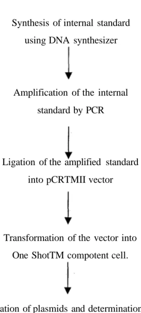
II. Cloning of the PCR amplified internal standard molecule
All cloning procedures (ligation, transformation) were carried out according to the manufacturer's recommendations. Flow chart of the experimental process is given in Figure 2.
6 Sinan SÜZEN, James M. PARRY
Synthesis of internal standard using DNA synthesizer
Amplification of the internal standard by PCR
Ligation of the amplified standard into pCRTMII vector
Transformation of the vector into One ShotTM compotent cell.
Isolation of plasmids and determination of the presence of insert by restriction digestion
Figure 2. Basic steps of producing the cloned internal standard molecule.
III. Quantitation of plasmids
After cloning, the plasmids which contain internal standard molecules were isolated using the QIA Prep-Spin Plasmid Purification Kit. The purified plasmids were then quantified using a Beckman DU-65 spectrophotometry by the measurement of optical density at 260 nm and were found to have concentration of 3.91 x 10¹⁰ copies per 1.
RSM assay with the internal standard molecule
1 g aliquots of genomic DNA extracted from N-methyl-N-nitrosourea (MNU) treated rats were digested using 12 units of HindIII restriction endonuclease with Taq buffer in a final volume of 20 1 overnight at 37°C. Known copy numbers, 100 copies, of the internal standard molecules were added to each of the digested mixtures. The mixtures were then amplified in a
Ankara Ecz. Fak. Derg 28 (1) 1-13, 1999 7
50 1 PCR reaction using 1,25 U Tag polymerase, 200 M of each dNTP, and 20 pmol of each primer. The reactions were overlaid with 70 1 of mineral oil and amplified on a Hybaid Omni Gene Thermal Cycler for 32 PCR cycles using the following thermocycling protocol:
a) one cycle of denaturation at 94°C for 2 minutes,
b) five cycles of denaturation at 94°C for 1 minute, annealing at the individual temperatures for 30 seconds, extension at 72°C for 30 seconds,
c) the appropriate number of cycles as indicated by denaturation at 94°C for 30 seconds, annealing at the individual temperatures for 30 seconds, extension at 72°C for 30 seconds.
17 1 of PCR products were then subjected to a second round of HindIII digestion at 37°C overnight. 10 J of the RSM products consisting of the amplified internal standard molecule were mixed with 2 1 of loading buffer and electrophoresed on 6% polyacrylamide gel and stained with silver.
Electrophoresis of resistant RSM products
The remaining resistant endonuclease recognition sequences were quantitated using the Automated Laser Fluorescent ALF DNA Sequencer (Pharmacia).
4 1 of resistant RSM products were mixed with 4 1 of loading dye solution (Pharmacia) and were loaded onto 6% 9:1 (acrylamide to bisacrylamide) acrylamide gel (Pharmacia Ready Mix) in 0.8 X TBE buffer system. Electrophoresis conditions were 1900 V, 150 mA, 36 W, 4 mW laser and 55°C for 1.5 hours.
The resistant RSM products and the amplified internal standard molecules were quantified on the ALF DNA Sequencer using the Fragment Manager Software Package. The Fragment Manager Software for ALF DNA Sequencer is designed specifically for high-resolution analysis and includes such features as fragment size calculation, peak or band quantitation.
RESULTS
In order to quantify the recognition sequences of resistant restriction endonuclease, the internal standard molecule was used for the RSM assay. The assay was performed on H-ras gene of exon 1 fragment of MNU treated rats.
After synthesizing, the internal standard was amplified with the same primer pair of the
H-ras gene exon 1 fragment. Restriction analysis of the internal standard molecules was carried
Sinan SÜZEN, James M. PARRY
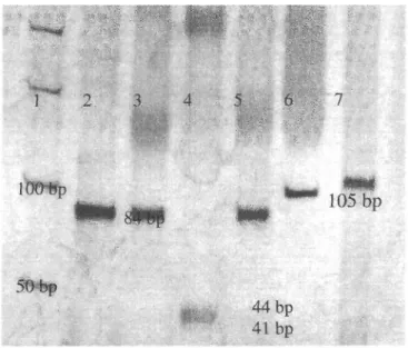
of the amplified products gave uncleaved bands, and Rsal restriction endonuclease digestion of the amplified products showed that the internal standard molecule was the same sequences as constructed. In addition, EcoRl restriction endonuclease digestion of plasmid molecules revealed that because the internal standard molecule was flanked on each side by EcoRl sites in the vector it was successfully inserted into the vector (Figure3).
Figure 3. Silver stained polyacrylamide gel shows restriction enzyme analysis of the amplified
internal standard molecules and plasmids. Lane 1 is gel marker. Lane 2 contains 84 basepairs (bp) product of the internal standard. Lane 3 and 5 contain the unrestricted internal standard molecules using HindII and Cfol restriction enzymes respectively. Lane 4 is Rsal restriction enzyme digestion fragments (41-44 bp) of the amplified internal standard molecules. Lane 6 shows the internal standard insert after EcoRl restriction enzyme digestion of the amplified plasmids. Lane 7 contains 105 bp of H-ras gene exon 1 amplification product.
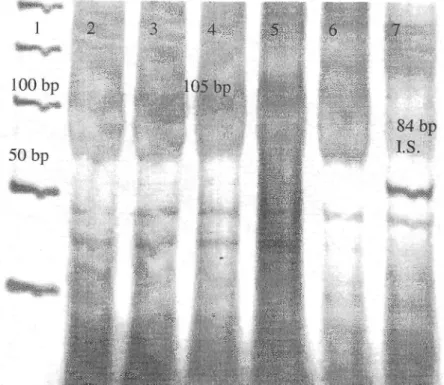
The internal standard included RSM assay was performed on the genomic DNA extracted from brain, kidney, liver, pancreas, spleen and testis on H-ras gene exon 1 fragment of HindIII restriction endonuclease recognition sequences. No resistant restriction enzyme recognition sequences were observed in the control untreated tissue samples. 5 resistant restriction enzyme sequences were detected in the DNA extracted from liver, pancreas and spleen in the MNU treated animals (Figure 4). Insensitive restriction enzyme sites were not detected in the DNA extracted from brain, kidney and testis.
Ankara Ecz. Fak. Derg 28 (1) 1-13, 1999
Figure 4. Silver stained 6% polyacrylamide gel shows the internal standard included RSM
assay analysis of the rat H-ras gene exon 1 fragment using HindIII restriction enzyme. Lane 1 is gel marker. Lanes 2-6 contain the resistant RSM assay products from the MNU treated rats. Lane 7 contains amplification products of the H-ras gene exon 1 fragment (105 bp) and the internal standard ( I S . ) molecules (84 bp).
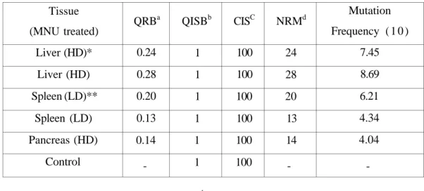
Resistant RSM products were quantitated on the ALF DNA Sequencer by comparing the amount of fluorescence in the internal standard and in the resistant RSM bands using the Fragment Manager Software Package. The quantity of the internal standard and resistant RSM products is shown in Table 1. The number of resistant RSM (NRM) molecules were calculated (12) as follows:
NRM = QRB X CIS / QISB
Where, QRB : quantity of resistant RSM band, CIS : copy number of internal standard, QISB : quantity of internal standard band.
10 Sinan SÜZEN, James M. PARRY
Table 1. The quantity data obtained of the internal standard and resistant RSM bands using the
Fragment Manager Software for ALF DNA Sequencer. The mutation frequency (MF) is equal to the (NRM) divided by the number of copies of the template in 1 g of genomic DNA (3.22 x
105). Tissue (MNU treated) Liver (HD)* Liver (HD) Spleen (LD)** Spleen (LD) Pancreas (HD) Control QRBa 0.24 0.28 0.20 0.13 0.14 -QISBb 1 1 1 1 1 1 CISC 100 100 100 100 100 100 NRMd 24 28 20 13 14 -Mutation Frequency ( 1 0 ) 7.45 8.69 6.21 4.34 4.04
-a: quantity of resistant RSM band, b: quantity of internal standard band,
c: copy number of internal standard, d: The number of resistant RSM molecules.
*: HD, high dose (60 mg/kg), **: LD, low dose (20 mg/kg).
DISCUSSION
The aim of this study was to develop a method to quantify resistant restriction endonuclease recognition sequences accurately by using the RSM assay. Quantification was achieved by using an internal standard molecule in the amplification step of the RSM assay.
There are some difficulties in quantitative amplification reactions as many factors are capable of varying with the ability of PCR to amplify small amount of nucleic acids exponentially. Thus, this exponential nature of DNA amplification may cause to unreliable quantitation in PCR because of tube to tube variations in the efficiency of amplification.
Despite the fact that there are sometimes unpredictable quantities of product obtained in PCR, the use of an internal standard sequence provides a control for the amplification. The internal standard may either have (1) different, or (2) identical primer requirements to the experimental target DNA. Although the former approach has the advantage that two pairs of primers are used in the reaction so that the experimental and internal standard targets do not
Ankara Ecz. Fak. Derg 28 (1) 1-13, 1999 1 1
compete for primers. This feature may also be seen as a disadvantage in that differences in primer efficiency between primer pairs may occur. The latter approach uses an internal standard which has the same primer requirements as the experimental target but differs in product site or by the presence of restriction site.
The internal standard molecule that was employed in quantification of H-ras gene was designed by deleting the HindIII restriction endonuclease recognition site present in the target sequence. Thus, a mutated internal standard molecule that was insensitive to HindIII restriction endonuclease digestion was used in the RSM assay. The internal standard molecule was 21 bases shorter than target fragment, hence, the identification of synthetic molecules from resistant RSM products was easily achieved by polyacrylamide gel electrophoresis. The same primer pair was used to amplify the internal standard molecule in order to amplify with equal efficiency with the target sequence. However, because of the shorter nature of the molecules, the preferential amplification of the internal standard in favour of the target genomic DNA sequences may create a problem. To overcome this problem and provide similar amplification features with the genomic target, the internal standard was cloned into a vector of 4 Kb. Thus, the synthetic molecule was not short and could be amplified under the same amplification conditions as the target sequences. The internal standard was included in the amplification of the RSM assay at approximately the same concentration as that expected for any mutated target restriction endonuclease recognition sequences.
The mutation frequency was ascertained for 5 resistant restriction endonuclease recognition sequences from the MNU treated rat tissues by running the samples on the ALF DNA Sequencer and quantitating the fluorescence of each RSM and internal standard band using the Fragment Manager Software. These insensitive RSM assay products including 2 livers, 2 spleens and 1 pancreas which had been observed in the HindIII restriction endonuclease recognition site of the H-ras gene exon 1 sequences. The calculated mutation frequencies were found to be 7.45 and 8.69 xl0-5 for liver, 4.34 and 6.21 x 10⁻⁵ for spleen and
4.03 x 10⁻⁵ for pancreas samples. No restriction endonuclease insensitive sequences were
observed on DNA extracted from untreated control animals. This indicates that the background mutation frequency was less than 5.8 x l0-7, assuming that 1 mg of mammalian genomic DNA
contains 3 x 10"5 copies of single copy gene (13). The average spontaneous mutation
frequencies is estimated to be 10-8 -10-10 mutations per base pair and these frequencies may
12 Sinan SÜZEN, James M. PARRY
mutation frequency may be in the order of 10-4-10-6. The MNU induced mutation frequency,
calculated to be 10-5 in this study, was consistent wit the expected figure.
In order to precisely quantify resistant restriction endonuclease recognition sequences, the internal standard molecules was included in the RSM assay in this study. The results presented in this paper suggest that the internal standard introduced into the RSM assay approach can provide accurate quantitation of small numbers of base changes in the restriction endonuclease recognition sequences and can be applied to any gene sequence of any tissue.
REFERENCES
1. Parry, J.M., Shamsher, M., Skibinski, D.O.F., "Restriction site mutation analysis, a
proposed methodology for the detection and study of DNA base changes following mutagen exposure" Mutagenesis, 5, 209-212 (1990).
2. Suzen, S., Jenkins, G.J.S., Parry, M., "Application of the restriction site mutation
technique to N-methyl-N-nitrosourea-induced mutations in the rat" Teratogen.
Carcinogen. Mutagen., 18, 171-182 (1998).
3. Jenkins, G.J.S., Mitchell, I.G., Parry, J.M., "Enhanced restriciton site mutation
(RSM) analysis of 1,2-dimethylhydrazine induced mutations, using endogenous p53 intron sequences" Mutagenesis, 12, 117-123 (1997).
4. Myers, B., Parry, J.M., "The application of the restriction site mutation assay to the study of the induction of mutations in the tissues of rodents" Mutagenesis, 9, 175-177 (1994).
5. Gilliand, G., Perrin, S., Bunn, N.F., "Competitive PCR for quantitation of mRNA" In:
PCR protocols: A guide in methods and applications. Innis, M.A., Gelfand, D.H., Sninsky, J.J. (Eds.), Academic Press, New York, p. 60-69 (1992).
6. Wang, M., Doyle, M.V., Mark, D.F., "Quantitation of mRNA by the polymerase chain
reaction" Proc. Natl. Acad. Sci. USA, 86, 9719-9721 (1992).
7. Katz, E.D., Bloch, W., Wages, J. Jr., "HPLC quantitation and identification of DNA
amplified by the polymerase chain reaction" Amplifications, 8, 10-13 (1992).
8. Higuchi, G.R., Fockler, C, Dollinger, G., Watson, R., "Kinetic PCR analysis:
Real-time monitoring of DNA amplification reactions" Biotechnology, 11, 1026- 1030 (1993).
9. Celi, S.F., Zenilman, E.M., Shudliner, R.A., "A rapid and versatile method to
Ankara Ecz. Fak. Derg 28 (1)1-13, 1999 13
10. Sambrook, J., Fritsch, E.F., Manniatis, T. Molecular Cloning A Laboratory Manual,
Cold Spring Harbor Laboratory Press, Second Edition, p.9.16-9.19 (1989).
11. Mead, D.A., Pey, N.K., Herrnstadt, C, Marcil, R.A., Smith, L.M. "A universal
method for the direct cloning of PCR amplified nucleic acid" Bio-Technol, 9, 657-663 (1991).
12. Kang, J., Kiihn, J.E., Schafer, P., Immelman, A., Henco, K. "Quantification of DNA
and RNA by PCR" in PCR 2. A Practical Approach, McPherson, M.J., Hames, B.D., Taylor, G.R. (Eds.) Oxford University Press, New York p.119-133 (1995).
13. Russel, P.J., Genetics, Fourth Edition, HarperCollins College Publishers, New York
(1996).
14. Cerutti, P., Hussain, P., Pourzand, C, Aguilar, F. "Mutagenesis of the H-ras