TURKISH JOURNAL of
DOI: 10.5152/TurkJOrthod.2017.17034
CASE REPORT
Treatment of Class II, Division 2 Malocclusion with
Miniscrew Supported En-Masse Retraction: Is
Deepbite Really an Obstacle for Extraction Treatment?
ABSTRACT
A 17-year-old female patient, whose chief complaint was her unpleasing smile, had skeletal and dental class II malocclusion, hypo-divergent facial type with a severely increased overbite. Among the treatment options, upper-first-premolar extractions followed by miniscrew-supported en-masse retraction was the treatment of choice. After the initial levelling and alignment, miniscrews with 1.5- to 1.4-mm diameter and 7-mm lenght, were installed between the roots of the second premolars and the first molars, bilaterally. En-masse retraction was achieved on a 0.016x0.022-inch stainless steel archwire with 7-mm long power hooks placed distal to the lat-eral incisors, and with nickel-titanium (NiTi) closed coil springs exerting 250-gr of force per side. At the end of the treatment, deepbite, incisor inclinations and interincisal angle were corrected, and Class II molar relationship with good intercuspation was achieved. Up-per 2-2, lower 3-3 retainers were bonded for retention. As a result, deepbite and Class II canine relationship was successfully corrected with simultaneous incisor intrusion and retraction using miniscrew-supported en-masse retraction.
Keywords: Orthodontic anchorage techniques, Class II malocclusion, division 2, orthodontic space closure
INTRODUCTION
It was the beginning of the new millennia when researchers presented solid evidence against the long-standing bias; that extraction therapy had major aesthetic drawbacks in patients with increased overbite and/or gingival exposure, as the incisors tend to extrude during retraction (1-3). Miniscrews were the secret ingredient in this new tratment recipe. In a clinical case report, Park and Kwon stated that maxillary anterior teeth showed bodily intrusion and retraction during miniscrew-supported en-masse retraction if an upward and backward force pass-ing near the center of resistance was used (1). Therefore, extraction therapy was no more a contraindication for deepbite patients when planned with the appropriate adjunct treatment mechanics.
The aim of this case report is to present the treatment planning and progress of a young adult patient with a skeletal Class 2 malocclusion and a severe deepbite.
CASE PRESENTATION
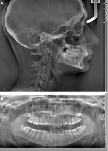
A 17-year-old female patient complained of her unpleasing smile. Diagnostic records revealed that she had a mild skeletal Class 2 malocclusion, bimaxillary retrusion, and hypodivergent facial type. She also had Angle Class II malocclusion with retrusive upper and lower incisors, mildly increased overjet, and severely increased overbite due to both upper- and lower-incisor overeruptions (Figure 1, 2). The Curve of Spee in the lower arch was deep-ened as expected. Skeletal maturational indicators showed that she had completed her growth. According to the dental casts analysis, 5.5 mm upper and 4.4 mm lower dental arch discrepancies were present.
Nilüfer İrem Tunçer, Ayça Arman Özçırpıcı
Department of Orthodontics, School of Dentistry Başkent University, Ankara, Turkey
Corresponding Author: Nilüfer İrem Tunçer, Department of Orthodontics, School of Dentistry Başkent University, Ankara, Turkey
E-mail: iremtuncher@gmail.com
©Copyright 2017 by Turkish Orthodontic Society - Available online at www.turkjorthod.org
Received: 26 July 2017 Accepted: 24 August 2017
84
Cite this article as: Tunçer Nİ, Özçırpıcı AA. Treatment of Class II, Division 2 Malocclusion with Miniscrew Supported En-Mase Retraction: Is Deepbite Really an Obstacle for Extraction Treatment? Turkish J Orthod 2017; 30: 84-8.
The following were the treatment options: mandibular advance-ment surgery, fixed functional appliance treatadvance-ment with maxil-lary molar distalization, and upper-first-premolar extractions fol-lowed by miniscrew-supported en-masse retraction. The patient was reluctant to accept the first two treatment options, and she decided to proceed with extraction therapy followed by minis-crew-supported en-masse retraction.
After the extractions, 0.018x0.025-inch incisor and canine brack-ets, and 0.022x0.028-inch premolar brackets and molar tubes with MBT prescription (VictoryTM Series, 3M Unitek, CA, USA)
were bonded on the upper arch. Wider brackets and tubes were used at the posterior section, and the second molars were not bonded until the end of retraction to ease the sliding of the arch-wire through the slots. The incisor brackets were not bonded incisally to let them express their predetermined torque values. After the maxillary dental arch was fully levelled and aligned, miniscrews 1.5- to 1.4-mm diameter and 7-mm lenght (AbsoAn-chor, Dentos, Daegu, Korea), were installed between the roots of the second premolars and the first molars bilaterally, and 6-8 mm apically to the archwire level. En-masse retraction was achieved on a 0.016x0.022-inch stainless steel archwire with 7-mm-long power hooks (Ortho Organizers, CA, USA) placed distal to the
lateral incisors. From these power hooks, nickel-titanium (NiTi) closed coil springs (Ormco Corp, CA, USA) were attached to the miniscrews and adjusted to exert 250-gr of force per side (Figure 3). Retraction was ended when canines reached Class I relation-ship.
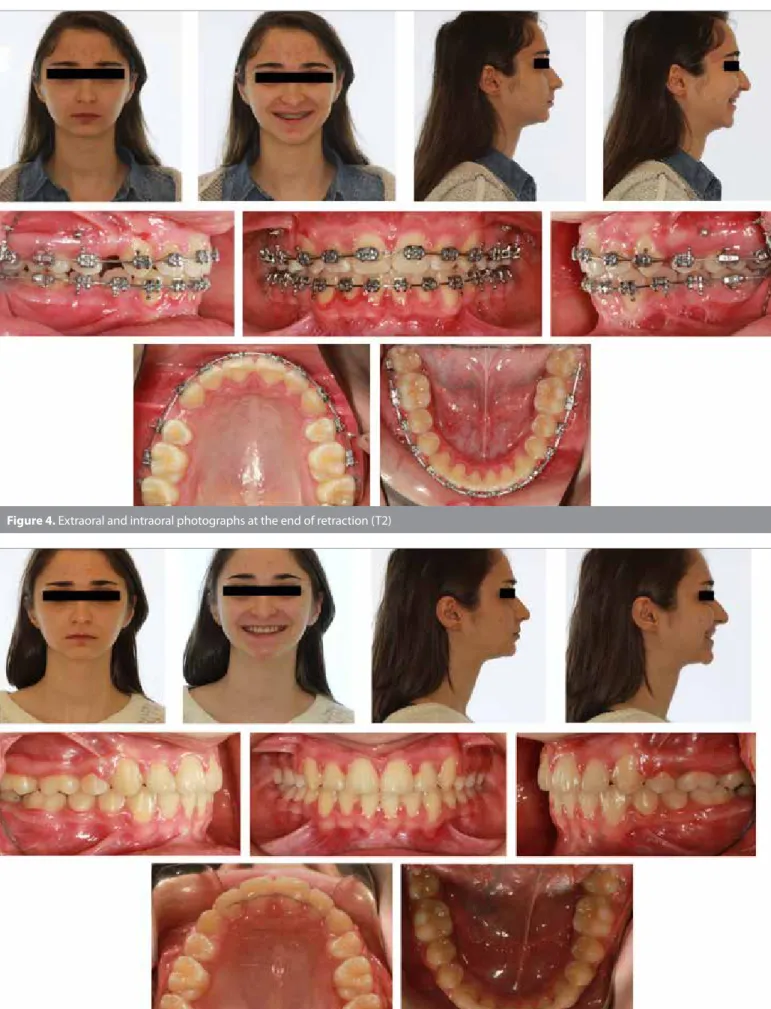
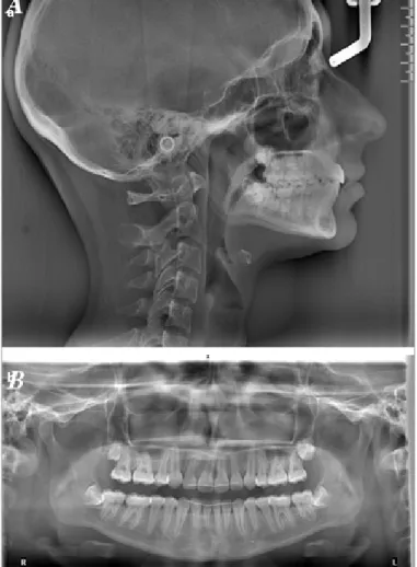
During en-masse retraction, molars moved distally and achieved a cusp-to-cusp relationship on the right side. At the end of the treatment, Class II molar relationship was preserved, and canines reached Class I canine relationship (Figure 4). Overbite and in-terincisal angles were overcorrected to provide retention of the deepbite. Supracrestal fiberotomies were performed around the formerly rotated teeth. Upper 2-2, lower 3-3 retainers were also bonded for retention (Figure 5, 6). The total treatment time was 29 months, without any significant root resorption.
DISCUSSION
Our aim in this case was to open the bite both with upper- and lower-incisor intrusions and still provide the patient with an aes-thetic smile against her decreased lower facial height. We were aware that the treatment planning needed compromise, because the upper-incisor exposure was almost ideal at the beginning of the treatment and would worsen with intrusion. However, this
85
was the best treatment approach we could offer the patient oth-er than surgoth-ery, given the sagittal and voth-ertical dimensions of the skeletal infrastructure.
Upper incisors intruded approximately 2.7 mm throughout the treatment period, 1 mm of which purely occurred during retrac-tion, as a result of the vertical component of the retraction force. The effect of intrusion can also be confirmed with the upward movement of the root apices of the incisors (−1.5 mm) (Figure 7). Yet, the numerical value of the intrusion degree can be assumed to be much higher, because the incisors kept intruding, while the retraction process itself was forcing them to extrude.
When evaluated sagittally, the anterior teeth tipped palatally by 6°. The amount of incisal edge and apical root movement was 4.5 mm and 1.3 mm, respectively. The Ia/Io ratio (the amount of apical root movement over incisal edge movement) was found to be 0.29 (2). These data, altogether, indicate that en-masse re-traction was primarily achieved by controlled tipping and partly by translation. This more parallel-like retraction pattern can be attributed to the level of force that is closer to the center of the resistance than conventional en-masse retraction.
The SNA angle decreased gradually during the treatment, where-as the SNB angle stayed almost the same, reducing the ANB an-gle. The A point moved 1.1 mm backwards as a result of 1.3 mm of apical root movement and 6° of palatal tipping (Figure 7). This movement of the A point can be explained as the result of alve-olar remodeling that occurred together with the change in the axial inclinations of incisors, which is also shown in a prospective study conducted by Al-Nimri et al. (4) on cases presenting Class
86
Turkish J Orthod 2017; 30: 84-8 Tunçer and Özçırpıcı. Miniscrew Supported En-Masse Retraction
Figure 3. Miniscrew-supported en-masse retraction mechanic. Figure 2. Pretreatment (T0) radiographic records; (a) lateral cephalometric radiograph, and (b) panoramic radiograph a
87
Figure 4. Extraoral and intraoral photographs at the end of retraction (T2)
II, division 2 malocclusion. Although a reduction in the promi-nence of soft tissue A point was not desired in this particular pa-tient, this type of tissue remodeling may especially be beneficial in bimaxillary protrusive or skeletal Class 2 cases with maxillary prognathia.
CONCLUSION
Miniscrews introduce versatility to the retraction mechanics by offering the opportunity of intruding the incisors, while retract-ing them in a more parallel fashion, without significant amount of root resorption or miniscrew loss.
Ethic Committee Approval: N/A.
Informed Consent: Written informed consent was obtained from the
patient.
Peer-review: Externally peer-reviewed.
Author Contributions: Concept - N.İ.T., A.A.Ö.; Design - N.İ.T., A.A.Ö.;
Su-pervision - N.İ.T., A.A.Ö.; Resources - N.İ.T., A.A.Ö.; Materials - N.İ.T., A.A.Ö.; Data Collection and/or Processing - N.İ.T.; Analysis and/or Interpretation - N.İ.T., A.A.Ö.; Literature Search - N.İ.T.; Writing Manuscript - N.İ.T.; Critical Review - N.İ.T., A.A.Ö.
Conflict of Interest: No conflict of interest was declared by the authors. Financial Disclosure: The authors declared that this study has received
no financial support. REFERENCES
1. Park HS, Kwon TG. Sliding mechanics with microscrew implant an-chorage. Angle Orthod 2004; 74: 703-10.
2. Upadhyay M, Yadav S, Patil S. Mini-implant anchorage for en-masse retraction of maxillary anterior teeth: a clinical cephalometric study. Am J Orthod Dentofacial Orthop 2008; 134: 803-10. [CrossRef] 3. Rajni N, Shetty KS, Prakash AT. To compare treatment duration,
anchor loss and quality of retraction using conventional enmasse sliding mechanics and enmasse sliding mechanics with using mi-cro-implants. Journal of Indian Orthodontic Society 2010; 44: 52-61. 4. Al-Nimri KS, Hazza'a AM, Al-Omari RM. Maxillary incisor proclination
effect on the position of point A in class II division 2 malocclusion. Angle Orthod 2009; 79: 880-4. [CrossRef]
88
Turkish J Orthod 2017; 30: 84-8 Tunçer and Özçırpıcı. Miniscrew Supported En-Masse Retraction
Figure 6. Posttreatment (T3) radiographic records; (a) lateral cephalometric radiograph, and (b) panoramic radiograph. a
b


