World Journal of
Gastrointestinal Oncology
World J Gastrointest Oncol 2018 October 15; 10(10): 282-366
S
Contents
Monthly Volume 10 Number 10 October 15, 2018
WJGO|www.wjgnet.com I October 15, 2018|Volume 10|Issue 10|
EDITORIAL
282 Optimizing outcomes for patients with gastric cancer peritoneal carcinomatosis
Leiting JL, Grotz TE
290 Inhibiting focal adhesion kinase: A potential target for enhancing therapeutic efficacy in colorectal cancer therapy
Jeong KY
REVIEW
293 Simultaneous curative resection of double colorectal carcinoma with synchronous bilobar liver metastases
De Raffele E, Mirarchi M, Cuicchi D, Lecce F, Ricci C, Casadei R, Cola B, Minni F
MINIREVIEWS
317 Histo-molecular oncogenesis of pancreatic cancer: From precancerous lesions to invasive ductal adenocarcinoma
Riva G, Pea A, Pilati C, Fiadone G, Lawlor RT, Scarpa A, Luchini C
328 Facing the challenge of venous thromboembolism prevention in patients undergoing major abdominal surgical procedures for gastrointestinal cancer
Mastoraki A, Mastoraki S, Schizas D, Patras R, Krinos N, Papanikolaou IS, Lazaris A, Liakakos T, Arkadopoulos N 336 Role of pre-transplant 18F-FDG PET/CT in predicting hepatocellular carcinoma recurrence after liver
transplantation
Yaprak O, Acar S, Ertugrul G, Dayangac M
ORIGINAL ARTICLE
Basic Study
344 miR-122-5p as a novel biomarker for alpha-fetoprotein-producing gastric cancer
Maruyama S, Furuya S, Shiraishi K, Shimizu H, Akaike H, Hosomura N, Kawaguchi Y, Amemiya H, Kawaida H, Sudo M, Inoue S, Kono H, Ichikawa D
Retrospective Study
351 Prognostic value of vascular endothelial growth factor receptor 1 and class III β-tubulin in survival for non-metastatic rectal cancer
Kong XQ, Huang YX, Li JL, Zhang XQ, Peng QQ, Tang LR, Wu JX
360 Predictive factors for lymph node metastasis and defining a subgroup treatable for laparoscopic lymph node dissection after endoscopic submucosal dissection in poorly differentiated early gastric cancer
Contents
ABOUT COVER
World Journal of Gastrointestinal Oncology
Volume 10 Number 10 October 15, 2018
Editorial Board Member of World Journal of Gastrointestinal Oncology, Jong Park, PhD, Associate Professor, Division of Cancer Prevention and Control, H. Lee Moffitt Cancer Center, College of Medicine, University of South Florida, Tampa, FL 33612, United States
World Journal of Gastrointestinal Oncology (World J Gastrointest Oncol, WJGO, online ISSN
1948-5204, DOI: 10.4251) is a peer-reviewed open access academic journal that aims to guide clinical practice and improve diagnostic and therapeutic skills of clinicians.
WJGO covers topics concerning carcinogenesis, tumorigenesis, metastasis, diagnosis,
prevention, prognosis, clinical manifestations, nutritional support, molecular mechanisms, and therapy of benign and malignant tumors of the digestive tract. The current columns of WJGO include editorial, frontier, diagnostic advances, therapeutics advances, field of
vision, mini-reviews, review, topic highlight, medical ethics, original articles, case report, clinical case conference (Clinicopathological conference), and autobiography. Priority publication will be given to articles concerning diagnosis and treatment of gastrointestinal oncology diseases. The following aspects are covered: Clinical diagnosis, laboratory diagnosis, differential diagnosis, imaging tests, pathological diagnosis, molecular biological diagnosis, immunological diagnosis, genetic diagnosis, functional diagnostics, and physical diagnosis; and comprehensive therapy, drug therapy, surgical therapy, interventional treatment, minimally invasive therapy, and robot-assisted therapy.
We encourage authors to submit their manuscripts to WJGO. We will give priority
to manuscripts that are supported by major national and international foundations and those that are of great clinical significance.
World Journal of Gastrointestinal Oncology (WJGO) is now indexed in Science Citation Index
Expanded (also known as SciSearch®), PubMed, and PubMed Central. The 2018 edition of
Journal Citation Reports® cites the 2017 impact factor for WJGO as 3.140 (5-year impact factor: 3.228), ranking WJGO as 39 among 80 journals in gastroenterology and hepatology (quartile in category Q2), and 114 among 222 journals in oncology (quartile in category Q3).
7901 Stoneridge Drive, Suite 501, Pleasanton, CA 94588, USA Telephone: +1-925-2238242
Fax: +1-925-2238243
E-mail: [email protected] Help Desk: http://www.f6publishing.com/helpdesk http://www.wjgnet.com
PUBLISHER
Baishideng Publishing Group Inc 7901 Stoneridge Drive,
Suite 501, Pleasanton, CA 94588, USA Telephone: +1-925-2238242 Fax: +1-925-2238243 E-mail: [email protected]
Help Desk: http://www.f6publishing.com/helpdesk http://www.wjgnet.com
PUBLICATION DATE October 15, 2018
COPYRIGHT
© 2018 Baishideng Publishing Group Inc. Articles published by this Open-Access journal are distributed under the terms of the Creative Commons Attribution Non-commercial License, which permits use, distribu-tion, and reproduction in any medium, provided the original work is properly cited, the use is non commer-cial and is otherwise in compliance with the license. SPECIAL STATEMENT
All articles published in journals owned by the Baishideng Publishing Group (BPG) represent the views and opinions of their authors, and not the views, opinions or policies of the BPG, except where other-wise explicitly indicated.
INSTRUCTIONS TO AUTHORS http://www.wjgnet.com/bpg/gerinfo/204 ONLINE SUBMISSION
http://www.f6publishing.com NAME OF JOURNAL
World Journal of Gastrointestinal Oncology
ISSN ISSN 1948-5204 (online) LAUNCH DATE February 15, 2009 FREQUENCY Monthly EDITORIAL BOARD MEMBERS
All editorial board members resources online at http:// www.wjgnet.com/1948-5204/editorialboard.htm EDITORIAL OFFICE
Jin-Lei Wang, Director
World Journal of Gastrointestinal Oncology
Baishideng Publishing Group Inc
EDITORS FOR
THIS ISSUE
Responsible Assistant Editor: Xiang Li Responsible Science Editor: Fang-Fang Ji Responsible Electronic Editor: Wen-Wen Tan Proofing Editorial Office Director: Jin-Lei Wang Proofing Editor-in-Chief: Lian-Sheng Ma
AIM AND SCOPE
Published online: October 15, 2018
Abstract
The last two decades have seen a paradigm shift in the selection of patients with hepatocellular carcinoma (HCC) for liver transplantation. Microvascular invasion and differentiation have been the most significant factors affecting post-transplant recurrence; however, because of inherent disadvantages of pre-transplant biopsy, histological criteria never gained popularity. Recently, the selection criteria evolved from morphological to biological criteria, such as biomarkers and response to loco-regional therapy. With the introduction of multi-modality imaging, combination of computed tomo-graphy with nuclear medicine imaging, particularly, 18F-fluorodeoxyglucose positron emission tomography fulfilled an unmet need and rapidly became a critical component of HCC management. This review article will focus on the use of 18F-fluorodeoxyglucose posi-tron emission tomography combined with computed tomography in the pre-transplant evaluation of HCC patients with special discussion on its ability to predict HCC recurrence after liver transplantation.
Key words: 18F-fluorodeoxyglucose positron emission tomography; Hepatocellular carcinoma; Recurrence; Liver transplantation
© The Author(s) 2018. Published by Baishideng Publishing
Group Inc. All rights reserved.
Core tip: The last two decades have seen a paradigm shift in the selection of patients with hepatocellular carcinoma (HCC) for liver transplantation. With the introduction of multimodality imaging, combination of computed tomography with nuclear medicine imaging
Onur Yaprak, Gokhan Ertugrul, Murat Dayangac, Medipol University Hospital, Center for Organ Transplantation, Istanbul 34214, Turkey
Sencan Acar, Atasehir Memorial Hospital, Center for Organ Transplantation, Istanbul 34758, Turkey
ORCID number: Onur Yaprak (0000-0003-1941-8290); Sencan
Acar (0000-0001-8086-0956); Gokhan Ertugrul (0000-0002- 8351-4220); Murat Dayangac (0000-0002-1240-7233).
Author contributions: Yaprak O designed the aim of this
minireview; Dayangac M, Yaprak O, Acar S and Ertugrul G contributed equally to this work, reviewed the references, generated the tables, and wrote the manuscript.
Conflict-of-interest statement: No conflict of interest.
Open-Access: This article is an open-access article which was
selected by an in-house editor and fully peer-reviewed by external reviewers. It is distributed in accordance with the Creative Commons Attribution Non Commercial (CC BY-NC 4.0) license, which permits others to distribute, remix, adapt, build upon this work non-commercially, and license their derivative works on different terms, provided the original work is properly cited and the use is non-commercial. See: http://creativecommons.org/ licenses/by-nc/4.0/
Manuscript source: Invited manuscript
Correspondence to: Onur Yaprak, MD, Associate Professor,
Medipol University Hospital, Center for Organ Transplantation, Goztepe Mah. Metin Sk. No.4, Bagcilar, Istanbul 34214, Turkey. [email protected]
Telephone: +90-53-22239566 Received: April 11, 2018
Peer-review started: April 11, 2018 First decision: April 23, 2018 Revised: May 27, 2018 Accepted: June 13, 2018 Article in press: June 14, 2018
336 Submit a Manuscript: http://www.f6publishing.com
DOI: 10.4251/wjgo.v10.i10.336
World J Gastrointest Oncol 2018 October 15; 10(10): 336-343
ISSN 1948-5204 (online)
October 15, 2018|Volume 10|Issue 10|
WJGO|www.wjgnet.com
MINIREVIEWS
Role of pre-transplant 18F-FDG PET/CT in predicting
hepatocellular carcinoma recurrence after liver
transplantation
fulfilled an unmet need and rapidly became a critical component of HCC management. This review article will focus on the use of 18F-fluorodeoxyglucose positron emission tomography in the pre-transplant evaluation of HCC patients with special discussion on its ability to predict HCC recurrence after liver transplantation.
Yaprak O, Acar S, Ertugrul G, Dayangac M. Role of pre-transplant 18F-FDG PET/CT in predicting hepatocellular carcinoma recur-rence after liver transplantation. World J Gastrointest Oncol 2018; 10(10): 336-343 Available from: URL: http://www. wjgnet.com/1948-5204/full/v10/i10/336.htm DOI: http://dx.doi. org/10.4251/wjgo.v10.i10.336
INTRODUCTION
Hepatocellular carcinoma (HCC) is the most common type of primary liver cancer. Currently, HCC is the sixth most common cancer with more than a half million new cases diagnosed annually, and it is the second leading cause of cancer-related mortality in the world[1]. The
global risk of HCC has been largely associated with hepatitis B and C virus infection. In addition, improved survival from cirrhosis and increasing rates of obesity and non-alcoholic fatty liver disease are expected to contribute to the ever-increasing incidence of HCC[2,3].
Because of the strong link between cirrhosis and HCC, liver transplantation (LT) is the best treatment option, since it removes the tumor and the underlying tumor-generating cirrhosis. Recently, HCC has been reported as the most common indication for LT in the United States[4].
Until the landmark study by Mazzaferro et al[5] in
1996, the liberal selection of HCC patients for LT resulted in high recurrence rates and poor survival. With the introduction of Milan criteria (MC), excellent long-term outcomes have been achieved that were not different from those of patients without HCC. The MC have been validated in several studies and widely accepted as the benchmark for selection of patients with HCC for de-ceased donor LT (DDLT). Subsequent studies searching for more liberal morphological criteria have shown that it was possible to extend the size and number of tumors without compromising post-transplant outcome[6-11] (Table
1). Despite being continually expanded, aforementioned morphological criteria have been criticized for a variety of reasons: they were restrictive and precluded numerous patients who otherwise would have benefited from LT with a low risk of HCC recurrence; they relied solely on tumor burden (defined as the size and number of tumors at a certain point) and excluded the factors related to tumor behavior (i.e., tumor differentiation, molecular markers, and response to bridging therapy); they depen-ded on imaging parameters that were inconsistent: in patients within MC, up to 40% had explant pathology that exceeded the MC, and in those beyond MC, up to
34% had explant pathology that was within the MC[12,13].
An earlier study investigating the correlation between pathologic and radiologic staging according to the mor-phological criteria have found that the accuracy of ima-ging classification for both Milan and (University of Cali fornia San Francisco (UCSF) criteria was only 60%[14].
In patients with HCC, vascular invasion has been defined as one of the major determinants of the out-come after LT[15]. Further studies have shown that tumor
differentiation has also been an independent predictor of recurrence and survival after the transplant[16,17]. Despite
initial hesitancy against the use of pre-transplant tumor biopsy, Toronto criteria have led the way to the use of histological criteria in selection of patients with HCC for LT[12]. However, pre-transplant tumor biopsy has not
gained popularity because of its limitations: In spite of the invasive biopsy procedures, the presence of vascular invasion and tumor differentiation may not be detec-ted reliably; the sensitivity of biopsy varies depending on location of the tumor, needle size, and tumor size. Moreover, preoperative needle biopsy may increase tumor seeding and post-transplant recurrence[18].
Never-theless, this was the beginning of a new era when there was a shift in selection criteria from morphological to the combination of biological and histomorphological criteria[19].
Meanwhile, major transplant centers in Asia star-ted to expand aggressively the morphological criteria with the addition of biomarkers to the patient selection process. While in the West, alpha-fetoprotein (AFP) has been traditionally used as a reference biomarker to screen and support the diagnosis of HCC; in the East, des-gamma-carboxy prothrombin (DCP) was introduc-ed as a significant marker for assessing the biological behavior of HCC, particularly in Japan. Shirabe et al[20]
reported that selection of HCC patients for LT might improve with the use of DCP measurement because pre-transplant DCP level has been shown to be a significant predictor of microvascular invasion (MVI).
The utilization of a combination of biological and mor-phological data has been a perfect fit for living donor LT (LDLT), which was not restricted by deceased donor organ allocation system. The Kyoto group reported their selection criteria to include no more than 10 tumors, all less than 5 cm in diameter with DCP levels less than 400 ng/mL[21], while the Kyushu group suggested more
extended criteria to include a tumor size of less than 5 cm and DCP levels less than 300 ng/mL with no limita-tion on the number of tumors[22]. Both centers achieved
outstanding post-transplant outcomes. The criteria that incorporated biomarkers with expanded morphological criteria are shown in Table 2[21-24].
As the selection criteria have been continuously ex-panded, search for new criteria to predict the biological behavior of HCC also continued. To this end, response to loco-regional therapy (LRT) has been suggested as a surrogate marker of tumor biology[19]. Bridging therapies
primarily focused on reducing the tumor burden and has Yaprak O et al. Role of 18F-FDG PET/CT in LT for HCC
been recommended to downstage the HCC patients who exceeded the morphological selection criteria to within the MC to become eligible for DDLT[25]. In addition, long
waiting times for DDLT and high dropout rates have led to an active approach to the treatment of HCC with LRT to prevent progression while awaiting LT. The LRTs have also been used in LDLT to exclude patients with unfavorable tumor behavior, such as the patients who are unresponsive to treatment or those with progression upon observation. The interval between therapy and LT was found to help in identifying the patients who have HCC with poor tumor biology with an increased risk of post-transplant recurrence[26].
Despite the ability of cross-sectional imaging studies to reliably diagnose HCC, neither computed tomography (CT), nor magnetic resonance imaging (MRI) have been instrumental as a marker of tumor biology[27] (Table 3).
With the introduction of multimodality imaging, combi-nation of CT with nuclear medicine imaging, particularly 18Ffluorodeoxyglucose positron emission tomography (18FFDGPET/CT), fulfilled an unmet need and rapidly became a critical component of HCC management[28].
This review article will focus on the use of 18F-FDG PET/
CT in the setting of LT for HCC with special discussion on its ability to predict HCC recurrence after LT.
18F-FDG PET/CT IMAGING IN HCC
The successful application of 18F-FDG to a growing number of oncological indications has led to the wi-despread use of 18F-FDG-PET/CT in the diagnosis, staging and follow-up of patients with distinct types of cancer. Oncological imaging using 18F-FDG is based on the principle of enhanced glucose metabolism in tumors as compared with normal tissues. However, in normal hepatic parenchyma, where the concentration of glucose-6-phosphatase is high, the rapid clearance of 18F-FDG leads to a reduced discrimination between normal tissue and well-differentiated HCC. Because of the fact that low-grade HCC exhibits a lower FDG avidity, the general reported false-negative rate of 18F-FDG-PET/CT approaches 50% in the imaging of HCC[29]. The
18F-FDG uptake in HCC ranges from 38% to 70% with an overall sensitivity of only about 60%[29-32].
In the liver, PET/CT positivity is determined by examining whether the FDG uptake in tumor is signi-ficantly higher than that in the surrounding liver par-enchyma. Standardized uptake values (SUV) of the lesions are calculated by plotting a circular region of interest (ROI) at the area of the maximum FDG uptake in the PET images. Numerous studies have defined PET/ CT positivity vs PET/CT negativity by using the maximum SUV (SUVmax) within ROI. In a retrospective study of 280 patients undergoing LDLT for HCC, Lee et al[33]
defined the SUVmax values for PET/CT positivity and negativity as 4.46 and 3.08, respectively (P < 0.001). However, SUV measurements are prone to be influenced by a variety of factors, including high glucose metabolism in the normal liver tissue, as well as the factors related with scanner and reconstruction parameters. Therefore,
338 WJGO|www.wjgnet.com
Table 1 Morphological criteria used in selection of patients with hepatocellular carcinoma for liver transplantation
Ref. Year Size and number
Milan[5] 1996 1 lesion ≤ 5 cm, or 2 to 3 lesions each ≤ 3
University of California San Francisco[6] 2001 1 lesion ≤ 6.5 cm, 2-3 lesions each ≤ 4.5 cm with total tumor diameter ≤ 8 cm
Tokyo University[8] 2008 Up to 5 tumors, each < 5 cm
Asan Medical Center[9] 2008 The largest tumor diameter < 5 cm, tumor number ≤ 6
Alberta[10] 2008 Total tumor volume < 115 cm
Valencia[11] 2008 Up to 3 tumors, each < 5 cm, and a cumulative tumor burden ≤ 10 cm
Up-to-seven[7] 2009 7 as the sum of the size of the largest tumor and total number of tumors
Table 2 The use of biomarkers with expanded morphological criteria
Ref. Year No. of patients Criteria Overall survival Within criteria Beyond criteria
Kyoto[21] 2007 136 Up to 10 tumors, all ≤ 5 cm; DCP ≤ 400 ng/mL 87% (5-yr) 37% (5-yr)
Kyushu[22] 2007 40 Any number, tumor diameter ≤ 5 cm; DCP < 300 ng/mL 77% (3-yr) 40% (3-yr)
Seoul[23] 2007 140 Any number, tumor diameter ≤ 5 cm; AFP ≤ 400 ng/mL 87% (5-yr) 23% (5-yr)
Hangzhou[24] 2008 195 Total tumor diameter ≤ 8 cm; or total tumor diameter > 8 cm and
grade Ⅰ/Ⅱ and AFP ≤ 400 ng/mL
71% (5-yr) 19% (5-yr)
Table 3 The criteria used for prediction of biological behavior of hepatocellular carcinoma in the pre-transplant setting
Biomarkers (AFP, DCP)[21-24]
The neutrophil-lymphocyte ratio[27]
Pre-transplant liver biopsy[12]
Response to loco-regional therapy[19]
Test of time (3-mo waiting period)[19,26]
Dynamic evaluation (tumor doubling time and change in AFP)[19]
FDG-PET scan
AFP: Alpha-fetoprotein; DCP: Des-gamma-carboxy prothrombin; FDG-PET: Fluorodeoxyglucose positron emission tomography.
October 15, 2018|Volume 10|Issue 10|
for HCC, although 11C-choline PET had a better detection rate for well-differentiated lesions and the addition of 11C-acetate to 18F-FDG-PET/CT significantly increased the overall sensitivity and specificity for the detection of HCC, the complementary role of 18F-FDG should not be underestimated as a marker of poorly differentiated tumor pathology[51-53].
CORRELATION BETWEEN 18F-FDG PET/
CT AND HISTOLOGICAL FINDINGS
In HCC, the growth rate and the activity of glycolytic enzymes are related[54]. Therefore, contrary to well
di-fferentiated HCC, poorly didi-fferentiated HCC cells have low glucose-6 phosphatase activity and high uptake of 18F-FDG[30]. Recent studies have suggested that
maximum standardized uptake values in 18F-FDG PET/CT imaging demonstrated strong correlation with histopathological characteristics of HCC, such as MVI and tumor grade[28,55-57]. The reported accuracy rate of
18F-FDG-PET/CT for detection of MVI invasion and tumor differentiation in HCC ranged between 68.3% to 88.1% and 57.4% to 71.4%, respectively[55].
Considering the risk of tumor seeding and limitations related to multifocality and microscopic heterogeneity within tumor, 18F-FDG-PET/CT is a more valuable tool in the prediction of tumor biology. The maximum stan-dardized uptake value (SUVmax) and ratio of tumor-to-normal liver SUVmax value (SUVmax T/L) have been recognized as objective indices for the definition of 18F-FDG-PET/CT positivity. In a recent study on 65 HCC patients who underwent 18F-FDG-PET/CT before LT, Lin et al[41] have found that the SUVmax T/L ratio was
an independent predictor of vascular invasion. The opti-mal cutoff values for SUVmax of the tumor and SUVmax T/L ratio for the prediction of HCC vascular invasion were 3.80 and 1.49, respectively. In another study that reviewed 18FFDGPET/CT findings of 34 patients with HCC who underwent LT, Bailly et al[40] reported that none
of the patients with SUVmax L/T ratio > 1.15 had well differentiated HCC.
A study from Seoul National University investigated the association of the gadoxetic acid-enhanced MR and many researchers suggested using either tumor
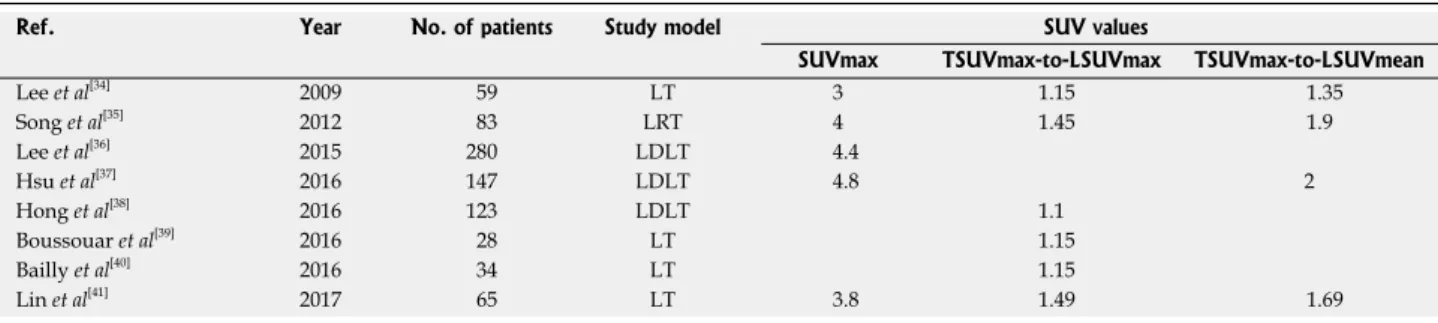
SUV-max to normal-liver SUVSUV-max (TSUVSUV-max/LSUVSUV-max) or tumor SUVmax to normal-liver SUVmean (TSUVmax/ LSUVmean) values instead of SUVmax to identify PET/ CT positivity[34-41] (Table 4).
While 18F-FDG-PET/CT has demonstrated substan-dard sensitivity in discovering new HCC, it has been useful in detecting extra-hepatic metastases, with de-tection rates reported as high as 100%[42,43].
18F-FDG-PET/CT has also been reported to detect post-treatment recurrences earlier and at higher rates than conventional imaging modalities[44]. The sensitivity of 18F-FDG-PET/
CT is size-dependent in both extra-hepatic metastases and recurrences. Sugiyama et al[42] reported a detection
rate of 83% for extra-hepatic metastases > 1 cm, which was only 13% for lesions ≤ 1 cm in diameter. In patients with post-transplant HCC recurrence, Kim et al[45] reported
that a detection rate of > 90% has been achieved for extra-hepatic metastases when the lesions were larger than 1 cm in diameter. However, 18F-FDG-PET/CT was not able to detect any of the extra-hepatic lesions under 1 cm and demonstrated a low detection rate of less than 10% for intrahepatic recurrences. They reported a detection rate of 100% in bone, 60% in the lungs, and 100% in lymph nodes. 18F-FDG-PET/CT has also been used in the evaluation of patients with unexplained AFP elevation after surgical or interventional treatment[46]
. In HCC patients presenting with portal vein thrombosis, 18F-FDG-PET/CT was found more valuable than con-ventional imaging studies in differential diagnosis of tumor thrombus[47,48].
Considering the limited role of 18F-FDG-PET/CT in the detection of HCC because of its low overall sensitivity, Ho et al[49] advocated for the use of 11C-acetate, which
showed better detection sensitivity of 87.3% compared to 47.3% using 18F-FDG. In another study from Hong Kong, which evaluated the accuracy of dual-tracer PET/ CT in HCC patients who underwent either partial hepa-tectomy or LT, the sensitivity of 11C-acetate PET/CT was significantly higher than those of 18FFDGPET/CT and contrast-enhanced CT for the detection of small HCCs (87.0% vs 17.4% and 43.5%, respectively)[50]. Recent
studies have concluded that in patients undergoing LT
Table 4 The standardized uptake values used to define clinically significant 18F-fluorodeoxyglucose positron emission tomography/ computed tomography positivity for hepatocellular carcinoma
Ref. Year No. of patients Study model SUV values
SUVmax TSUVmax-to-LSUVmax TSUVmax-to-LSUVmean
Lee et al[34] 2009 59 LT 3 1.15 1.35 Song et al[35] 2012 83 LRT 4 1.45 1.9 Lee et al[36] 2015 280 LDLT 4.4 Hsu et al[37] 2016 147 LDLT 4.8 2 Hong et al[38] 2016 123 LDLT 1.1 Boussouar et al[39] 2016 28 LT 1.15 Bailly et al[40] 2016 34 LT 1.15 Lin et al[41] 2017 65 LT 3.8 1.49 1.69
SUV: Standardized uptake values; TSUVmax: Tumor SUVmax; LSUVmax: Normal-liver SUVmax.
340
the 18FFDGPET/CT findings with the MVI in patients who underwent LT for HCC[58]. Multivariate analysis
revealed that peritumoral enhancement and the ratio of tumor maximum standardized uptake value (SUV) to normal liver mean SUV (TSUVmax/LSUVmean) ≥
1.2 had a statistically significant association with MVI, with an odds ratio of 10.6 and 14.2, respectively. With regard to predicting MVI, the sensitivity and specificity was 35.7% and 93.3% for MRI and 64.3% and 86.7% for PET/CT, respectively. For the prediction of MVI, a sensitivity of 78.6% and a specificity of 80% were achi eved when both imaging modalities were combined.
CORRELATION BETWEEN 18F-FDG PET/
CT AND MORPHOLOGICAL CRITERIA
As the selection criteria for LT shifted towards biologi-cal criteria, MC as the current gold standard and other morphological criteria have been challenged with a number of studies using 18F-FDG PET/CT. Kornberg et
al[59] was the first to investigate the prognostic value of
preoperative 18F-FDG PET/CT in liver transplant candi-dates with HCC. They concluded that PET/CT negative patients with HCC beyond MC might achieve excellent post-transplant disease-free survival (DFS). In a more re-cent study, they combined the pre-transplant 18F-FDG-PET/CT assessments with Up-to-seven criteria[60]. Among
116 patients with HCC who underwent 18F-FDG-PET/CT prior to LT, 5-year DFS was comparable between patients within Up-to-seven criteria (n = 85) and those beyond Up-to-seven criteria with negative PET/CT (n = 16) (81.0% vs 87.1%, P = 0.5).
A Japanese multicenter study including 182 LDLT reci-pients from 16 Japanese LT centers investigated the significance of pretransplant 18FFDGPET/CT at a much larger scale. While patients beyond MC had a signifi-cantly higher recurrence rate at 5 years compared with those within MC (38% vs 7%, P < 0.001), a subgroup of “beyond MC” patients with negative PET/CT and low AFP (< 115 ng/mL) showed similar recurrence rate with
“within MC” patients (19%, P = 0.1)[61]. Similar data were
recently published by the Taiwan group who combined pre-transplant PET/CT results with UCSF criteria for predicting the risk of post-transplant HCC recurrence. In a group of 147 patients with HCC who underwent 18F-FDG-PET/CT and proceeded to LDLT, patients within UCSF criteria and those beyond UCSF criteria with a low FDG uptake had similar post-transplant recurrence rates (3.6% vs 11.1%)[37].
Another study from Korea investigated the clinical impact of 18F-FDG-PET/CT in patients undergoing LDLT for advanced HCC, where more than half of the patients were beyond MC. In patients beyond either MC (n = 147) or UCSF (n = 136) criteria, PET/CT negative patients had 5-year DFS rates of 73.3% and 72.8%, respectively. Despite the fact that these figures were significantly lower than those of patients within MC (89.8%), the outcome is highly acceptable when the discussion shifts from “zero recurrence” towards targeting 50% 5-year survival as an acceptable goal in advanced HCC[33].
ROLE OF 18F-FDG PET/CT IN
PREDICTING POST-TRANSPLANT HCC
RECURRENCE
Seoul National University Hospital was the first to report the effectiveness of pre-transplant 18F-FDG-PET/CT to predict post-transplant HCC recurrence[28]. Further
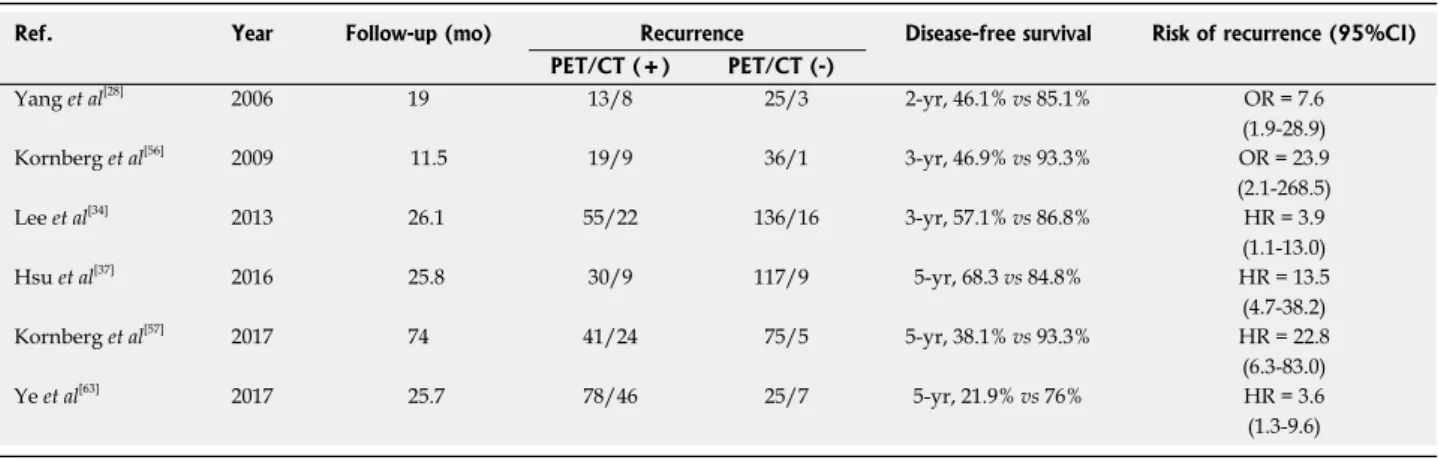
studies have shown that a high 18F-FDG uptake on pre-transplant PET/CT was a strong predictive factor for MVI and tumor recurrence after LT[56,33,62] (Table 5).
In a cohort of 116 liver transplant patients with HCC, Kornberg et al[60] reported a 5-year DFS rate of 93.3%
in PET/CT negative patients vs 38.1% in PET/CT positive patients. PET/CT positive patients showed a recurrence rate of 58.5%, while only 6.7% of the PET/CT negative patients had recurrence. Ye et al[63] also investigated the
clinical value of pre-transplant PET/CT in the selection and prognostic prediction of patients with advanced
WJGO|www.wjgnet.com
Table 5 The use of 18F-fluorodeoxyglucose positron emission tomography/computed tomography in predicting post-transplant hepatocellular carcinoma recurrences
Ref. Year Follow-up (mo) Recurrence Disease-free survival Risk of recurrence (95%CI) PET/CT (+) PET/CT (-) Yang et al[28] 2006 19 13/8 25/3 2-yr, 46.1% vs 85.1% OR = 7.6 (1.9-28.9) Kornberg et al[56] 2009 11.5 19/9 36/1 3-yr, 46.9% vs 93.3% OR = 23.9 (2.1-268.5) Lee et al[34] 2013 26.1 55/22 136/16 3-yr, 57.1% vs 86.8% HR = 3.9 (1.1-13.0) Hsu et al[37] 2016 25.8 30/9 117/9 5-yr, 68.3 vs 84.8% HR = 13.5 (4.7-38.2) Kornberg et al[57] 2017 74 41/24 75/5 5-yr, 38.1% vs 93.3% HR = 22.8 (6.3-83.0) Ye et al[63] 2017 25.7 78/46 25/7 5-yr, 21.9% vs 76% HR = 3.6 (1.3-9.6) PET/CT: Positron emission tomography/computed tomography.
October 15, 2018|Volume 10|Issue 10|
HCC in the LT setting. Patients with a positive 18F-FDG-PET/CT had significantly increased risk of posttranspl ant recurrence compared to PET/CT negative patients (59.0% vs 28.0%, P = 0.007). In patients with positive PET/CT, they reported a significantly lower 5year DFS rate than that of patients with negative PET/CT (76.0%
vs 21.9%, P < 0.001). In another study investigating
the role of PET/CT as a prognostic factor for early HCC recurrence after LT, Lee et al[62] have shown that median
SUVmax of PET/CT-positive tumors in the early, late, and no recurrence groups was 5.2, 3.7, and 3.2, respe-ctively. They concluded that preoperative 18F-FDG-PET/ CT was an independent and significant prognostic factor for early tumor recurrence after LT for HCC.
Hong et al[38] further developed the concept,
hy-pothesizing that the combination of 18F-FDG PET/CT positivity and serum AFP level might improve the predic-tion of post-LT outcome for patients with HCC. Using cut-off values of 200 ng/mL for AFP and 1.1 for SUVmax T/L ratio for the definition of “highrisk” HCC, they found that the rate of MVI and poor differentiation was 33% and 92%, respectively in the high-risk group. They reported 5-year DFS rates of 49.1% vs 93.4% in PET/CT positive
vs negative patients and 47.7% vs 88.3% in high AFP vs
low AFP patients. In the high-risk group (n = 12), 5-year DFS rate was only 8.4%.
CONCLUSION
In patients with HCC, LT is the best treatment option. The selection criteria for LT have been shifting from mor-phological to the combination of biological and histomor-phological criteria. When combined with serum markers, 18F-FDG-PET/CT represents the “new generation” of bio-logical criteria, which has the potential to further improve the prediction of tumor behavior and to provide a better risk stratification model for HCC.
REFERENCES
1 Mittal S, El-Serag HB. Epidemiology of hepatocellular carcino-ma: consider the population. J Clin Gastroenterol 2013; 47 Suppl: S2-S6 [PMID: 23632345 DOI: 10.1097/MCG.0b013e3182872f29] 2 El-Serag HB, Kanwal F. Epidemiology of hepatocellular carcinoma
in the United States: where are we? Where do we go? Hepatology 2014; 60: 1767-1775 [PMID: 24839253 DOI: 10.1002/hep.27222] 3 McGlynn KA, London WT. The global epidemiology of
hepato-cellular carcinoma: present and future. Clin Liver Dis 2011; 15: 223-243, vii-vix [PMID: 21689610 DOI: 10.1016/j.cld.2011.03.006] 4 Yang JD, Larson JJ, Watt KD, Allen AM, Wiesner RH, Gores GJ,
Roberts LR, Heimbach JA, Leise MD. Hepatocellular Carcinoma Is the Most Common Indication for Liver Transplantation and Placement on the Waitlist in the United States. Clin Gastroenterol
Hepatol 2017; 15: 767-775.e3 [PMID: 28013117 DOI:
10.1016/j.c-gh.2016.11.034]
5 Mazzaferro V, Regalia E, Doci R, Andreola S, Pulvirenti A, Bozzetti
F, Montalto F, Ammatuna M, Morabito A, Gennari L. Liver transplan-tation for the treatment of small hepatocellular carcinomas in patients with cirrhosis. N Engl J Med 1996; 334: 693-699 [PMID: 8594428 DOI: 10.1056/NEJM199603143341104]
6 Yao FY, Ferrell L, Bass NM, Watson JJ, Bacchetti P, Venook A, Ascher NL, Roberts JP. Liver transplantation for hepatocellular
carcinoma: expansion of the tumor size limits does not adversely impact survival. Hepatology 2001; 33: 1394-1403 [PMID: 11391528 DOI: 10.1053/jhep.2001.24563]
7 Mazzaferro V, Llovet JM, Miceli R, Bhoori S, Schiavo M, Mariani L, Camerini T, Roayaie S, Schwartz ME, Grazi GL, Adam R, Neuhaus P, Salizzoni M, Bruix J, Forner A, De Carlis L, Cillo U, Burroughs AK, Troisi R, Rossi M, Gerunda GE, Lerut J, Belghiti J, Boin I, Gugenheim J, Rochling F, Van Hoek B, Majno P; Metroticket Investigator Study Group. Predicting survival after liver transplantation in patients with hepatocellular carcinoma beyond the Milan criteria: a retrospective, exploratory analysis. Lancet Oncol 2009; 10: 35-43 [PMID: 19058754 DOI: 10.1016/S1470-2045(08)70284-5]
8 Sugawara Y, Kokudo N. Surgical treatment of hepatocellular
carcin-oma: comparison of resection and transplantation. Oncology 2008;
75 Suppl 1: 119-123 [PMID: 19092281 DOI: 10.1159/000173433]
9 Lee SG, Hwang S, Moon DB, Ahn CS, Kim KH, Sung KB, Ko GY, Park KM, Ha TY, Song GW. Expanded indication criteria of living donor liver transplantation for hepatocellular carcinoma at one large-volume center. Liver Transpl 2008; 14: 935-945 [PMID: 18581465 DOI: 10.1002/lt.21445]
10 Toso C, Trotter J, Wei A, Bigam DL, Shah S, Lancaster J, Grant DR, Greig PD, Shapiro AM, Kneteman NM. Total tumor volume predicts risk of recurrence following liver transplantation in patients with hepatocellular carcinoma. Liver Transpl 2008; 14: 1107-1115 [PMID: 18668667 DOI: 10.1002/lt.21484]
11 Silva M, Moya A, Berenguer M, Sanjuan F, López-Andujar R, Pareja E, Torres-Quevedo R, Aguilera V, Montalva E, De Juan M, Mattos A, Prieto M, Mir J. Expanded criteria for liver transplantation in patients with cirrhosis and hepatocellular carcinoma. Liver Transpl 2008; 14: 1449-1460 [PMID: 18825681 DOI: 10.1002/lt.21576] 12 DuBay D, Sandroussi C, Sandhu L, Cleary S, Guba M, Cattral
MS, McGilvray I, Ghanekar A, Selzner M, Greig PD, Grant DR. Liver transplantation for advanced hepatocellular carcinoma using poor tumor differentiation on biopsy as an exclusion criterion.
Ann Surg 2011; 253: 166-172 [PMID: 21294289 DOI: 10.1097/
SLA.0b013e31820508f1]
13 Bruix J, Llovet JM. Prognostic prediction and treatment strategy in hepatocellular carcinoma. Hepatology 2002; 35: 519-524 [PMID: 11870363 DOI: 10.1053/jhep.2002.32089]
14 Sotiropoulos GC, Malagó M, Molmenti E, Paul A, Nadalin S, Brokalaki E, Kühl H, Dirsch O, Lang H, Broelsch CE. Liver trans-plantation for hepatocellular carcinoma in cirrhosis: is clinical tumor classification before transplantation realistic? Transplanta
tion 2005; 79: 483-487 [PMID: 15729176 DOI: 10.1097/01.
TP.0000152801.82734.74]
15 Hemming AW, Cattral MS, Reed AI, Van Der Werf WJ, Greig PD, Howard RJ. Liver transplantation for hepatocellular carcinoma. Ann
Surg 2001; 233: 652-659 [PMID: 11323504 DOI: 10.1097/0000065
8-200105000-00009]
16 Roayaie S, Schwartz JD, Sung MW, Emre SH, Miller CM, Gondolesi GE, Krieger NR, Schwartz ME. Recurrence of hepa-tocellular carcinoma after liver transplant: patterns and prognosis.
Liver Transpl 2004; 10: 534-540 [PMID: 15048797 DOI: 10.1002/
lt.20128]
17 Cillo U, Vitale A, Bassanello M, Boccagni P, Brolese A, Zanus G, Burra P, Fagiuoli S, Farinati F, Rugge M, D’Amico DF. Liver transplantation for the treatment of moderately or well-differentiated hepatocellular carcinoma. Ann Surg 2004; 239: 150-159 [PMID: 14745321 DOI: 10.1097/01.sla.0000109146.72827.76]
18 Clavien PA, Lesurtel M, Bossuyt PM, Gores GJ, Langer B, Perrier A; OLT for HCC Consensus Group. Recommendations for liver transplantation for hepatocellular carcinoma: an international consensus conference report. Lancet Oncol 2012; 13: e11-e22 [PMID: 22047762 DOI: 10.1016/S1470-2045(11)70175-9] 19 Cillo U, Giuliani T, Polacco M, Herrero Manley LM, Crivellari G,
Vitale A. Prediction of hepatocellular carcinoma biological behavior in patient selection for liver transplantation. World J Gastroenterol 2016; 22: 232-252 [PMID: 26755873 DOI: 10.3748/wjg.v22.i1.232] 20 Shirabe K, Itoh S, Yoshizumi T, Soejima Y, Taketomi A, Aishima S,
Maehara Y. The predictors of microvascular invasion in candidates
50: 682-687 [PMID: 19372474 DOI: 10.2967/jnumed.108.060574]
35 Song MJ, Bae SH, Yoo IeR, Park CH, Jang JW, Chun HJ, Choi BG, Lee HG, Choi JY, Yoon SK. Predictive value of 18
F-fluoro-deoxyglucose PET/CT for transarterial chemolipiodolization of he-patocellular carcinoma. World J Gastroenterol 2012; 18: 3215-3222 [PMID: 22783045 DOI: 10.3748/wjg.v18.i25.3215]
36 Lee SD, Lee B, Kim SH, Joo J, Kim SK, Kim YK, Park SJ. Proposal of new expanded selection criteria using total tumor size and (18)F-fluorodeoxyglucose - positron emission tomography/computed tomography for living donor liver transplantation in patients with hepatocellular carcinoma: The National Cancer Center Korea criteria. World J Transplant 2016; 6: 411-422 [PMID: 27358787 DOI: 10.5500/wjt.v6.i2.411]
37 Hsu CC, Chen CL, Wang CC, Lin CC, Yong CC, Wang SH, Liu YW, Lin TL, Lee WF, Lin YH, Chan YC, Wu YJ, Eng HL, Cheng YF. Combination of FDG-PET and UCSF Criteria for Predicting HCC Recurrence After Living Donor Liver Transplantation.
Transplantation 2016; 100: 1925-1932 [PMID: 27306534 DOI:
10.1097/TP.0000000000001297]
38 Hong G, Suh KS, Suh SW, Yoo T, Kim H, Park MS, Choi Y, Paeng JC, Yi NJ, Lee KW. Alpha-fetoprotein and (18)F-FDG positron emission tomography predict tumor recurrence better than Milan criteria in living donor liver transplantation. J Hepatol 2016; 64: 852-859 [PMID: 26658686 DOI: 10.1016/j.jhep.2015.11.033] 39 Boussouar S, Itti E, Lin SJ, Decaens T, Evangelista E, Chiaradia M,
Chalaye J, Baranes L, Calderaro J, Laurent A, Pigneur F, Duvoux C, Azoulay D, Costentin C, Rahmouni A, Luciani A. Functional imaging of hepatocellular carcinoma using diffusion-weighted MRI and (18)F-FDG PET/CT in patients on waiting-list for liver transplantation. Cancer Imaging 2016; 16: 4 [PMID: 26883745 DOI: 10.1186/s40644-016-0062-8]
40 Bailly M, Venel Y, Orain I, Salamé E, Ribeiro MJ. 18F-FDG PET in Liver Transplantation Setting of Hepatocellular Carcinoma: Predicting Histology? Clin Nucl Med 2016; 41: e126-e129 [PMID: 26545024 DOI: 10.1097/RLU.0000000000001040]
41 Lin CY, Liao CW, Chu LY, Yen KY, Jeng LB, Hsu CN, Lin CL, Kao CH. Predictive Value of 18F-FDG PET/CT for Vascular In-vasion in Patients With Hepatocellular Carcinoma Before Liver Transplantation. Clin Nucl Med 2017; 42: e183-e187 [PMID: 28114226 DOI: 10.1097/RLU.0000000000001545]
42 Sugiyama M, Sakahara H, Torizuka T, Kanno T, Nakamura F, Futatsubashi M, Nakamura S. 18F-FDG PET in the detection of extrahepatic metastases from hepatocellular carcinoma. J
Gastroenterol 2004; 39: 961-968 [PMID: 15549449 DOI: 10.1007/
s00535-004-1427-5]
43 Yoon KT, Kim JK, Kim DY, Ahn SH, Lee JD, Yun M, Rha SY, Chon CY, Han KH. Role of 18F-fluorodeoxyglucose positron emission tomography in detecting extrahepatic metastasis in pre-treatment staging of hepatocellular carcinoma. Oncology 2007; 72 Suppl 1: 104-110 [PMID: 18087190 DOI: 10.1159/000111715] 44 Paudyal B, Oriuchi N, Paudyal P, Tsushima Y, Iida Y, Higuchi T,
Hanaoka H, Miyakubo M, Takano A, Ishikita T, Endo K. Early diagnosis of recurrent hepatocellular carcinoma with 18F-FDG PET after radiofrequency ablation therapy. Oncol Rep 2007; 18: 1469-1473 [PMID: 17982632 DOI: 10.3892/or.18.6.1469]
45 Kim YK, Lee KW, Cho SY, Han SS, Kim SH, Kim SK, Park SJ. Usefulness 18F-FDG positron emission tomography/computed tomography for detecting recurrence of hepatocellular carcinoma in posttransplant patients. Liver Transpl 2010; 16: 767-772 [PMID: 20517911 DOI: 10.1002/lt.22069]
46 Chen YK, Hsieh DS, Liao CS, Bai CH, Su CT, Shen YY, Hsieh JF, Liao AC, Kao CH. Utility of FDG-PET for investigating unexplained serum AFP elevation in patients with suspected hepatocellular car-cinoma recurrence. Anticancer Res 2005; 25: 4719-4725 [PMID: 16334166]
47 Hanajiri K, Mitsui H, Maruyama T, Kondo Y, Shiina S, Omata M, Nakagawa K. 18F-FDG PET for hepatocellular carcinoma pre-senting with portal vein tumor thrombus. J Gastroenterol 2005; 40: 1005-1006 [PMID: 16261443 DOI: 10.1007/s00535-005-1667-z] 48 Kurtovic J, Van Der Wall H, Riordan SM. FDG PET for
discri-342 for liver transplantation with hepatocellular carcinoma-with special reference to the serum levels of des-gamma-carboxy prothrombin.
J Surg Oncol 2007; 95: 235-240 [PMID: 17323337 DOI: 10.1002/
jso.20655]
21 Kaido T, Ogawa K, Mori A, Fujimoto Y, Ito T, Tomiyama K, Takada Y, Uemoto S. Usefulness of the Kyoto criteria as expanded selection criteria for liver transplantation for hepatocellular carcinoma.
Surgery 2013; 154: 1053-1060 [PMID: 24074704 DOI: 10.1016/
j.surg.2013.04.056]
22 Uchiyama H, Itoh S, Yoshizumi T, Ikegami T, Harimoto N, Soejima Y, Harada N, Morita K, Toshima T, Motomura T, Maehara Y. Living donor liver transplantation for hepatocellular carcinoma: results of prospective patient selection by Kyushu University Criteria in 7 years. HPB (Oxford) 2017; 19: 1082-1090 [PMID: 28888776 DOI: 10.1016/j.hpb.2017.08.004]
23 Kwon CH, Kim DJ, Han YS, Park JB, Choi GS, Kim SJ, Joh JW, Lee SK. HCC in living donor liver transplantation: can we expand the Milan criteria? Dig Dis 2007; 25: 313-319 [PMID: 17960066 DOI: 10.1159/000106911]
24 Zheng SS, Xu X, Wu J, Chen J, Wang WL, Zhang M, Liang TB, Wu LM. Liver transplantation for hepatocellular carcinoma: Hang-zhou experiences. Transplantation 2008; 85: 1726-1732 [PMID: 18580463 DOI: 10.1097/TP.0b013e31816b67e4]
25 Heimbach JK, Kulik LM, Finn RS, Sirlin CB, Abecassis MM, Roberts LR, Zhu AX, Murad MH, Marrero JA. AASLD guidelines for the treatment of hepatocellular carcinoma. Hepatology 2018; 67: 358-380 [PMID: 28130846 DOI: 10.1002/hep.29086]
26 Pomfret EA, Washburn K, Wald C, Nalesnik MA, Douglas D, Russo M, Roberts J, Reich DJ, Schwartz ME, Mieles L, Lee FT, Florman S, Yao F, Harper A, Edwards E, Freeman R, Lake J. Report of a national conference on liver allocation in patients with hepatocellular carcinoma in the United States. Liver Transpl 2010;
16: 262-278 [PMID: 20209641 DOI: 10.1002/lt.21999]
27 Halazun KJ, Hardy MA, Rana AA, Woodland DC 4th, Luyten EJ, Mahadev S, Witkowski P, Siegel AB, Brown RS Jr, Emond JC. Negative impact of neutrophil-lymphocyte ratio on outcome after liver transplantation for hepatocellular carcinoma. Ann
Surg 2009; 250: 141-151 [PMID: 19561458 DOI: 10.1097/SLA.
0b013e3181a77e59]
28 Yang SH, Suh KS, Lee HW, Cho EH, Cho JY, Cho YB, Yi NJ, Lee KU. The role of (18)F-FDG-PET imaging for the selection of liver transplantation candidates among hepatocellular carcinoma pa-tients. Liver Transpl 2006; 12: 1655-1660 [PMID: 16964589 DOI: 10.1002/lt.20861]
29 Khan MA, Combs CS, Brunt EM, Lowe VJ, Wolverson MK, Solomon H, Collins BT, Di Bisceglie AM. Positron emission tomo-graphy scanning in the evaluation of hepatocellular carcinoma.
J Hepatol 2000; 32: 792-797 [PMID: 10845666 DOI: 10.1016/
S0168-8278(00)80248-2]
30 Torizuka T, Tamaki N, Inokuma T, Magata Y, Sasayama S, Yonekura Y, Tanaka A, Yamaoka Y, Yamamoto K, Konishi J. In vivo assessment of glucose metabolism in hepatocellular carcinoma with FDG-PET. J Nucl Med 1995; 36: 1811-1817 [PMID: 7562048] 31 Talbot JN, Fartoux L, Balogova S, Nataf V, Kerrou K, Gutman
F, Huchet V, Ancel D, Grange JD, Rosmorduc O. Detection of hepatocellular carcinoma with PET/CT: a prospective comparison of 18F-fluorocholine and 18F-FDG in patients with cirrhosis or chronic liver disease. J Nucl Med 2010; 51: 1699-1706 [PMID: 20956466 DOI: 10.2967/jnumed.110.075507]
32 Blechacz B, Gores GJ. Positron emission tomography scan for a hepatic mass. Hepatology 2010; 52: 2186-2191 [PMID: 20967825 DOI: 10.1002/hep.24002]
33 Lee SD, Kim SH, Kim SK, Kim YK, Park SJ. Clinical Impact of 18F-Fluorodeoxyglucose Positron Emission Tomography/Computed Tomography in Living Donor Liver Transplantation for Advanced Hepatocellular Carcinoma. Transplantation 2015; 99: 2142-2149 [PMID: 25905981 DOI: 10.1097/TP.0000000000000719]
34 Lee JW, Paeng JC, Kang KW, Kwon HW, Suh KS, Chung JK, Lee MC, Lee DS. Prediction of tumor recurrence by 18F-FDG PET in liver transplantation for hepatocellular carcinoma. J Nucl Med 2009;
WJGO|www.wjgnet.com October 15, 2018|Volume 10|Issue 10|
mination between tumor extension and blood thrombus as a cause for portal vein thrombosis in hepatocellular carcinoma: important role in exclusion of transplant candidacy. Clin Nucl Med 2005; 30: 408-410 [PMID: 15891293 DOI: 10.1097/01.rlu.0000162606.83862.a7] 49 Ho CL, Yu SC, Yeung DW. 11C-acetate PET imaging in
hepato-cellular carcinoma and other liver masses. J Nucl Med 2003; 44: 213-221 [PMID: 12571212]
50 Cheung TT, Ho CL, Lo CM, Chen S, Chan SC, Chok KS, Fung JY, Yan Chan AC, Sharr W, Yau T, Poon RT, Fan ST. 11C-acetate and 18F-FDG PET/CT for clinical staging and selection of patients with hepatocellular carcinoma for liver transplantation on the basis of Milan criteria: surgeon’s perspective. J Nucl Med 2013; 54: 192-200 [PMID: 23321459 DOI: 10.2967/jnumed.112.107516]
51 Park JW, Kim JH, Kim SK, Kang KW, Park KW, Choi JI, Lee
WJ, Kim CM, Nam BH. A prospective evaluation of 18F-FDG and 11C-acetate PET/CT for detection of primary and metastatic hepatocellular carcinoma. J Nucl Med 2008; 49: 1912-1921 [PMID: 18997056 DOI: 10.2967/jnumed.108.055087]
52 Wu HB, Wang QS, Li BY, Li HS, Zhou WL, Wang QY. F-18 FDG in conjunction with 11C-choline PET/CT in the diagnosis of hepatocellular carcinoma. Clin Nucl Med 2011; 36: 1092-1097 [PMID: 22064078 DOI: 10.1097/RLU.0b013e3182335df4] 53 Yamamoto Y, Nishiyama Y, Kameyama R, Okano K, Kashiwagi
H, Deguchi A, Kaji M, Ohkawa M. Detection of hepatocellular carcinoma using 11C-choline PET: comparison with 18F-FDG PET.
J Nucl Med 2008; 49: 1245-1248 [PMID: 18632827 DOI: 10.2967/
jnumed.108.052639]
54 Sweeney MJ, Ashmore J, Morrıs HP, Weber G. Comparatıve bio-chemıstry hepatomas. IV. isotope studies of glucose and fructose metabolism in liver tumors of different growth rates. Cancer Res 1963; 23: 995-1002 [PMID: 14050771]
55 Lee SD, Kim SH. Role of positron emission tomography/computed tomography in living donor liver transplantation for hepatocellular carcinoma. Hepatobiliary Surg Nutr 2016; 5: 408-414 [PMID: 27826555 DOI: 10.21037/hbsn.2016.08.01]
56 Kornberg A, Freesmeyer M, Bärthel E, Jandt K, Katenkamp K, Steenbeck J, Sappler A, Habrecht O, Gottschild D, Settmacher U. 18F-FDG-uptake of hepatocellular carcinoma on PET predicts microvascular tumor invasion in liver transplant patients. Am J
Transplant 2009; 9: 592-600 [PMID: 19191771 DOI: 10.1111/
j.1600-6143.2008.02516.x]
57 Kornberg A, Küpper B, Tannapfel A, Büchler P, Krause B, Witt U, Gottschild D, Friess H. Patients with non-[18
F]fludeoxyglucose-avid advanced hepatocellular carcinoma on clinical staging may achieve long-term recurrence-free survival after liver transplanta-tion. Liver Transpl 2012; 18: 53-61 [PMID: 21850692 DOI: 10.1002/lt.22416]
58 Ahn SY, Lee JM, Joo I, Lee ES, Lee SJ, Cheon GJ, Han JK, Choi BI. Prediction of microvascular invasion of hepatocellular car-cinoma using gadoxetic acid-enhanced MR and (18)F-FDG PET/ CT. Abdom Imaging 2015; 40: 843-851 [PMID: 25253426 DOI: 10.1007/s00261-014-0256-0]
59 Kornberg A, Küpper B, Thrum K, Katenkamp K, Steenbeck J, Sappler A, Habrecht O, Gottschild D. Increased 18F-FDG uptake of hepatocellular carcinoma on positron emission tomography independently predicts tumor recurrence in liver transplant patients.
Transplant Proc 2009; 41: 2561-2563 [PMID: 19715974 DOI:
10.1016/j.transproceed.2009.06.115]
60 Kornberg A, Witt U, Schernhammer M, Kornberg J, Ceyhan GO,
Mueller K, Friess H, Thrum K. Combining 18F-FDG positron emission tomography with Up-to-seven criteria for selecting suitable liver transplant patients with advanced hepatocellular carcino-ma. Sci Rep 2017; 7: 14176 [PMID: 29074969 DOI: 10.1038/ s41598-017-14430-9]
61 Takada Y, Kaido T, Shirabe K, Nagano H, Egawa H, Sugawara Y, Taketomi A, Takahara T, Wakabayashi G, Nakanishi C, Kawagishi N, Kenjo A, Gotoh M, Toyoki Y, Hakamada K, Ohtsuka M, Akamatsu N, Kokudo N, Takeda K, Endo I, Takamura H, Okajima H, Wada H, Kubo S, Kuramitsu K, Ku Y, Ishiyama K, Ohdan H, Ito E, Maehara Y, Honda M, Inomata Y, Furukawa H, Uemoto S, Yamaue H, Miyazaki M, Takada T; LTx-PET study group of the Japanese Society of Hepato-Biliary-Pancreatic Surgery and the Japanese Liver Transplantation Society. Significance of preoperative fluorodeoxyglucose-positron emission tomography in prediction of tumor recurrence after liver transplantation for hepatocellular carcinoma patients: a Japanese multicenter study. J Hepatobiliary
Pancreat Sci 2017; 24: 49-57 [PMID: 27806426 DOI: 10.1002/
jhbp.412]
62 Lee SD, Kim SH, Kim YK, Kim C, Kim SK, Han SS, Park SJ. (18)F-FDG-PET/CT predicts early tumor recurrence in living donor liver transplantation for hepatocellular carcinoma. Transpl Int 2013; 26: 50-60 [PMID: 23106431 DOI: 10.1111/j.1432-2277.2012.01572.x] 63 Ye YF, Wang W, Wang T, Yu J, Geng L, Yu SF, Yan S, Zheng SS.
Role of 18F fludeoxyglucose positron emission tomography in the selection of liver transplantation candidates in patients with hepatocellular carcinoma. Hepatobiliary Pancreat Dis Int 2017; 16: 257-263 [PMID: 28603093 DOI: 10.1016/S1499-3872(17)60011-0]
P- Reviewer: Chen JN, Chok KSH, Chiu KW, Tallon-Aguilar L, Xu X,
Yeo W S- Editor: Ji FF L- Editor: Filipodia
E- Editor: Tan WW