INTRODUCTION
Preventive measures, use of fluoride agents and improvement of oral hygiene, have verified effective in reducing caries prevalence. However, posterior teeth have not uniform tooth surfaces as pit and fissure surfaces. The morphology of the occlusal surface in posterior teeth obstructs the mechanical cleaning and reduces the effects of preventive measures. Studies have demonstrated that the use of pit and fissure sealants is an effective way for preventing caries1-6).
Light-cured resin sealants used the most common and conventionally. The polymerization of light-cured sealants can be achieved with many light sources (e.g. quartz tungsten halogen, plasma arc and light emitting diode (LED)). Current sealants are most commonly cured with a quartz tungsten halogen curing lights (QTHs), which has a peak absorption at a maximum of 465 nm. QTHs have some limitations, such as relatively short working life span, high operating temperatures and decreased light output over time3,7,8). LEDs can be operated for thousands of hours without a significant reduction in light output. The reduced temperature associated with LEDs also can prevent degradation of light guides and does not pose a threat to the pulpal tissue. Several studies, however, have shown that LEDs with relatively low irradiances may result in insufficiently cured composites and, therefore, adversely effects mechanical properties of the restorations. The light output of the first-generation LEDs required improvement to match with the cure produced by QTHs.
A number of second-generation LEDs with high power light sources are now available. The results of studies have shown that these are capable of curing CPQ-initiated composites in half the radiation time of their predecessor. The curing kinetics of photo polymerized dental sealants using LEDs showed that the second-generation LEDs reached conversion similar to control in only 10 s2,3,8). The third-generation of LED curing units contains multiple diodes (violet/blue diodes, polywave), and recently a third-generation LED curing unit was marketed (Valo, Ultradent, South Jordan, UT, USA), which is claimed to reach irradiances of up to 3,200 mW/cm2 depending on the chosen mode.
These time saving approaches could have significant clinical implications, provided that there is no adverse effect on the fissure sealant’s marginal integrity. The purpose of this in vitro study was to evaluate the effect of different generation light emitting diodes and quartz tungsten halogen curing units used for various sealants curing on the microleakage and microhardness values.
MATERIALS AND METHODS Microleakage section
Sample size calculations were performed using PASS software (NCSS, LLC, Kaysville, UT, USA). It is estimated at about 180 teeth with a 90% confidence interval level and a standard error of 5%. Caries-free, extracted for orthodontic reasons human mandibular third molars were collected from healthy children ages 15–20. Participants signed the informed consent form. Each tooth was stored in 0.12% thymol solution immediately after extraction and for the same amount of time. Organic remnants were removed off of teeth,
Effect of new light curing units on microleakage and microhardness of resin
sealants
Mehmet BANI1 and Resmiye Ebru TIRALI2
1 Department of Pediatric Dentistry, Faculty of Dentistry, Gazi University, Emek, Ankara, Turkey 2 Department of Pediatric Dentistry, Faculty of Dentistry, Başkent University, Bahçelievler, Ankara, Turkey
Corresponding author, Mehmet BANI; E-mail: [email protected]
To determine new developed light curing units with shorter curing times effects on microleakage and microhardness values for resin fissure sealants. Resin filled sealant (UltraSeal-XT), resin unfilled sealant (Delton Type-II) and ormocer-based sealant (Admira-Seal) were light cured with a quartz-tungsten-halogen (QTH), two LED light and a high power LED. Two hundred and forty extracted human molars were randomly allocated into four groups according to used light-curing unit and three subgroups were formed for three different fissure sealant materials. Specimens were immersed in 0.5% basic fuchsin for 24 h, sectioned and examined under a stereomicroscope, and scored for marginal microleakage. Knoop hardness number (KHN) readings were measured after 48 h. Statistical analyses of test were found in significant difference both microleakage and microhardness values between the various light curing units. The time saving approaches in the curing light were determined higher microhardness, although it was found in higher microleakage.
Keywords: Microleakage, Microhardness, Fissure sealant, Light curing unit
Color figures can be viewed in the online issue, which is avail-able at J-STAGE.
Received Oct 23, 2015: Accepted Feb 16, 2016
Table 1 Light curing used in the study
Light curing Curing unit Curing time Manufacturer (mW/cmIntensity2) Peak Wavelength(nm)
Hilux QTH 20 s Benlioğlu Dental, Ankara, Turkey 600 410–500 Elipar Free Light 2 LED 10 s 3M ESPE, St Paul, MN, USA 1,000 430–480 Valo Standart
Power mode (SP) LED 10 s Ultradent, South Jordan, UT, USA 1,000 440–480 Valo Xtra
Power mode (XP)
Plasma
LED 3 s Ultradent 3,200 440–480
Table 2 Fissure sealant used in the study Fissure
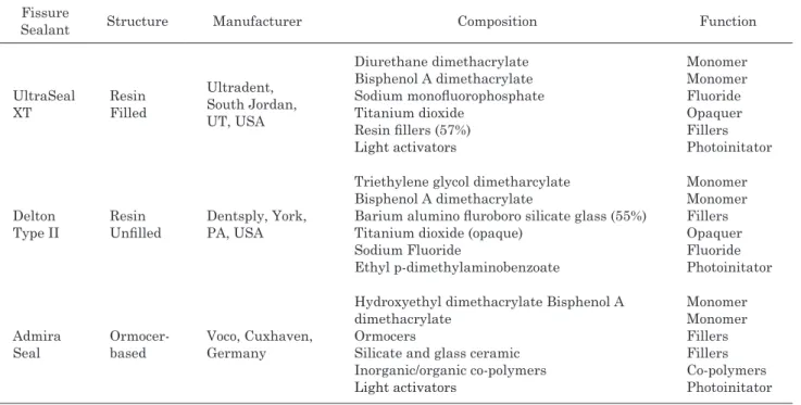
Sealant Structure Manufacturer Composition Function
UltraSeal XT Resin Filled Ultradent, South Jordan, UT, USA Diurethane dimethacrylate Bisphenol A dimethacrylate Sodium monofluorophosphate Titanium dioxide Resin fillers (57%) Light activators Monomer Monomer Fluoride Opaquer Fillers Photoinitator Delton Type II Resin Unfilled Dentsply, York, PA, USA
Triethylene glycol dimetharcylate Bisphenol A dimethacrylate
Barium alumino fluroboro silicate glass (55%) Titanium dioxide (opaque)
Sodium Fluoride Ethyl p-dimethylaminobenzoate Monomer Monomer Fillers Opaquer Fluoride Photoinitator Admira Seal Ormocer-based Voco, Cuxhaven, Germany
Hydroxyethyl dimethacrylate Bisphenol A dimethacrylate
Ormocers
Silicate and glass ceramic Inorganic/organic co-polymers Light activators Monomer Monomer Fillers Fillers Co-polymers Photoinitator
and the occlusal surfaces were cleaned with a slow speed bristle brush with a continuous irrigation of water coolant. The teeth were then examined under a dissecting microscope to discard those with caries, any visible hypoplasia, extraction damage, or microcracks. Teeth with irregular occlusal morphology were also excluded. The condition laboratory was under the daylight 63% relative humidity at room temperature. In the study, three various fissure sealant materials and four curing units were used (Tables 1 and 2).
Accordingly, 240 teeth were selected and assigned randomly into 1 of 4 experimental groups:
In group 1 (Hilux), pits and fissures were etched with 37% phosphoric acid (Vococid, Voco, Cuxhaven, Germany) for 30 s, rinsed with air-water spray for 15 s, and air-dried for 10 s. A bonding resin (Clearfil SE Bond, Kuraray, Okayama, Japan) was applied on the fissures
according to the manufacturer’s instructions and light with a conventional QTH curing unit (Hilux, Benlioğlu Dental, Ankara, Turkey). A radiometer was utilized for detecting the curing unit’s performance. Thereafter, three subgroup was formed by 20 teeth according to applied different fissure sealants mentioned in Table 2. Light curing was performed for 30 s with the same QTH curing unit, and kept distance standard 2 mm.
In group 2 (Elipar) acid-etch and bonding procedures were performed as in group 1, with the adhesive resin being light cured for 10 s with another LED curing unit (Elipar Free Light 2, 3M ESPE, St. Paul, MN, USA). Three subgroups were formed as with group 1 and light cured with the Elipar curing unit for 10 s, and kept distance standard 2 mm.
In group 3 (Valo SP), acid-etch and bonding procedures were performed as in groups 1 and 2, with
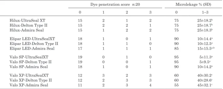
Table 3 Dye penetration score, percentage of microleakage and standard deviation for the various curing lights and sealant materials
Dye penetration score n:20 Microlekage % (SD)
0 1 2 3 0 1–3 Hilux-UltraSeal XT Hilux-Delton Type II Hilux-Admira Seal 15 15 15 2 2 1 1 2 2 2 1 2 75 75 75 25±18.2b 25±18.7b 25±18.3b Elipar LED-UltraSealXT Elipar LED-Delton Type II Elipar LED-Admira Seal
18 18 17 1 1 1 0 1 1 1 0 1 90 90 85 10±14.4a 10±12.5a 15±15.5a,b Valo SP-UltraSealXT Valo SP-Delton Type II Valo SP-Admira Seal
19 19 18 0 0 1 1 0 0 0 1 1 95 95 90 5±11.3a 5±9.5a 10±14.2a Valo XP-UltraSealXT Valo XP-Delton Type II Valo XP-Admira Seal
12 12 11 3 2 2 2 3 3 3 3 4 60 60 55 40±30.2c 40±28.6c 45±32.1c
a,b,c: Statistically significant differences among groups are expressed (p>0.05).
the adhesive resin being light cured for 10 s with another LED curing unit (Valo Standart Power, Ultradent). Three subgroups were formed as with groups 1 and 2 and light cured with the Valo SP curing unit for 10 s, and kept distance standard 2 mm.
In group 4 (Valo XP), acid-etch and bonding procedures were performed as in groups 1, 2 and 3, with the adhesive resin being light cured for 3 s with another LED curing unit (Valo Xtra Power, Ultradent). Three different fissure sealants were applied as with groups 1, 2 and 3 and light cured with the Valo XP curing unit for 3 s, and kept distance standard 2 mm.
Specimens were stored in deionized water at 37°C for 24 h, after which thermal cycling in deionized water was performed at 5±2 to 55±2°C for 500 cycles with a dwell time of 30 s and a transfer time of 10 s. Thereafter, the teeth were kept in distilled water at 37°C for 4 weeks before dye-penetration procedures. The water (pH:7) was changed every week.
Prior to dye penetration, the apices were sealed with sticky wax and the samples were coated with 2 consecutive layers of nail varnish up to 1 mm from the sealant margins. Samples were then immersed in 0.5% basic fuchsine solution (Wako Pure Chemical Industry, Osaka, Japan) for 24 h. After thoroughly rinsing with distilled water, the samples were air-dried and embedded in epoxy resin (Struers, Copenhagen, Denmark). Five parallel longitudinal sections were made through the occlusal surfaces using a low-speed diamond saw (Isomet, Buehler, Lake Bluff, IL, USA) in the bucco-lingual direction. One calibrated researcher examined all sections under a stereomicroscope at ×16 magnification (Wild Type 308700, Heerbrugg, Switzerland). He was blind to the groups.
For each section, the following criteria was used to rate dye penetration scores: score 0=no dye penetration;
1=dye penetration restricted to the outer half of the sealant; 2=dye penetration into the inner half of the sealant; and 3=dye penetration into the underlying fissure. The microleakage score of each tooth was obtained by calculating the mean score of 5 slices each. Statistical evaluation of microleakage scores between the test groups was performed using the Kruskal-Wallis test with significance set at p=0.05.
Microhardness section
Three various sealant materials and above-described of the four curing units were investigated by using for each of LCU and material (Tables 1 and 2). All specimen were prepared by a single operator, with care taken to avoid bubble formation.
The sealant material was placed into a 2-mm-thick silicone mold with a 4-mm internal diameter. The mold was placed between 2 glass slides. Sample size calculations were performed using PASS software (NCSS, LLC). It is estimated at about 180 specimens with a 90% confidence interval level and a standard error of 5%. One hundred eighty specimens were prepared and assigned randomly into1 of 4 experimental groups. Each specimen was cured for the appropriate amount of time from the top surface only. The specimens were stored in the dark under 100% relative humidity at 37°C for 48 h. Five Knoop Hardness Number (KHN) readings were made at least 1 mm from the edge of each top and bottom surface after an indenter dwell time of 15 s and a load of 50 g (Microhardness Tester FM-700, Mitutoyo G3, Mitutoyo, Tokyo, Japan). A Bottom/Top (B/T) KHN ratio was calculated for each specimen. Top and bottom surface hardness and B/T KHN means were determined for each group. Microhardness scores between the test groups were performed using the Kruskal-Wallis test. The level of significance was set at p=0.05.
Fig. 1 View of the dye penetration scores 0, 1, 2 and 3.
a; score 0=no dye penetration, b; score 1=dye penetration restricted to the outer half of the sealant, c; score 2=dye penetration into the inner half of the sealant, d; score 3=dye penetration into the underlying fissure.
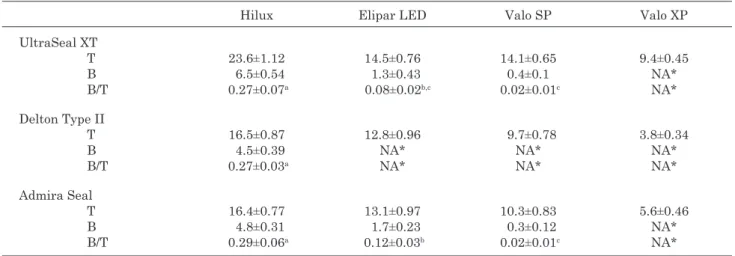
Table 4 Means and standard deviations of top (T) and bottom (B) hardness and hardness ratio (B/T) for the various curing lights and sealant materials
Hilux Elipar LED Valo SP Valo XP UltraSeal XT T B B/T 23.6±1.12 6.5±0.54 0.27±0.07a 14.5±0.76 1.3±0.43 0.08±0.02b,c 14.1±0.65 0.4±0.1 0.02±0.01c 9.4±0.45 NA* NA* Delton Type II T B B/T 16.5±0.87 4.5±0.39 0.27±0.03a 12.8±0.96 NA* NA* 9.7±0.78 NA* NA* 3.8±0.34 NA* NA* Admira Seal T B B/T 16.4±0.77 4.8±0.31 0.29±0.06a 13.1±0.97 1.7±0.23 0.12±0.03b 10.3±0.83 0.3±0.12 0.02±0.01c 5.6±0.46 NA* NA* *NA=not applicable.
a,b,c: Statistically significant differences among groups are expressed (p>0.05).
RESULTS Microleakage section
Total 240 teeth scores were analyzed in the results, it were included to each light curing units and fissure
sealant subgroups 20 samples. The means and standard deviations of microleakage for the various light curing units and sealant materials are shown in Table 3. Also, Table 3 compares the results of the tooth scores with no microleakage (score 0) with those where microleakage
was present (scores 1, 2 and 3) (Fig. 1). The results showed that the various sealing material had not a statistically significant influence on the microleakage (p>0.05). Statistical analyses of these scores with a Kruskal-Wallis test resulted in significant difference between the various light curing units. Microleakage values demonstrated respectively the highest Valo XP, Hilux, Elipar and Valo SP groups. Valo XP exhibited statistically significantly higher microleakage, although Valo SP and Elipar were found in lowest microleakage. Valo SP had demonstrated a significantly lower leakage according to Hilux in all the sealant, however Elipar was excluding Admira Seal (p>0.05).
Microhardness section
Total 180 specimens scores were analyzed in the results, it was included to each light curing units and fissure sealant subgroups 15 samples. The means and standard deviations of microhardnes for the various light curing units and sealant materials are shown in Table 4. There was a significant difference among the groups in terms of the microhardness ratio (p<0.005). Microhardness values showed respectively the highest Valo XP, Valo SP, Elipar and Hilux group.
The results showed that the Delton Type II presented a significantly higher hardness ratio than the other fissure sealants.
DISCUSSION
The pit and fissure sealants’ effectiveness for reducing caries risk have been demonstrated by many studies9). Despite a lot of advantages for light cured resin sealants, gap formation between tooth and material interfaces due to polymerization shrinkage is considered one of the most important disadvantage10). Various attempts have been made for reducing operating time for anxious, uncooperative patients by newly developed light curing units11,12). Although studies stated that decreasing the curing time had no effect on the microleakage of the sealants2,13), there is a scarcity of published data regarding their effects on the marginal integrity and degree of conversion.
Today, a majority of light curing units is based on the light-emitting diode (LED) technology with a single high-powered diode. Second-generation LED curing units typically reach irradiances of 1,200 to 1,500 mW/ cm2. The third-generation of LED curing units contains multiple diodes, and recently a third-generation LED curing unit was marketed (VALO, Ultradent), which is claimed to reach irradiances of up to 3,200 mW/cm2 depending on the chosen mode14). Dental visible light curing materials generally contain a diketone-type photoinitiator that absorbs light in the 400- to 500-nm range covered by the blue light from the visible spectrum. The studies showed that the emission peak of blue LED at 465 nm coincided with the absorption peak of camphoroquinone (CQ) at 467 nm8,15), but when the resin material contains new photointiators that absorb light energy in lower regions of visible spectrum
early generation LED lights failed when compared with QTH16).
Interestingly, rapid cure did not have an adverse effect on Valo XP group’s microleakage values. It is possible that, when upper part of the sealant polymerized adequately, it gained retention from the good adaptation. Short curing time might not allow the composite resin flow to form resin tags but this may not be valid for resin sealants. Nevertheless a higher stress level between the layers of resin material due to inadequately curing may increase the flexure under masticatory forces should not be forgotten.
For clinical success of light cured resin sealants adequate polymerization is always necessary. The higher degree of conversion for resin material provides increased mechanical properties. Knoop Hardness Number shows relative degree of conversion for resin materials17). Unfilled resin sealant (Delton) showed a significant higher hardness ratio as previous studies2,18) can be explained by light’s more easier passage from unfilled resin material.
In the present study, the lowest micro hardness values found for Hilux group. This was an unexpected result but light units spectrum is not the only factor that effect polymerization quality of the resin fissure sealants. Different (filler/matrix) compositions of light curing dental materials may have a more pronounced influence on material properties than radiant exposure19).
Higher amount of filler particles provides less porosity20), better wear resistance21), good shear-bond strength22) and similar retention scores as conventional ones20). For the ormocer-based sealant tested, relatively high viscosity that cause greater thickness may explain the not statically significant but higher microleakage values. It is important for the clinician to understand that the polymerization process varies from different types of curing units and different resin materials. Further studies are needed to understand the nature of complex relationship between resin sealant’s polymerization and different light curing technology.
CONCLUSION
Sealant microleakage and microhardness were effected by the type of light curing device technology. Valo XP exhibited statistically significantly higher microhardness, although it was found in statistically significantly higher microleakage. Valo SP showed lowest microleakage and higher microhardness values. UltraSeal XT and Delton Type II were showed similar microleakage, but Delton Type II presented a significantly higher hardness ratio than the other fissure sealants. This paper adds to the literature results of an in vitro study investigating whether the microleakage and microhardness of a different sealant is influenced either a quartz-tungsten halogen (QTH), two LED light and a high power LED. This study demonstrated that sealant microleakage and microhardness were effected by the type of light curing device technology.
ACKNOWLEDGMENTS All authors deny any conflicts of interest.
REFERENCES
1) Shah S, Roebuck EM, Nugent Z, Deery C. In vitro microleakage of a fissure sealant polymerized by either a quartz tungsten halogen curing light or a plasma arc curing light. Int J Paediatr Dent 2007; 17: 371-377.
2) Duangthip D, Ballungpattama S, Sitthisettapong T. Effect of light curing methods on microleakage and microhardness of different resin sealants. J Dent Child (Chic) 2011; 78: 88-95. 3) Cehreli SB, Arikan S, Gulsahi K, Arhun N, Arman A, Sargon
M. Effect of LED curing on marginal integrity of an ormocer-based sealant. J Dent Child (Chic) 2009; 76: 53-57.
4) Bohanan HM, Bader JD. Future impact of public health and preventive methods on the incidence of dental decay. J Can Dent Assoc 1984; 50: 229-233.
5) Ripa LW. Sealants revisited: An update of the effectiveness of pit and fissure sealants. Caries Res 1993; 27: 77-82.
6) Rethman J. Trends in preventive care: Caries risk assessment and indications for sealants. J Am Dent Assoc 2000; 131: 85-115.
7) Tarle Z, Meniga A, Knezevic A, Sutalo J, Ristic M, Pichler G. Composite conversion and temperature rise using a conventional, plasma arc, and experimental blue LED curing unit. J Oral Rehabil 2002; 29: 662-667.
8) Jandt KD, Mills RW, Blackwell GB, Ashworth SH. Depth of cure and compressive strength of dental composites cured with blue light emitting diodes (LEDs). Dent Mater 2000; 16: 41-47.
9) Ahovuo-Saloranta A, Hiiri A, Nordblad A, Worthington H, Makela M. Pit and fissure sealants for preventing dental decay in the permanent teeth of children and adolescents. Cochrane Database Syst Rev 2004; 3: CD001830.
10) Yap AU, Wang HB, Siow KS, Gan LM. Polymerization shrinkage of visible light cured composites. Oper Dent 2000; 25: 98-103.
11) Frost T, Norevall LI, Persson M. Bond strengt hand clinical efficiency for two light guide sizes in orthodontic bracket bonding. Br J Orthod 1997; 24: 35-40.
12) James JW, Miller BH, English JD, Tadlock LP, Buschang PH. Effects of high-speed curing devices on shear bond strength and microleakage of orthodontic brackets. Am J Orthod Dentofacial Orthop 2003; 123: 555-561.
13) Bodrumlu E, Topuz O, Uzun Ö. Comparison of halogen versus LED light-cured temporary endodontic filling materials for sealing. Minerva Stamatol 2013; 62: 193-198.
14) Flury S, Lussi A, Hickel R, Ilie N. Light curing through glass ceramics with a second- and a third-generation LED curing unit: effect of curing mode on the degree of conversion of dual-curing resin cements. Clin Oral Investig 2013; 17: 2127-2137.
15) Mills RW, Uhl A, Jandt KD. Optical power outputs, spectra and dental composite depths of cure, obtained with blue light emitting diode (LED) and halogen light curing units (LCUs). Br Dent J 2002; 193: 459-463.
16) Ikemura K, Endo T. A review of the development of radical photopolymerization initiators used for designing light-curing dental adhesives and resin composites. Dent Mater J 2010; 29: 481-501.
17) Pilo R, Cardash HS. Post-irradiation polymerization of different anterior and posterior visible light activated resin composites. Dent Mater 1992; 8: 299-304.
18) Mills RW, Jandt KD, Ashworth SH. Dental composite depth of cure with halogen and blue light emitting diode technology. Br Dent J 1999; 186: 388-391.
19) Hadis M, Leprince JG, Shortall AC, Devaux J, Leloup G, Palin WM. High irradiance curing and anomalies of exposure reciprocity law in resin-based materials. J Dent 2011; 39: 549-557.
20) Oba AA, Sönmez IŞ, Ercan E, Dülgergil T. Comparison of retention rates of fissure sealants using two flowable restorative materials and a conventional resin sealant: two-year follow-up. Med Princ Pract 2012; 21: 234-237.
21) Ciamponi AL. Assessment “in vitro” of the microleakage in enamel/sealant interface: Influence of contamination, using the primer, and different sealants [thesis]. Sao Paulo, Brazil: School of Dentistry, University of Sao Paulo; 1995.
22) Eminkahyagil N, Gokalp S, Baseren M, Korkmaz Y, Karabulut E. Sealant and composite bond strength to enamel with antibacterial/self-etching adhesives. Int J Pediatr Dent 2005; 15: 274-281.