Review Paper
Review of evoked and event-related delta responses in the human brain
Bahar Güntekin
⁎
, Erol Ba
şar
Brain Dynamics, Cognition and Complex Systems Research Center, Istanbul Kültür University, Istanbul 34156, Turkey
a b s t r a c t
a r t i c l e i n f o
Available online 7 February 2015 Keywords: EEG Delta Evoked oscillations Event-related oscillations Emotion
In the last decade, the brain's oscillatory responses have invaded the literature. The studies on delta (0.5–3.5 Hz) oscillatory responses in humans upon application of cognitive paradigms showed that delta oscillations are relat-ed to cognitive processes, mainly in decision making and attentional processes.
The present manuscript comprehensively reviews the studies on delta oscillatory responses upon cognitive stim-ulation in healthy subjects and in different pathologies, namely Alzheimer's disease, Mild Cognitive Impairment (MCI), bipolar disorder, schizophrenia and alcoholism. Further delta oscillatory response upon presentation of faces, facial expressions, and affective pictures are reviewed. The relationship between pre-stimulus delta activity and post-stimulus evoked and event-related responses and/or oscillations is discussed. Cross-frequency cou-plings of delta oscillations with higher frequency windows are also included in the review.
The conclusion of this review includes several important remarks, including that delta oscillatory responses are involved in cognitive and emotional processes. A decrease of delta oscillatory responses could be a general elec-trophysiological marker for cognitive dysfunction (Alzheimer's disease, MCI, bipolar disorder, schizophrenia and alcoholism). The pre-stimulus activity (phase or amplitude changes in delta activity) has an effect on post-stimulus EEG responses.
© 2016 The Authors. Published by Elsevier B.V. This is an open access article under the CC BY license (http://creativecommons.org/licenses/by/4.0/).
1. Introduction
The superposition of evoked and induced oscillations in different fre-quency windows gives rise to Evoked Potentials (EPs) and/or Event-Related Potentials (Başar, 1980, 1998, 1999). In his book EEG-Brain Dynamics,Başar (1980)described the relationship between spontane-ous EEG and the characteristics of evoked potentials in the frequency domain and in the time domain. In the frequency domain, it was commented that spontaneous EEG and the EPs have common frequency bands, and, upon stimulation, spectral regularization of the EEG occurs. In the time domain, oscillatory responses could have damped oscillatory character; amplitude enhancement could occur in most of the response components. Time locking among responses to successive stimuli gives rise to induced activity. The correlation between the response and the EEG prior to stimulus was also described. After thirty-four years of re-search in the area of brain oscillatory responses, all the properties of the brain oscillatory responses mentioned above have been supported by many researchers working in this area (Lakatos et al., 2008, 2013; Besle et al., 2011; Cravo et al., 2013; Gomez-Ramirez et al., 2011;
Henry and Obleser, 2012; Kösem et al., 2014; Stefanics et al., 2010; Jones et al., 2006; Bernat et al., 2007; Ishii et al., 2009).
Delta oscillatory responses are defined as EEG oscillatory responses in the 0.5–3.5 Hz frequency range. In the human brain,Başar and Stampfer (1985)andStampfer and Başar (1985)were among thefirst researchers who demonstrated the importance of delta responses in cognitive processes. Currently, publications support the view of these authors that delta responses are involved in cognitive processes, mainly in decision-making and attentional processes.
To our knowledge, there are two previous reviews on delta oscilla-tions (Harmony, 2013; Knyazev, 2012).Knyazev (2012)reviewed delta oscillations with the thesis that delta oscillation manifests the most ancient oscillatory mode in comparison to higher frequencies. This review is different from the other two in that we mainly focus on the delta oscillatory responses rather than spontaneous EEG delta activ-ity. Our review includes studies on event-related delta responses upon cognitive and emotional paradigms. We primarily focus on the human Evoked and/or Event-Related delta responses during sensory, cognitive, or emotional processes. We further will emphasize the role of delta re-sponses as a candidate general electrophysiological marker in cognitive dysfunctions as Alzheimer's disease, mild cognitive impairment, bipolar disorder, schizophrenia, and alcoholism. Cross-frequency couplings of delta oscillations with higher frequency windows and the relation of pre-stimulus delta activity with post-stimulus EEG responses will be also discussed.
⁎ Corresponding author at: Istanbul Kultur University, Brain Dynamics, Cognition and Complex Systems Research Center, Ataköy Campus, Bakırköy, 34156 Istanbul, Turkey. Tel.: +90 212 498 4393; fax: +90 212 498 4546.
E-mail address:[email protected](B. Güntekin).
http://dx.doi.org/10.1016/j.ijpsycho.2015.02.001
0167-8760/© 2016 The Authors. Published by Elsevier B.V. This is an open access article under the CC BY license (http://creativecommons.org/licenses/by/4.0/).
Contents lists available atScienceDirect
International Journal of Psychophysiology
2. Generators of delta activity
Research has shown several generators of delta oscillations in human brain. Long-lasting hyperpolarizations generated by pyramidal neurons yield EEG delta oscillations (Steriade, 1993; Steriade and Buzsaki, 1990; Steriade et al., 1990). Glial cells have also been reported to produce slow oscillations (Amzica, 2002; Amzica and Steriade, 2000). Two intrinsic currents of thalamocortical cells generate delta oscilla-tions (Steriade, 1993). Delta oscillations have also been recorded from nucleus accumbens (Leung and Yim, 1993), dopaminergic neurons in the ventral tegmental area (Grace, 1995), ventral pallidum (Lavin and Grace, 1996), and brain stem (Lambertz and Langhorst, 1998). 3. Analysis of delta oscillatory responses
There are several types of oscillatory activity in the brain
1) Spontaneous EEG oscillations are those recorded without any exter-nal physical stimulation.
2) Evoked oscillations are evoked upon application of a pure sensory stimulation, i.e. upon any visual or auditory stimulation.
3) Event-related oscillations are elicited by application of a stimulation containing a task or strategy, i.e. oddball P300 response.
Table 1summarizes some of the system theory methods used to an-alyze the spontaneous EEG, Evoked, and Event-Related oscillations. As we also mentioned in our recent review (Güntekin and Başar, 2014), there are several mathematical tools to analyze the evoked/event-related spectra. One of the most commonly used is the Fast Fourier Transform (FFT). First, in order to perform Fourier analysis of brain responses, an averaging procedure is applied to the data; following arti-fact rejection, selective averaging is performed. The averaged potentials (EP and/or ERP) are then analyzed via FFT and, according to the cut-off frequencies of evoked power spectra, digitalfiltering is applied to EPs or ERPs (e.g. for delta, 0.5–3.5 Hz; for theta, 4–7 Hz; for alpha, 8–13 Hz; for beta, 14–30 Hz; and for gamma, 28–48 Hz filters are applied). A grand average is also applied by performing averaging across subjects. It is possible to conduct statistical analysis of evoked/event-related power measures and to analyze the peak-to-peak amplitudes offiltered oscilla-tory responses (Başar, 1998, 1999).
When FFT or any other mathematical tool is applied to EPs or ERPs, the phase-locked activity is primarily represented in the transform. However, oscillatory responses could instead include time-locked activ-ity, which is termed induced activity (Başar, 1998, 1999). In order to see the induced activity, it is possible to apply FFT or any other mathemat-ical tool (e.g. wavelet transform) to the single epochs, and then the evoked and induced activities (total power) are analyzed by averaging the FFTs of these epochs. In order to obtain only the induced power, evoked power is excluded from the total power (Herrmann et al., 2004). Event-related phase consistency across trials is an important meth-od allowing researchers to see how phase information varies between trials. Kolev et al. (1998) used single-sweep wave identification (SSWI) histograms to analyze phase-locking. Tallon-Baudry et al. (1996)defined a method called phase-locking factor, andDelorme and Makeig (2004)called this method inter-trial phase coherence (ITPC).Lachaux et al. (1999)defined a method called phase-locking sta-tistics, in which responses to repeated stimuli were used to identify la-tencies at which the phase difference between the signals varies little across trials and between two electrode sites.
The analysis of spontaneous EEG coherence, evoked coherence and event-related coherence between two electrode pairs is an important tool to understand functional connectivity between two different elec-trode pairs (Bendat and Piersol, 1967; Lopes da Silva et al., 1980; Petsche and Etlinger, 1998; Rappelsberger et al., 1982). Successful use of Evoked and/or Event-Related coherences has been reported previ-ously for healthy subjects (Güntekin and Başar, 2010; Qassim et al., 2013), Alzheimer's patients (Başar et al., 2010; Güntekin et al., 2008), and bipolar disorder patients (Özerdem et al., 2010, 2011) upon appli-cation of the oddball paradigm.
4. Relation between P300 and delta oscillatory responses
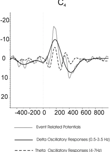
In neuroscience, references to the target response during the oddball strategy are often associated with the delta response. This is only par-tially the case. The delta response is the damped oscillatory response, which is obtained following digitalfiltering in the frequency window of 0.5–3.5 Hz (black curve inFig. 1). However, the target response also contains other oscillatory responses in theta, alpha, beta and gamma frequency ranges (Başar-Eroğlu and Başar, 1991; Başar-Eroğlu et al., 1991, 1993, 2001; Demiralp et al., 1999, 2001; Güntekin et al., 2013; Karakaş et al., 2000; Kolev et al., 1997; Sakowitz et al., 2001; Schürmann et al., 1997; Spencer and Polich, 1999; Öniz and Başar, 2009; Yordanova et al., 2000). Delta response, which is the most ample signal with a negative peak around 200 ms and positive peak around 400 ms, is superimposed with the theta response from which the second oscillation coincides with the delta response. Therefore, the unfiltered P300 response shows a sharp positive peak around 400 ms following the target stimuli. Due to this analysis, delta response gains importance in all studies in which a cognitive load is presented. This cognitive load, in turn, is correlated with attention, perception, learning, and memory. Further, with hearing and visual threshold signals, the only remaining component is the delta response, which allows assigning a perceptual decision to the delta response (Parnefjord and Başar, 1999).
We emphasize the analytic description inFig. 1because, in the present review, it will be shown that all types of cognitive deficits are accompanied by a reduction of the delta response. Accordingly, the description of the delta response gains high importance in all types of impairments related to cognitive deficits. The analysis of oddball exper-iments should indicate the interplay between delta and theta responses, as well as the alpha prolongation for the achievement of a cognitive pro-cess. However, in order to present an understandable and transparent interpretation of the P300 process, the consideration of the delta re-sponse alone highly facilitates the preliminary steps in electrophysio-logical understanding of cognitive processes.
Table 1
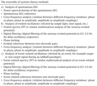
The ensemble of systems theory methods. The ensemble of systems theory methods a) Analysis of spontaneous EEG
• Power spectral density of the spontaneous EEG • Spontaneous EEG coherence
• Cross frequency analysis (relation between different frequency windows: phase to phase, phase to amplitude, amplitude to amplitude couplings)
b) Analysis of evoked oscillations (elicited by simple light, tone signal, etc.) • Evoked spectra (FFT or similar mathematical analysis of the sensory-evoked
potential)
• Digital filtering (digital filtering of the sensory-evoked potential in 0.5–3.5 Hz for delta oscillatory responses)
• Phase-locking
• Evoked coherence between two electrode pairs
• Cross frequency analysis (relation between different frequency windows: phase to phase, phase to amplitude, amplitude to amplitude couplings)
c) Analysis of event-related oscillations (elicited by an event, for example target or non-target signal during an oddball paradigm)
• Event-related spectra (FFT or similar mathematical analysis of an event-related potential)
• Digital filtering (digital filtering of the sensory-evoked potential in 0.5–3.5 Hz for delta oscillatory responses)
• Phase-locking
• Event-related coherence between two electrode pairs
• Cross frequency analysis (relation between different frequency windows: phase to phase, phase to amplitude, amplitude to amplitude couplings)
Beginning in 1984 with a series of experiments, Başar and his group analyzed the relationship between P300 and delta oscillatory responses. Başar et al. (1984)analyzed delta responses during omitted stimuli. In their experiment, they stimulated the subjects by repetitive acoustical tone bursts in which every third or fourth signal was omitted. These au-thors reported that, in delta and theta frequency ranges, the EEG prior to omitted stimulation showed, in most of the trials, a phase-reordering: the P300-wave seems to be a continuation of the ordered and stationary pre-EEG. Further, they commented that in some cases, the P300-wave depicts an amplitude enhancement against delta and theta components of pre-EEG.
Stampfer and Başar (1985)andBaşar and Stampfer (1985)were among thefirst researchers to analyze delta oscillatory responses upon oddball paradigm. These authors reported that N100 peaks of both non-target and target ERP were formed mainly by 4–7 Hz and 7–17 Hz oscillations. The large positive peak around 200 ms (P200) was formed mainly by 1–2.5 Hz activity.
Schürmann et al. (1995)showed that by selective averaging, the am-plitude of P300 response increased when single trials with high en-hancement in delta frequency range were averaged. P300 responses were also contributed to by theta and gamma oscillatory responses (Başar-Eroğlu and Başar, 1991; Demiralp et al., 1999, 2001; Karakaş et al., 2000; Kolev et al., 1997; Spencer and Polich, 1999; Yordanova et al., 2000).
Başar-Eroğlu et al. (1992)reported that target responses showed characteristic P300 complexes in the time domain and were accompa-nied by a delta peak with a center frequency of 2 Hz. In a single trial analysis of P300 delta responses,Schürmann et al. (1995)reported that delta responses were higher for target stimulations in comparison to non-target stimulation.Schürmann et al. (2001)later showed that delta responses were found in single trails closely resembling the un fil-tered P300 response. Induced delta responses in perception of auditory stimuli near the hearing threshold were reported byParnefjord and Başar (1999), which could possibly be correlates of signal detection and decision-making.
Using wavelet decomposition,Demiralp et al. (1999)showed that increased positivity of the 4th delta (0.5–4 Hz) coefficient after stimula-tion was the most important feature of the P300 response. Later, Demiralp et al. (2001)reported that by averaging the sweeps with a positive delta coefficient in the 310–430 ms time window a clear P300 wave could be obtained.Demiralp et al. (2001)also showed that in-creased amplitude of the delta response and inin-creased amplitudes and prolongation of theta and alpha responses were observed in the target responses in comparison to non-target responses.
By time frequency analysis of oddball P300,Kolev et al. (1997) re-ported that the delta component is most probably associated with P3b. On the other hand, in a recent studyPrada et al. (2014)showed that bursts of delta power were associated with the novelty P300 but not with target P3b. These authors showed that nP3 potentials were strongly associated with delta oscillatory responses during both novel sounds and tonal task-switch cues. Further, they demonstrated that delta oscillatory responses did not correlate with P3b amplitude (Prada et al., 2014). Mathes et al. (2012) showed that enhanced single-trial amplitude of delta activity contributed to the larger P3 for the modified probes rather than identical probes. In their study, identi-cal probes were the ones that were previously encoded, while modified probes were the ones that were modified in color, in shape, or in both color and shape.
Jones et al. (2006), using the method of S-transform decomposition (time–frequency analysis), demonstrated that the early part of the P300 wave was formed by theta oscillations, and the main part of the P300 waveform was formed by delta oscillations. In a relevant broad analysis, Bernat et al. (2007)analyzed delta, theta and alpha frequencies during visual oddball paradigm in a very large group of subjects (2068 sub-jects) using principal component analysis (PCA) decomposition. Their study, with a different methodology and large number of subjects, con-firmed the previous work that the major operating rhythms of P300 are mainly the delta and theta oscillations (Başar et al., 2001; Başar-Eroğlu et al., 1992; Demiralp et al., 1999, 2001; Karakaş et al., 2000; Kolev et al., 1997; Spencer and Polich, 1999; Yordanova et al., 2000). In a MEG study,Ishii et al. (2009)also showed that, during auditory oddball paradigm, delta responses increased, and these authors concluded that frontocentral–parietal delta synchronization was functionally engaged in the auditory attention and memory updating processes (see also the analytic description inFig. 1). In a recent study by analysis of event-related coherences upon auditory oddball paradigm, we have shown that, not only in local circuit but also in long-range connections, delta os-cillations are involved in P300 responses (Güntekin and Başar, 2010). Fur-thermore, we have shown that target stimulation elicited higher delta and theta coherence values than non-target stimulation.Qassim et al. (2013) confirmed the results byGüntekin and Başar (2010)using the method of wavelet coherence and upon stimulation of visual oddball paradigm. These authors also showed that target stimulation elicited higher delta and theta coherence values than non-target stimulation.
The relation between delta responses and ERP components were not just reported for oddball paradigms. The researchers also showed that delta and theta responses contribute to the Error Related Negativity (Bernat et al., 2005andHall et al., 2007), the feedback negativity (Bernat et al., 2011, 2012andNelson et al., 2011) and the N2 and P3 during go/no-go task (Harper et al., 2014).
Fig. 1. ERP, delta (0.5–3.5 Hz) oscillatory response and theta (4–7 Hz) oscillatory response of C4electrode upon application of visual oddball paradigm. Gray line represent event
re-lated potential (average of twenty-five epochs filtered between 0.5–30 Hz). Black line rep-resents delta oscillatory responses (average of twenty-five epochs filtered between 0.5– 3.5 Hz) and dashed line represents theta oscillatory responses (average of twenty-five epochsfiltered between 4–7 Hz).
5. Delta oscillatory responses as a general electrophysiological marker for cognitive dysfunction
The role of delta oscillations during cognitive processes is also sup-ported by studies analyzing event-related delta oscillations of different cognitively impaired patient groups during cognitive stimulations. In several studies, cognitively impaired subject groups (Mild cognitive impairment (MCI), Alzheimer's disease (AD), Schizophrenia (SCZ), Bipolar disorder (BP)) showed reduced amplitudes of delta oscillatory responses during cognitive paradigms. Reduction of delta responses during aging upon presentation of cognitive stimulation was also re-ported during visual oddball paradigm (Emek-Savaş et al., 2016) and during go/no-go tasks (Schmiedt-Fehr and Başar-Eroğlu, 2011). On the other hand, elderly subjects showed higher delta coherence upon presentation of auditory oddball paradigm (Maurits et al., 2006).
Yener et al. (2008, 2012)andCaravaglios et al. (2008)showed that delta oscillatory responses were reduced in Alzheimer's disease upon application of visual and auditory oddball paradigms. The subject groups ofYener et al. (2008, 2012)were untreated Alzheimer's disease patients and patients treated with a cholinesterase inhibitor. These au-thors showed that both patient groups had reduced delta oscillatory re-sponses at central electrodes upon application of target stimulation during visual and auditory oddball paradigms.Yener et al. (2008, 2012) asked the subjects to mentally count the targets, while Caravaglios et al. (2008)asked the subjects to press a button during tar-get tones of auditory oddball paradigm. One of the important results fromCaravaglios et al. (2008)was the relation between pre-stimulus delta activity and post-stimulus delta response during target stimula-tion. The healthy subject group had lower values of pre-stimulus delta activity and higher values of post-stimulus activity. On the other hand, this was not the case for Alzheimer's disease patients. The patient group had high pre-stimulus delta activity and, after stimulus, post-stimulus delta response did not increase, but rather stayed almost the same as pre-stimulus activity (see also the analytic description in Fig. 1). In the spontaneous EEG research, increased delta and theta activ-ity and decreased alpha activactiv-ity of Alzheimer's patients have been re-ported (Dierks et al., 1993; Huang et al., 2000; Jelic et al., 2000; Jeong, 2004; Babiloni et al., 2006, 2009a,b). Although spontaneous delta activ-ity is increased in Alzheimer's disease, delta response is reduced during a cognitive task. What could be the reason for these results? According toBaşar (1980) Rahn and Başar (1993a,b), andBaşar et al. (1998), there is an inverse relationship between pre-stimulus activity and post-stimulus activity. According toBaşar (1980), rather than being indepen-dent of the pre-stimulus EEG, evoked potentials arise from the ampli-tude enhancement in the frequency bands of pre-stimulus EEG. The results ofBaşar (1980),Rahn and Başar (1993a,b), andBaşar et al. (1998)showed that an inverse relationship between pre-stimulus ac-tivity in a certain frequency band and the response amplitude is charac-teristic of a resonance phenomenon. Increased delta activity in the spontaneous EEG of AD patients leads to decreased event-related delta responses during cognitive load. A tentative approach could be the in-verse relationship between EEG and event-related response. According-ly, it can be supposed that the already increased pre-stimulus delta oscillations lead the network to be busy; therefore, an increased delta response cannot be expected in such a busy network.
In Alzheimer's disease, event-related delta coherence of AD patients was reduced in comparison to healthy subjects upon application of visu-al oddbvisu-all paradigm (Başar et al., 2010; Güntekin et al., 2008). On the other hand, there were no differences between AD subjects and healthy subjects during a sensory visual stimulation (Başar et al., 2010). The re-sults of these two studies also showed that delta oscillations govern cognitive processes and are reduced in long-range connections of AD patients.
In Mild Cognitive Impairment patients, reduced delta responses were also reported in comparison to healthy controls for both visual (Yener et al., 2013) and auditory oddball paradigms (Kurt et al., 2014).
Furthermore, this decrease was reported to be gradual. There was con-tinuity of the decrease in delta responses among healthy subjects to MCI and AD (Yener and Başar, 2013). In this special issue, we also present that the decrease of delta response is correlated with the loss of volume and gray matter density of frontal lobes in MCI patients (Yener et al., 2016-in this issue).
In Spontaneous EEG recordings, not only in AD (Babiloni et al., 2009a,b; Valladares-Neto et al., 1995) but also in other pathologies, increased delta activity was reported. Increase of spontaneous EEG delta activity was reported for Attention Deficit Hyperactivity Disorder (ADHD) (Clarke et al., 2001a,b; Dupuy et al., 2014; Markovska-Simoska and Pop-Jordanova, 2010; Matousek et al., 1984), depression (Bjørk et al., 2008; Gatt et al., 2008; Korb et al., 2008; Saletu et al., 2010), Parkinson's disease (Neufeld et al., 1988, 1994; Soikkeli et al., 1991; Zijlmans et al., 1998), bipolar disorder (Chen et al., 2008; El-Badri et al., 2001) and schizophrenia (Alfimova and Uvarova, 2003, 2008; Bates et al., 2009; Begic et al., 2000; Boutros et al., 2008; Fehr et al., 2001, 2003; Karson et al., 1987). Controversially, in studies analyzing delta responses upon cognitive stimulation, de-creased delta responses were reported. As mentioned in the above par-agraph, this could be due to the inverse relationship between pre-stimulus activity and post-pre-stimulus activity (Rahn and Başar (1993a, b);Başar et al., 1998).
Roschke and Fell (1997) showed decreased delta response in schizophrenia patients during target stimulation of auditory oddball paradigm.Ford et al. (2008)analyzed event delta responses during au-ditory oddball paradigm, whereasErgen et al. (2008)analyzed event delta responses during visual oddball paradigm, and both authors re-ported a decrease of delta responses in schizophrenia patients in com-parison to healthy controls.Ergen et al. (2008)reported decreased evoked phase-locked delta responses to target stimulation for schizo-phrenia patients in comparison to healthy controls, whereas no such difference was obtained for total activity.Bates et al. (2009) and Doege et al. (2010a,b)analyzed evoked and induced delta responses in schizophrenia patients and healthy controls during Go/No-Go para-digm.Bates et al. (2009)reported that patients revealed lower values of evoked and induced delta response in comparison to healthy controls in a visual Go/No-Go paradigm. The patients revealed less evoked delta response than controls for correct–reject trials. The results ofErgen et al. (2008)andBates et al. (2009)both showed that, rather than induced or total activity, evoked delta response was more deteriorated in schizo-phrenia patients.Doege et al. (2010a)reported decreased delta re-sponses in schizophrenia patients in comparison to healthy controls during auditory Go/No-Go paradigm, consistent with Bates et al. (2009)who showed reduced delta responses for schizophrenia patients during visual Go/No-Go paradigm.
In spontaneous EEG, researchers showed increased synchronization of delta oscillations (Chen et al., 2008) and greater power in delta activ-ity in patients with bipolar disorder compared with controls (El-Badri et al., 2001). There are few studies analyzing event-related delta oscilla-tions in bipolar disorder patients.Atagün et al. (2014)showed that pa-tients with bipolar disorder had reduced evoked delta responses during simple auditory tones and reduced event-related delta responses during target stimulation of auditory oddball paradigm in comparison to healthy controls. Further,Atagün et al. (2014) showed that this difference was most pronounced during target stimulation and over temporal–paritetal (TP7, TP8) and occipital electrodes (O1, O2).
In Spontaneous EEG, Parkinson's patients showed a slowing of delta and theta oscillations or increased power of delta and theta oscillations in comparison to healthy controls (Neufeld et al., 1988, 1994; Bonanni et al., 2008; Olde Dubbelink et al., 2013; Serizawa et al., 2008; Soikkeli et al., 1991; Pugnetti et al., 2010; Zijlmans et al., 1998).Morita et al. (2009)reported that the slowing of these rhythms was more obvious over the progression of the illness. There are few studies on Parkinson's disease and event-related oscillations. To our knowledge, among these studies, only one analyzed the event-related delta oscillations during
cognitive load. In the 250–600 ms time window,Dushanova et al. (2009)found decreased delta responses in Parkinson's patients in com-parison to healthy controls whileSchmiedt et al. (2005)andEllfolk et al. (2006)showed differences in theta and alpha frequency ranges be-tween the patient group and healthy controls.Dushanova et al. (2010) reported differences in beta and gamma frequency ranges between Parkinson's patients and healthy controls.
In alcoholics, event-related delta responses were also reported to be different than controls. During Go/No-Go paradigm, alcoholics had sig-nificant reductions in delta responses in comparison to healthy controls for both Go and No-Go trials (particularly in the No-Go) (Kamarajan et al., 2004).Jones et al. (2006)showed that alcoholics had lower delta oscillatory responses than healthy controls during the visual odd-ball paradigm as well. Adolescent offspring of alcoholics also showed re-duced delta oscillatory responses than age-matched controls upon target stimulation of visual oddball paradigm (Rangaswamy et al., 2007). Genetic mouse models of high alcohol preference studies also showed that delta oscillatory responses are reduced in alcohol-preferring mice in comparison to non-alcohol-preferring mice (Criado and Ehlers, 2009).
In a recent study,Burwell et al. (2014)showed that delta and theta phase-locking and evoked energy were related to P3 amplitude and re-ductions in both delta and theta phase-locking, and evoked energy were associated with externalizing spectrum diagnosis, including childhood disruptive disorders, adult antisociety and substance dependence. Learning disabled children also showed reduced delta responses in comparison to healthy age-matched controls (Fernández et al., 2012).
To our knowledge, there is only one group of subjects that showed in-creased delta responses in comparison to healthy controls. In Asperger Syndrome, increased delta responses were reported during visual recog-nition of Chinese and English sentences over parietal–occipital regions. (Tsai et al., 2013).
6. Delta oscillatory responses upon stimulation of emotional processes
Delta oscillatory responses are usually increased in the frontal and central locations during cognitive load. However, the research on face perception, facial expression perception, and affective picture process-ing showed that the increase of delta oscillations in frontal regions is not a general phenomenon. In all these face and affective picture pro-cesses, delta oscillations increased in the temporal–parietal–occipital lo-cations rather than frontal lolo-cations. This could show that, depending on the stimulation, the generators of delta oscillations may vary.Başar et al. (2006, 2007)showed that during perception of grandmother and anonymous faces, occipital delta responses were higher in compar-ison to simple light stimulation. Further, the same group also showed that during perception of the faces of a loved person/known and appre-ciated person as well as an unknown person, delta responses over pos-terior locations increased in comparison to simple light stimulation (Başar et al., 2008). In the perception of facial expressions, occipital delta oscillatory responses were also higher than frontal delta oscillato-ry responses (Balconi and Lucchiari, 2006; Güntekin and Başar, 2009). According to these studies, it seems that an increase of delta responses in occipital regions could be specific to the perception of face and facial expression processes. However, before coming to strong conclusions, caution is needed. Is the increased occipital delta response to faces and facial expression stimuli unique for only face/facial expression par-adigms, or does delta response also increase in recognition of different objects?
The research also showed that delta oscillatory responses were dif-ferentiated during perception of known and unknown faces.Sakihara et al. (2012)reported that a familiar face elicited a higher delta response than unfamiliar faces over parietal and left-temporal areas.Başar et al. (2008)showed that delta responses were higher upon presentation of a face of a“loved person” in comparison to a “known and appreciated
person” or an “unknown person”. Supersensory stimulation of faces elicited higher delta responses in comparison to baseline EEG delta activity (Wilkinson et al., 2012). Thatcherized compared to non-Thatcherized faces also elicited higher delta response (Gersenowies et al., 2010). These results could show that delta oscillatory responses are also involved in the processes of episodic memory.
Delta oscillatory responses are also involved in the perception of fa-cial expression paradigms. The research on the brain oscillatory re-sponses during facial expression paradigms has shown that emotional stimulation (both angry and happy facial expressions) elicited higher delta responses than neutral facial expressions, especially for subjects who have more emotional involvement (Knyazev et al., 2009).Zhang et al. (2013)also showed that positive and negative IAPS pictures elicit-ed higher delta responses than neutral pictures, and delta responses de-creased between the age of 12 years and early adulthood. Neglectful mothers had lower delta power than control mothers upon presenta-tion of IAPS pictures (León et al., 2014). In a recent review, after reviewing all the literature on brain oscillatory responses during emotional paradigms, we have concluded that, while beta and gamma oscillatory responses are mostly related to valence, delta and theta responses are mostly related to arousal (Güntekin and Başar, 2014). This view is also supported by research performed using IAPS pictures.Klados et al. (2009)andBalconi et al. (2009a,b)showed that highly arousing pictures elicited higher delta responses than less arous-ing pictures.Klados et al. (2009)also showed that female subjects had higher delta response than male subjects. However, we have previously shown that female subjects had higher delta responses in comparison to male subjects even in a simple visual stimulation (Güntekin and Başar, 2007). Accordingly, it seems that female subjects had higher delta re-sponses than male subjects during visual stimulations. Whether this dif-ference also exists in the auditory and/or somatosensory stimulation remains uncertain; further research is needed at this point.
7. Cross frequency analysis-delta oscillations with higher frequency windows
The calculation of cross frequency couplings can be performed ac-cording to the analysis of phase–phase relations, phase–amplitude rela-tions and amplitude–amplitude relations of different frequency bands (Başar-Eroğlu et al., 1992; Canolty et al., 2006; Chrobak and Buzsaki, 1998; Darvas et al., 2009; Demiralp et al., 2007; Jensen and Colgin, 2007; Handel and Haarmeier, 2009; Palva et al., 2005; Schack and Weiss, 2005; Schack et al., 2002; Schutter and Knyazev, 2012; Tass et al., 1998).
Higher delta–beta couplings in the spontaneous EEG were reported during anxiety (Knyazev et al., 2006; Knyazev, 2011; Miskovic et al., 2010, 2011).Miskovic et al. (2011)showed that after successful behav-ioral therapy, delta–beta couplings in the clinical group normalized by the post-treatment assessment. Testosterone values were reported to be related to delta–beta coupling, with higher basal testosterone values related to reduced delta–beta coupling (Miskovic and Schmidt, 2009), and reduced delta–beta coupling was also reported after testosterone administration (Schutter and van Honk, 2004). On the other, hand higher cortisol values were related to increased delta–beta coupling (Schutter and van Honk, 2005), and, after cortisol administration, in-creased delta–beta coupling was also reported (van Peer et al., 2008).
Putman (2011)showed that delta–beta coherence was related to re-duced attentional avoidance of threat. According to this author, there was a negative association between delta–beta coherence and automat-ic, anxiety-driven attentional avoidance of threatening pictorial stimuli. In the rat brain using the method of phase–amplitude couplings, López-Azcárate et al. (2013) showed that the amplitudes of low gamma, high gamma and high frequency activities (−50 Hz, −80 Hz, −150 Hz) were coupled to the phase of delta activity in the cortico-basal ganglia network. Furthermore, they found that the dopaminergic system has a strong effect on this specific organization of delta–beta
coupling. Administration of apomorphine modulated the frequency from delta to theta; on the other hand, haloperidol administration, which has the inverse effect on the dopaminergic system in comparison to apomorphine, lowered the frequency of delta oscillations ( López-Azcárate et al., 2013).
Cohen et al. (2009)analyzed cross-frequency couplings in human medial frontal cortex while subjects were playing a decision-making game. These authors reported that amplitudes of alpha and beta oscilla-tions were synchronized with the phase of delta and theta oscillaoscilla-tions. The research on the relationship between delta oscillations and higher frequency windows has mostly been performed using spontane-ous EEG recordings. In the future, the research on the relationship be-tween delta oscillations and higher frequency windows upon different stimulations is needed in order to come to strong conclusions. 8. Relation between pre-stimulus delta activity and oscillatory responses
There is an important amount of work suggesting that pre-stimulus delta activity influences post-stimulus responses and ERP components. The pioneering work on this issue goes back to the work ofBaşar et al. (1984)andBaşar and Stampfer (1985). These authors reported that fixed stimulus intervals led to phase-reordering in the delta and alpha frequency bands, and cortical negativity at the stimulation point was seen in the ongoing EEG. In recent years, Barry and co-workers pub-lished series of experiments showing the relationship between stimulus onset phase of delta oscillations and post-stimulus EEG responses.Barry et al. (2003)showed that cortical negativity occurred in the delta range on stimulus onset during auditory oddball paradigm with afixed inter-stimulus interval. Later, the same group also showed that preferred brain states still occurred even during the inter-stimulus interval, was notfixed, and ranged between 1000 ms to 1300 ms (Barry et al., 2006).Barry et al. (2010)showed that preferred brain states also existed during Go/No-Go paradigms (SeeBarry, 2013for further infor-mation on preferred brain states).
Fiebelkorn et al. (2013)showed that low delta oscillations play an important role in perceptual outcomes and influence visual low thresh-old target detection. These authors further showed that there was a re-lation between phase of delta oscilre-lations and beta frequency band depending on the good or bad phase of delta oscillations. Good phase of delta oscillations was linked to the phase of beta oscillations in the 25 Hz frequency range, while the bad phase of delta oscillations was linked to the phase of gamma oscillations in the 35 Hz frequency win-dow (in their study,“good” delta phase means delta phase during the highest visual target detection;“bad” delta phase means delta phase during the lowest visual target detection).
There has been an increase of research on delta phase-reordering thatBaşar et al. (1984)andBaşar and Stampfer (1985)pointed out under a different name— “delta entrainment”. Recent methodical studies on delta entrainment have been performed using visual, audito-ry, and visual–auditory stimulations in monkeys (Lakatos et al., 2008, 2013) and in humans (Besle et al., 2011; Cravo et al., 2013; Gomez-Ramirez et al., 2011; Henry and Obleser, 2012; Kösem et al., 2014; Stefanics et al., 2010).Lakatos et al. (2008)showed that the phase entrainment of delta oscillations occurred during the visual and auditory attended stimulations only in the visual cortex of macaque monkey brains.Stefanics et al. (2010)showed that expected target stimulations with high confidence coincide with the negative-going phase of delta oscillations at fronto-central locations. These authors also showed that the phase of delta band oscillations at target onset was correlated with reaction times. In a MEG study,Kösem et al. (2014)showed that as perception of simultaneity shifted, audiovisual stimuli changed the phase of delta oscillations on stimulus onset. There is clear evidence that stimulations lead to phase-reordering of on-going delta oscillations, which could also be called phase entrainment (Başar and Stampfer, 1985; Lakatos et al., 2008) and delta phase
entrainment also predicts behavioral performance (Besle et al., 2011; Cravo et al., 2013; Gomez-Ramirez et al., 2011; Henry and Obleser, 2012; Kösem et al., 2014; Stefanics et al., 2010). Decreased delta phase entrainment in schizophrenia and dyslexia has also been reported. The weaker delta entrainment and decreased delta responses upon applica-tion of cognitive stimulaapplica-tion in schizophrenia should be taken together. It seems that properties of delta oscillatory dynamics such as delta en-trainment on stimulus onset or increase of delta oscillatory responses during cognitive load could be both affected generally in cognitively im-paired patients. If this is the case, weaker delta phase entrainment would also be seen in MCI patients, Alzheimer's disease, bipolar disorder patients, and alcoholics, in whom decreased delta responses during cog-nitive stimulation have been reported. Further research is needed on this issue. For more detailed information on delta phase entrainment, see the recent review byCalderone et al. (2014).
9. Concluding remarks
1. Delta oscillatory responses are involved in cognitive processes. A number of studies show the relationship between increased delta re-sponses and increased cognitive demands. Not only in oddball para-digms but also in different cognitive parapara-digms, increased delta response oscillations have been reported. Delta oscillatory responses are involved in attention, perception, signal detection, and decision-making. Induced delta responses in perception of auditory stimuli near the hearing threshold were reported byParnefjord and Başar (1999), which could possibly be correlating signal detection and decision-making.
2. The studies on delta responses and different cognitive impairment patients also support the idea that delta responses are involved in cognitive processes. The cognitively impaired patient groups had lower delta responses than healthy subjects.
3. The literature showed decreased delta oscillatory responses in MCI patients, Alzheimer's disease, bipolar disorder, schizophrenia, and al-coholism in comparison to age-matched healthy controls. According-ly, delta oscillatory responses are a candidate electrophysiological marker for cognitive dysfunction (seeSection 5). The search for elec-trophysiological markers for a specific disease should include the re-sults of other frequencies (theta, alpha, beta and gamma). These studies should also include the results of different brain dynamics methodologies (i.e. combined results of evoked power spectrums, phase-locking factors, and event related coherences). As an example, both bipolar disorder and schizophrenia patients had reduced delta oscillatory responses, but in bipolar disorder increased beta oscilla-tions were reported (Özerdem et al., 2008), while no suchfinding was reported for schizophrenia.
4. The major operating rhythms of P300 are mainly the delta and theta oscillations (Başar et al., 2001; Başar-Eroğlu et al., 1992; Demiralp et al., 1999, 2001; Karakaş et al., 2000; Kolev et al., 1997; Spencer and Polich, 1999; Yordanova et al., 2000) (see also the analytic de-scription inFig. 1). There are also studies trying to understand the re-lation of delta oscillatory responses with different components of P300. However, there are no consistent results regarding whether or not delta oscillations are related to P3b. According toKolev et al. (1997), delta component is most probably associated with P3b. On the other hand, a recent study byPrada et al. (2014)showed that bursts of delta power were associated with the novelty P300 but not with target P3b. Further research is needed on this subject. 5. Delta oscillatory responses are also related with face perception,
fa-cial expression perception, and affective picture processes. Research has shown that during perception of a loved person, delta oscillatory responses increase in the frontal areas in comparison to the percep-tion of an unknown person (Başar et al., 2008). Known face percep-tion also elicited higher delta responses than unknown faces (Sakihara et al. (2012)) over parietal and left-temporal areas. In the facial expression paradigms, researchers showed that emotional
pictures (both angry and happy facial expressions) elicited higher delta responses than neutral facial expressions (Knyazev et al., 2009). 6. We have recently reviewed that delta and theta oscillations are mostly related to arousal, while beta and gamma oscillatory re-sponses are mostly related to valence (Güntekin and Başar, 2014). This view is supported by the research ofKnyazev et al. (2009)in the facial expression paradigms as mentioned above, as well as for the IAPS pictures.Klados et al. (2009)andBalconi et al. (2009a,b) showed that highly arousing pictures (both positive and negative pictures) elicited higher delta responses than low arousing (neutral) pictures.
7. Female subjects had higher delta oscillatory responses than male subjects for even simple visual sensory stimulation (Güntekin and Başar, 2007). In the perception of facial expression paradigms, female subjects had higher delta responses than male subjects (Klados et al., 2009).
8. Delta oscillations have cross-frequency couplings with higher frequency windows in spontaneous EEG as well as during sensory/ cognitive stimulation (seeSection 7). The phase-reordering of delta oscillations on stimulus onset was reported before byBaşar and Stampfer (1985). Lakatos et al. (2008) confirmed that phase-reordering by visual and auditory stimulation in the monkey brain existed. The research on this issue has begun to have new attraction. There is clear evidence showing that stimulations lead to phase-reordering of ongoing delta oscillations, which could also be called phase entrainment (Başar and Stampfer, 1985; Lakatos et al., 2008), and delta phase entrainment also predicts behavioral performance (Besle et al., 2011; Cravo et al., 2013; Gomez-Ramirez et al., 2011; Henry and Obleser, 2012; Kösem et al., 2014; Stefanics et al., 2010). 9. Although there is evidence that delta oscillations are related to cogni-tive processes such as attention, perception, signal detection, and decision-making, caution is needed. According to Başar's theory (Başar, 1998, 1999, in review), different tasks and the functions that they elicit are represented by a different configuration of oscilla-tory dynamics. Due to this characteristic, the same frequency range is used to perform not just one but multiple functions, and each func-tion is represented in the brain by the superposifunc-tion of the oscilla-tions in various frequency ranges. The superposition principle indicates synergy between the alpha, beta, gamma, theta, and delta oscillations during performance of sensory–cognitive tasks. Thus, ac-cording to the superposition principle, integrative brain function is obtained through the combined action of multiple oscillations.
References
Alfimova, M., Uvarova, L., 2003.Cognitive peculiarities in relatives of schizophrenic and schizoaffective patients: heritability and resting EEG-correlates. Int. J. Psychophysiol. 49 (3), 201–216.
Alfimova, M.V., Uvarova, L.G., 2008.Changes in EEG spectral power on perception of neu-tral and emotional words in patients with schizophrenia, their relatives, and healthy subjects from the general population. Neurosci. Behav. Physiol. 38, 533–540.
Amzica, F., 2002.In vivo electrophysiological evidences for cortical neuron–glia interactions during slow (b1 Hz) and paroxysmal sleep oscillations. J. Physiol. Paris 96, 209–219.
Amzica, F., Steriade, M., 2000.Neuronal and glial membrane potentials during sleep and paroxysmal oscillations in the neocortex. J. Neurosci. 20, 6648–6665.
Atagün, M.I., Güntekin, B., Maşalı, B., Tülay, E., Başar, E., 2014.Decrease of event-related delta oscillations in euthymic patients with bipolar disorder. Psychiatry Res. Neuro-imaging 223 (1), 43–48.
Babiloni, C., Binetti, G., Cassarino, A., Dal Forno, G., Del Percio, C., Ferreri, F., Ferri, R., Frisoni, G., Galderisi, S., Hirata, K., Lanuzza, B., Miniussi, C., Mucci, A., Nobili, F., Rodriguez, G., Luca Romani, G., Rossini, P.M., 2006.Sources of cortical rhythms in adults during physiological aging: a multi-centric EEG study. Hum. Brain Mapp. 27 (2), 162–172.
Babiloni, C., Frisoni, G.B., Del Percio, C., Zanetti, O., Bonomini, C., Cassetta, E., Pasqualetti, P., Miniussi, C., De Rosas, M., Valenzano, A., Cibelli, G., Eusebi, F., Rossini, P.M., 2009a.
Ibuprofen treatment modifies cortical sources of EEG rhythms in mild Alzheimer's disease. Clin. Neurophysiol. 120 (4), 709–718.
Babiloni, C., Ferri, R., Binetti, G., Vecchio, F., Frisoni, G.B., Lanuzza, B., Miniussi, C., Nobili, F., Rodriguez, G., Rundo, F., Cassarino, A., Infarinato, F., Cassetta, E., Salinari, S., Eusebi, F., Rossini, P.M., 2009b.Directionality of EEG synchronization in Alzheimer's disease subjects. Neurobiol. Aging 30, 93–102.
Balconi, M., Lucchiari, C., 2006.EEG correlates (event-related desynchronization) of emotional face elaboration: a temporal analysis. Neurosci. Lett. 392 (1–2), 118–123.
Balconi, M., Brambilla, E., Falbo, L., 2009a.BIS/BAS, cortical oscillations and coherence in response to emotional cues. Brain Res. Bull. 80, 151–157.
Balconi, M., Falbo, L., Brambilla, E., 2009b.BIS/BAS responses to emotional cues: self re-port, autonomic measure and alpha band modulation. Personal. Individ. Differ. 47, 858–863.
Barry, R.J., 2013.Preferred pre-stimulus EEG states affect cognitive event-related poten-tials. Suppl. Clin. Neurophysiol. 62, 55–65.
Barry, R.J., de Pascalis, V., Hodder, D., Clarke, A.R., Johnstone, S.J., 2003.Preferred EEG brain states at stimulus onset in afixed interstimulus interval auditory oddball task, and their effects on ERP components. Int. J. Psychophysiol. 47 (3), 187–198.
Barry, R.J., Rushby, J.A., Smith, J.L., Clarke, A.R., Croft, R.J., 2006.Dynamics of narrow-band EEG phase effects in the passive auditory oddball task. Eur. J. Neurosci. 24 (1), 291–304.
Barry, R.J., de Blasio, F., Rushby, J.A., Clarke, A.R., 2010.Brain dynamics in the auditory Go/ NoGo task as a function of EEG frequency. Int. J. Psychophysiol. 78 (2), 115–128.
Başar, E., 1980.EEG–Brain Dynamics. Relation between EEG and Brain Evoked Potentials. Elsevier, Amsterdam (412 pp.).
Başar, E., 1998.Brain Function and Oscillations. I. Brain Oscillations: Principles and Approaches. Springer, Berlin, Heidelberg.
Başar, E., 1999.Brain function and oscillations. II. Integrative brain function. Neurophysi-ology and Cognitive Processes. Springer, Berlin, Heidelberg.
Başar, E., 2006w.The theory of the whole-brain-work. Int. J. Psychophysiol. 60 (2), 133–138 (in review).
Başar, E., Stampfer, H.G., 1985.Important associations among EEG-dynamics, event-related potentials, short-term memory and learning. Int. J. Neurosci. 26, 161–180.
Başar, E., Başar-Eroğlu, C., Rosen, R., Schütt, A., 1984.A new approach to endogenous event-related potentials in man: relation between EEG and P300-wave. Int. J. Neurosci. 24, 1–21.
Başar, E., Rahn, E., Demiralp, T., Schürmann, M., 1998.Spontaneous EEG theta activity con-trols frontal visual evoked potential amplitudes. Electroencephalogr. Clin. Neurophysiol. 108 (2), 101–109.
Başar, E., Başar-Eroğlu, C., Karakaş, S., Schürmann, M., 2001.Gamma, alpha, delta, and theta oscillations govern cognitive processes. Int. J. Psychophysiol. 39, 241–248.
Başar, E., Güntekin, B., Öniz, A., 2006.Principles of oscillatory brain dynamics and a trea-tise of recognition of faces and facial expressions. Prog. Brain Res. 159, 43–63.
Başar, E., Özgören, M., Öniz, A., Schmiedt, C., Başar-Eroğlu, C., 2007.Brain oscilla-tions differentiate the picture of one's own grandmother. Int. J. Psychophysiol. 64, 81–90.
Başar, E., Schmiedt-Fehr, C., Öniz, A., Başar-Eroğlu, C., 2008.Brain oscillations evoked by the face of a loved person. Brain Res. 1214, 105–115.
Başar, E., Güntekin, B., Tülay, E., Yener, G.G., 2010.Evoked and event related coherence of Alzheimer patients manifest differentiation of sensory–cognitive networks. Brain Res. 1357, 79–90.
Başar-Eroğlu, C., Başar, E., 1991.A compound P300–40 Hz response of the cat hippocam-pus. Int. J. Psychophysiol. 60, 227–237.
Başar-Eroğlu, C., Başar, E., Schmielau, F., 1991.P300 in freely moving cats with intracranial electrodes. Int. J. Neurosci. 60 (3–4), 215–226.
Başar-Eroğlu, C., Başar, E., Demiralp, T., Schürmann, M., 1992.P300-response: possible psychophysiological correlates in delta and theta frequency channels: a review. Int. J. Psychophysiol. 13, 161–179.
Başar-Eroğlu, C., Strüber, D., Stadler, M., Kruse, P., Başar, E., 1993.Multistable visual per-ception induces a slow positive EEG wave. Int. J. Neurosci. 73 (1–2), 139–151.
Başar-Eroğlu, C., Demiralp, T., Schürmann, M., Başar, E., 2001.Topological distribution of oddball‘P300’ responses. Int. J. Psychophysiol. 39 (2–3), 213–220.
Bates, A.T., Kiehl, K.A., Laurens, K.R., Liddle, P.F., 2009.Low-frequency EEG oscillations associated within formation processing in schizophrenia. Schizophr. Res. 115, 222–230.
Begic, D., Hotujac, L., Begic, N.J., 2000.Quantitative EEG in F-positive and F-negative schizophrenia. Acta Psychiatr. Scand. 101, 307–311.
Bendat, J.S., Piersol, A.G., 1967.Measurement and Analysis of Random Data. Wiley, New York (390 pp.).
Bernat, E.M., Williams, W.J., Gehring, W.J., 2005.Decomposing ERP time–frequency ener-gy using PCA. Clin. Neurophysiol. 116, 1314–1334.
Bernat, E.M., Malone, S.M., Williams, W.J., Patrick, C.J., Iacono, W.G., 2007.Decomposing delta, theta, and alpha time–frequency ERP activity from a visual oddball task using PCA. Int. J. Psychophysiol. 64, 62–74.
Bernat, E.M., Nelson, L.D., Steele, V.R., Gehring, W.J., Patrick, C.J., 2011.Externalizing psy-chopathology and gain–loss feedback in a simulated gambling task: dissociable com-ponents of brain response revealed by time–frequency analysis. J. Abnorm. Psychol. 120, 352–364.
Bernat, E.M., Nelson, L.D., Baskin-Sommers, A., 2012.Time–frequency theta and delta measures index separable components of feedback-processing. Int. J. Psychophysiol. 85, 341.
Besle, J., Schevon, C.A., Mehta, A.D., Lakatos, P., Goodman, R.R., McKhann, G.M., Emerson, R.G., Schroeder, C.E., 2011.Tuning of the human neocortex to the temporal dynamics of attended events. J. Neurosci. 31, 3176–3185.
Bjørk, M.H., Sand, T., Bråthen, G., Linaker, O.M., Morken, G., Nilsen, B.M., Vaaler, A.E., 2008.
Quantitative EEGfindings in patients with acute, brief depression combined with otherfluctuating psychiatric symptoms: a controlled study from an acute psychiatric department. BMC Psychiatry 8, 89.
Bonanni, L., Thomas, A., Tiraboschi, P., Perfetti, B., Varanese, S., Onofrj, M., 2008.EEG comparisons in early Alzheimer's disease, dementia with Lewy bodies and Parkinson's disease with dementia patients with a 2-year follow-up. Brain 131 (3), 690–705.
Boutros, N.N., Arfken, C., Galderisi, S., Warrick, J., Pratt, G., Iacono, W., 2008.The status of spectral EEG abnormality as a diagnostic test for schizophrenia. Schizophr. Res. 99 (1–3), 225–237.
Burwell, S.J., Malone, S.M., Bernat, E.M., Iacono, W.G., 2014.Does electroencephalogram phase variability account for reduced P3 brain potential in externalizing disorders? Clin. Neurophysiol. 125 (10), 2007–2015.
Calderone, D.J., Lakatos, P., Butler, P.D., Castellanos, F.X., 2014.Entrainment of neural oscillations as a modifiable substrate of attention. Trends Cogn. Sci. 18 (6), 300–309.
Canolty, R.T., Edwards, E., Dalal, S.S., Soltani, M., Nagarajan, S.S., Kirsch, H.E., Berger, M.S., Barbaro, N.M., Knight, R.T., 2006.High gamma power is phase-locked to theta oscilla-tions in human neocortex. Science 313, 1626–1628.
Caravaglios, G., Costanzo, E., Palermo, F., Muscoso, E.G., 2008.Decreased amplitude of au-ditory event-related delta responses in Alzheimer's disease. Int. J. Psychophysiol. 70 (1), 23–32.
Chen, S.S., Tu, P.C., Su, T.P., Hsieh, J.C., Lin, Y.C., Chen, L.F., 2008.Impaired frontal synchro-nization of spontaneous magnetoencephalographic activity in patients with bipolar disorder. Neurosci. Lett. 445, 174–178.
Chrobak, J.J., Buzsaki, G., 1998.Gamma oscillations in the entorhinal cortex of the freely behaving rat. J. Neurosci. 18, 388–398.
Clarke, A.R., Barry, R.J., McCarthy, R., Selikowitz, M., 2001a.Electroencephalogram differ-ences in two subtypes of attention-deficit/hyperactivity disorder. Psychophysiology 38, 212–221.
Clarke, A.R., Barry, R.J., McCarthy, R., Selikowitz, M., 2001b.Age and sex effects in the EEG: differences in two subtypes of attention-deficit/hyperactivity disorder. Clin. Neurophysiol. 112, 815–826.
Cohen, M.X., Elger, C.E., Fell, J., 2009.Oscillatory activity and phase-amplitude coupling in the human medial frontal cortex during decisionmaking. J. Cogn. Neurosci. 21, 390–402.
Cravo, A.M., Rohenkohl, G., Wyart, V., Nobre, A.C., 2013.Temporal expectation enhances contrast sensitivity by phase entrainment of low-frequency oscillations in visual cor-tex. J. Neurosci. 33, 4002–4010.
Criado, J.R., Ehlers, C.L., 2009.Event-related oscillations as risk markers in genetic mouse models of high alcohol preference. Neuroscience 163 (2), 506–523.
Darvas, F., Miller, K.J., Rao, R.P.N., Ojemann, J.G., 2009.Nonlinear phase–phase cross-frequency coupling mediates communication between distant sites in human neo-cortex. J. Neurosci. 29, 426–435.
Delorme, A., Makeig, S., 2004.EEGLAB: an open source toolbox for analysis of single-trial EEG dynamics including independent component analysis. J. Neurosci. Methods 134, 9–21.
Demiralp, T., Yordanova, J., Kolev, V., Ademoğlu, A., Devrim, M., Samar, V.J., 1999.Time– frequency analysis of single-sweep event-related potentials by means of fast wavelet transform. Brain Lang. 66, 129–145.
Demiralp, T., Ademoglu, A., Istefanopulos, Y., Başar-Eroğlu, C., Başar, E., 2001.Wavelet analysis of oddball P300. Int. J. Psychophysiol. 39 (2–3), 221–227.
Demiralp, T., Bayraktaroglu, Z., Lenz, D., Junge, S., Busch, N.A., Maess, B., Ergen, M., Herrmann, C.S., 2007.Gamma amplitudes are coupled to theta phase in human EEG during visual perception. Int. J. Psychophysiol. 64, 24–30.
Dierks, T., Ihl, R., Frolich, L., Maurer, K., 1993.Dementia of the Alzheimer type: effects on the spontaneous EEG described by dipole sources. Psychiatry Res. 50 (3), 51–162.
Doege, K., Jansen, M., Mallikarjun, P., Liddle, E.B., Liddle, P.F., 2010a.How much does phase resetting contribute to event-related EEG abnormalities in schizophrenia? Neurosci. Lett. 481 (1), 1–5.
Doege, K., Kumar, M., Bates, A.T., Das, D., Boks, M.P., Liddle, P.F., 2010b.Time and frequen-cy domain event related electrical activity associated with response control in schizo-phrenia. Clin. Neurophysiol. 121 (10), 1760–1771.
Dupuy, F.E., Clarke, A.R., Barry, R.J., Selikowitz, M., McCarthy, R., 2014.EEG and electroder-mal activity in girls with attention-deficit/hyperactivity disorder. Clin. Neurophysiol. 125 (3), 491–499.
Dushanova, J., Philipova, D., Nikolova, G., 2009.Event-related synchronization/ desynchronization during discrimination task conditions in patients with Parkinson's disease. Cell. Mol. Neurobiol. 29, 971–980.
Dushanova, J., Philipova, D., Nikolova, G., 2010.Beta and gamma frequency-range abnor-malities in parkinsonian patients under cognitive sensorimotor task. J. Neurol. Sci. 293, 51–58.
El-Badri, S.M., Ashton, C.H., Moore, P.B., Mursh, V.R., Ferrier, I.N., 2001. Electro-physiological and cognitive function in young euthymic patients with bipolar affec-tive disorder. Bipolar Disord. 3, 79–87.
Ellfolk, U., Karrasch, M., Laine, M., Pesonen, M., Krause, C.M., 2006.Event-related desynchronization/synchronization during an auditory–verbal working memory task in mild Parkinson's disease. Clin. Neurophysiol. 117 (8), 1737–1745.
Emek-Savaş, D.D., Güntekin, B., Yener, G.G., Başar, E., 2016.Decrease of delta oscillatory responses is associated with increased age in healthy elderly.“Brain Oscillations in a New Take-off State” special issue. Int. J. Psychophysiol 103, 103–109.
Ergen, M., Marbach, S., Brand, A., Başar-Eroğlu, C., Demiralp, T., 2008.P3 and delta band responses in visual oddball paradigm in schizophrenia. Neurosci. Lett. 440 (3), 304–308.
Fehr, T., Kissler, J., Moratti, S., Wienbruch, C., Rockstroh, B., Elbert, T., 2001.Source distri-bution of neuromagnetic slow waves and MEG delta activity in schizophrenic pa-tients. Biol. Psychiatry 50, 108–116.
Fehr, T., Kissler, J., Wienbruch, C., Moratti, S., Elbert, T., Watzl, H., Rockstroh, B., 2003.
Source distribution of neuromagnetic slow-wave activity in schizophrenic and de-pressive patients. Clin. Neurophysiol. 114 (11), 2052–2060.
Fernández, T., Harmony, T., Mendoza, O., López-Alanís, P., Marroquín, J.L., Otero, G., Ricardo-Garcell, J., 2012.Event-related EEG oscillations to semantically unrelated words in normal and learning disabled children. Brain Cogn. 80 (1), 74–82.
Fiebelkorn, I.C., Snyder, A.C., Mercier, M.R., Butler, J.S., Molholm, S., Foxe, J.J., 2013.Cortical cross-frequency coupling predicts perceptual outcomes. NeuroImage 69, 126–137.
Ford, J.M., Roach, B.J., Hoffman, R.S., Mathalon, D.H., 2008.The dependence of P300 amplitude on gamma synchrony breaks down in schizophrenia. Brain Res. 1235, 133–142.
Gatt, J.M., Kuan, S.A., Dobson-Stone, C., Paul, R.H., Joffe, R.T., Kemp, A.H., Gordon, E., Schofield, P.R., Williams, L.M., 2008.Association between BDNF Val66Met polymor-phism and trait depression is mediated via resting EEG alpha band activity. Biol. Psychol. 79, 275–284.
Gersenowies, J., Marosi, E., Cansino, S., Rodriguez, M., 2010.EEG power spectral measurements comparing normal and“thatcherized” faces. Int. J. Neurosci. 120, 570–579.
Gomez-Ramirez, M., Kelly, S.P., Molholm, S., Sehatpour, P., Schwartz, T.H., Foxe, J.J., 2011.
Oscillatory sensory selection mechanisms during intersensory attention to rhythmic auditory and visual inputs: a human electrocorticographic investigation. J. Neurosci. 31, 18556–18567.
Grace, A.A., 1995.The tonic/phasic model of dopamine system regulation: its relevance for understanding how stimulant abuse can alter basal ganglia function. Drug Alcohol Depend. 37, 111–129.
Güntekin, B., Başar, E., 2007.Brain oscillations are highly influenced by gender differences. Int. J. Psychophysiol. 65, 294–299.
Güntekin, B., Başar, E., 2009.Facial affect manifested by multiple oscillations. Int. J. Psychophysiol. 71, 31–36.
Güntekin, B., Başar, E., 2010.A new interpretation of P300 responses upon analysis of co-herences. Cogn. Neurodyn. 4, 107–118.
Güntekin, B., Başar, E., 2014.A review of brain oscillations in perception of faces and emo-tional pictures. Neuropsychologia 58, 33–51.
Güntekin, B., Saatçi, E., Yener, G.G., 2008.Decrease of evoked delta, theta and alpha coher-ences in Alzheimer patients during a visual oddball paradigm. Brain Res. 1235, 109–116.
Güntekin, B., Emek-Savaş, D.D., Kurt, P., Yener, G.G., Başar, E., 2013.Beta oscillatory re-sponses in healthy subjects and subjects with mild cognitive impairment. Neuroimage Clin. 3, 39–46.
Hall, J.R., Bernat, E.M., Patrick, C.J., 2007.Externalizing psychopathology and the error-related negativity. Psychol. Sci. 18, 326–333.
Handel, B., Haarmeier, T., 2009.Cross-frequency coupling of brain oscillations indicates the success in visual motion discrimination. NeuroImage 45, 1040–1046.
Harmony, T., 2013.The functional significance of delta oscillations in cognitive processing. Front. Integr. Neurosci. 7, 83.
Harper, J., Malone, S.M., Bernat, E.M., 2014.Theta and delta band activity explain N2 and P3 ERP component activity in a go/no-go task. Clin. Neurophysiol. 125 (1), 124–132.
Henry, M.J., Obleser, J., 2012.Frequency modulation entrains slow neural oscillations and optimizes human listening behavior. Proc. Natl. Acad. Sci. U. S. A. 109 (49), 20095–20100.
Herrmann, C.S., Munk, M.H., Engel, A.K., 2004.Cognitive functions of gamma-band activ-ity: memory match and utilization. Trends Cogn. Sci. 8, 347–355.
Huang, C., Wahlund, L.O., Dierks, T., Julin, P., Winblad, B., Jelic, V., 2000.Discrimination of Alzheimer's disease and mild cognitive impairment by equivalent EEG sources: a cross-sectional and longitudinal study. Clin. Neurophysiol. 11, 1961–1967.
Ishii, R., Canuet, L., Herdman, A., Gunji, A., Iwase, M., Takahashi, H., Takeda, M., 2009.
Cortical oscillatory power changes during auditory oddball task revealed by spatially filtered magnetoencephalography. Clin. Neurophysiol. 120 (3), 497–504.
Jelic, V., Johansson, S.E., Almkvist, O., Shigeta, M., Julin, P., Nordberg, A., Winblad, B., Wahlund, L.O., 2000.Quantitative electroencephalography in mild cognitive impairment: longitudinal changes and possible prediction of Alzheimer's disease. Neurobiol. Aging 21, 533–540.
Jensen, O., Colgin, L.L., 2007.Cross-frequency coupling between neuronal oscillations. Trends Cogn. Sci. 11, 267–269.
Jeong, J., 2004.EEG dynamics in patients with Alzheimer's disease. Clin. Neurophysiol. 115 (7), 1490–1505.
Jones, K.A., Porjesz, B., Chorlian, D., Rangaswamy, M., Kamarajan, C., Padmanabhapillai, A., Begleiter, H., 2006.S-transform time–frequency analysis of P300 reveals deficits in in-dividuals diagnosed with alcoholism. Clin. Neurophysiol. 117 (10), 2128–2143.
Kamarajan, C., Porjesz, B., Jones, K.A., Choi, K., Chorlian, D.B., Padmanabhapillai, A., Rangaswamy, M., Stimus, A.T., Begleiter, H., 2004.The role of brain oscillations as functional correlates of cognitive systems: a study of frontal inhibitory control in al-coholism. Int. J. Psychophysiol. 51, 155–180.
Karakaş, S., Erzengin, O.U., Başar, E., 2000.A new strategy involving multiple cognitive paradigms demonstrates that ERP components are determined by the superposition of oscillatory responses. Clin. Neurophysiol. 111, 1719–1732.
Karson, C.N., Coppola, R., Morihisa, J.M., Weinberger, D.R., 1987.Computed electroenceph-alographic activity mapping in schizophrenia. The resting state reconsidered. Arch. Gen. Psychiatry 44, 514–517.
Klados, M.A., Frantzidis, C., Vivas, A.B., Papadelis, C., Lithari, C., Pappas, C., Bamidis, P.D., 2009.A framework combining delta event-related oscillations (EROs) and synchroni-sation effects. Comput. Intell. Neurosci. 16 (Article ID 549419).
Knyazev, G.G., 2011.Cross-frequency coupling of brain oscillations: an impact of state anxiety. Int. J. Psychophysiol. 80 (3), 236–245.
Knyazev, G.G., 2012.EEG delta oscillations as a correlate of basic homeostatic and motiva-tional processes. Neurosci. Biobehav. Rev. 36 (1), 677–695.
Knyazev, G.G., Schutter, D.J.L.G., van Honk, J., 2006.Anxious apprehension increases cou-pling of delta and beta oscillations. Int. J. Psychophysiol. 61 (2), 283–287.
Knyazev, G.G., Slobodskoj-Plusnin, J.Y., Bocharov, A.V., 2009.Event-related delta and theta synchronization during explicit and implicit emotion processing. Neuroscience 164, 1588–1600.