Less than 5% of all head and neck tumours are formed by salivary gland tumours. Pleomorphic adenomas (PAs) (benign mixed tumour) constitute approximately 70% of all salivary gland tumours (1, 2). PAs usually originate from the parotid glands and submandibular glands (3). Less commonly, PAs occur from the minor glands, especially in the lips and hard-palate. PAs can also be originated in other sites such as the oral cavity, pharynx, sinuses, laryngeal area, and tracheal mucosa, where minor glands are present. PAs have rarely been found in the nasopharynx (1).
Generally, the presenting symptoms and treatment of PAs are based on the physical properties like the size and originat-ing area of the tumour. Patients most frequently present with obstructive symptoms, including difficulty breathing, chronic sinusitis, nasal and postnasal discharge (1). Other symptoms include epistaxis, otalgia, headache, and serous otitis media (1). The primary treatment of this tumour is complete surgical
resection. The nasopharynx is not easy to access with suitable surgical methods because of the inadequate exposure. Trans-mandibular, transmaxillary, transpalatal and transpterygoid approaches have been used for larger tumours. The transnasal approach using an endoscope is indicated for selected small tu-mours (2). In this article, we describe a PA in the nasopharynx of a patient who presented with serous otitis media and whose tumour was completely excised via endoscopic visualisation.
CASE PRESENTATION
The patient was male and 62 years-old. He had a sense of fullness, hearing loss and mild otalgia in his right ear for three months. There was no otologic disease or surgery in his pre-vious history. A normal right auditory canal but an opaque and bulging tympanic membrane was detected by otoscopy. Background: Pleomorphic adenomas (PAs) are the
most common type of benign salivary gland tumours and usually originate from the major salivary glands. PA originating from the nasopharynx has rarely been reported in the literature.
Case Report: A 62 year-old male presented with right
aural fullness and subjective hearing loss. Otoscopic examination revealed an opaque and bulging tympanic membrane. Nasal endoscopic examination revealed a smooth mass arising from the right wall of the naso-pharynx, obstructing the opening of the right
Eusta-chian tube. The MRI showed a 1.3x2x1.3 cm sharply outlined and lobar mass in the right lateral wall of the nasopharynx. The tumour was meticulously detached from its attachment site and excised via the transnasal route under endoscopic visualisation.
Conclusion: We reported pleomorphic adenoma in the
nasopharynx in a patient who presented with serous oti-tis media that successfully resected with transnasal en-doscopic surgery and discussed the treatment methods.
Keywords: Endoscopy, nasopharynx, pleomorphic
ad-enoma
Management of Pleomorphic Adenoma in the Nasopharynx:
A Case Report
1Department of Otorhinolaryngology, Bakirkoy Dr.Sadi Konuk Training and Research Hospital, İstanbul, Turkey 2Department of Otorhinolaryngology, Selcuk University Faculty of Medicine, Konya, Turkey
Zahide Mine Yazıcı
1, Yakup Yeğin
1, Ömer Erdur
2, Mustafa Çelik
1, Fatma Tülin Kayhan
1Address for Correspondence: Dr. Ömer Erdur, Department of Otorhinolaryngology, Selcuk University Faculty of Medicine, Konya, Turkey Phone: +90 505 371 79 08 e-mail: [email protected]
Received: 11.06.2014 Accepted: 08.10.2014 • DOI: 10.5152/balkanmedj.2015.15503 Available at www.balkanmedicaljournal.org
Cite this article as:
Yazıcı ZM, Yeğin Y, Çelik M, Kayhan FT, Erdur Ö. Management of pleomorphic adenoma in the nasopharynx: a case report. Balkan Med J 2015;32:118-20.
Case Report | 118 Copyright 2015 © Trakya University Faculty of Medicine Balkan Med J 2015;32:118-20
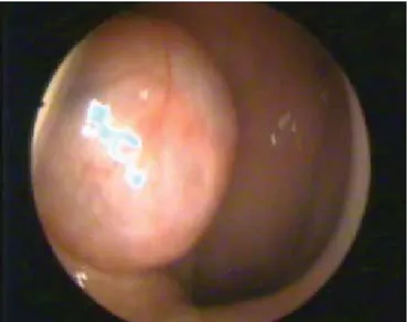
The tympanometric evaluation of the patient revealed a type B tympanogram. Given this finding, serous otitis media was considered. Nasal endoscopic examination revealed a cystic and smooth mass originating from the right side of the naso-pharynx. Also, it was enlarged to the Rosenmuller fossa and caused obstruction of the right Eustachian tube (Figure 1). The MRI showed a 1.3x2x1.3 cm sharply outlined lobar mass in the right side of the nasopharynx, spreading to the anterior of the fossa of Rosenmuller (Figure 2a, b).
By histological analysis of the biopsy, the lesion was diagnosed as PA. The area that the PA originated from was seen in the right anterolateral side of the nasopharynx with the aid of 0-degree and 30-degree endoscopes (Karl Storz, Germany). The lesion was to-tally excised from its originated area with the help of the
electro-cautery and forceps by the nasal endoscopic way. The Eustachian tube orifice was carefully inspected to determine the possibility of oedema and cautery inflammation. A shepard ventilation tube was inserted to the right tympanic membrane. No complication was developed. The postoperative diagnosis of the completely excised tumour histologically confirmed PA. Endoscopic and otoscopic examination of the patient revealed no abnormalities (Figure 3). One year after the surgery, control MRI showed no residual mass in the nasopharynx.
DISCUSSION
Pleomorphic adenoma constitutes the majority of benign tumours of all of the salivary glands (3). PAs are generally
FIG. 1. Nasopharyngeal endoscopic examination of tumor
FIG. 3. Postoperative endoscopic examination of tumor at 1 year follow-up period
FIG. 2. a, b. Axial and coronal MRI images showing a mass in the right side of the nasopharynx (arrow)
a b
119
Yazici et al. Nasopharyngeal Pleomorphic Adenoma
found in major salivary glands and rarely in the nasopharynx (3). To our knowledge, only a few cases of nasopharyngeal pleomorphic adenoma have been described in the English lit-erature (4).When a PA arises from the nasopharynx, the major symptom is nasal congestion due to obstruction of the nasal airway. Other symptoms, such as epistaxis, otalgia, dysphagia, hearing loss, mucopurulan rhinorrhea, and serous otitis media, may also be reported. In our case, the major symptoms were hearing loss and ear fullness. Due to the unusual localisation of the tumour, lymphoma, angiofibroma, haemangioma, ham-artoma, non-epithelial tumours, and various cysts should be considered in the differential diagnosis of PA (1).
The lesion usually appears with a smooth surface, an elastic and rubbery structure upon physical examination and no de-struction of the mucosa of the surrounding adjacent structure. In addition, a PA does not bleed easily. Radiological evalua-tion and endoscopic examinaevalua-tion are very useful in determin-ing the most appropriate surgical treatment. An imagdetermin-ing as-sessment alone cannot differentiate a PA from other low-grade malignant tumours of the nasopharynx. An incisional biopsy or fine needle aspiration biopsy is needed for the initial diagnosis before the main treatment of the tumour. However, fine needle aspiration is not possible for a mass is in the nasopharynx (5). Total surgery excision is not simple because of the location of the tumour in the nasopharynx, which is adjacent to important structures, such as the Eustachian tube and the cranial nerves. Thus, selecting the correct surgery is important to avoid com-plications. In the present case, we first performed a biopsy. Following the biopsy results, we totally removed the entire lesion via a transnasal endoscopic approach. Microscopically, epithelial, myoepithelial and stromal elements usually form the histologic structure of the PAs (2).Myxoid, cellular and mixed type are the main histological subgroups of PA.
The diagnosis of PA in the nasopharynx is not difficult. Complete excision is necessary for tumours with histologi-cally clear margins (5). Although the surgical approach is not easy, en bloc resection of a PA in the nasopharynx is feasible. Complications like bleeding and Eustachian tube dysfunction after operation can occur and are usually due to blind resec-tion. For nasopharyngeal PAs, the rate of malignant degenera-tion is not known exactly, but degeneradegenera-tion to carcinoma ex pleomorphic adenoma were reported in some case reports (3).
External approach to the nasopharynx may lead to signifi-cant postoperative morbidity. Therefore, we prefer to use an endoscopic approach to excise the tumour. The use of endo-scopic techniques provides a large surgical field and excellent visualisation, thereby avoiding surgical morbidity. Endoscopy also prevents blind resection and the destruction of adjacent structures. In the present case, the endoscopic approach was preferred to remove the tumour from the nasopharynx, and en-doscopy also helped to identify the attachment site of the lesion.
Careful follow-up of PA is necessary to ensure that an in-sufficient excision is diagnosed at an early stage. Endoscopic examination must be combined with imaging. In our patient, the control imaging and examination were clear. Combining the endoscopic approach with robotic surgery is another inter-esting possibility (6). Because of the potential malignant de-generation of recurrences, the surgery for recurrent PAs must be radical and wide.
Ethics Committee Approval: N/A.
Informed Consent: Written informed consent was obtained from the patient who participated in this study.
Peer-review: Externally peer-reviewed.
Author contributions: Concept - Z.M.Y., F.T.K., Y.Y., O.E.; De-sign - Z.M.Y., Y.Y., M.C., O.E., Y.Y.; Supervision - Z.M.Y., F.T.K.; Resource - Z.M.Y., Y.Y., M.C.; Materials - Z.M.Y., Y.Y., M.C.; Data Collection&/or Processing - Z.M.Y., Y.Y., M.C.; Analysis&/or In-terpretation - Z.M.Y., F.T.K., O.E.; Literature Search - Z.M.Y., Y.Y., M.C., O.E.; Writing - Z.M.Y., Y.Y., O.E.; Critical Reviews - Z.M.Y., F.T.K., Y.Y., O.E.
Conflict of Interest: No conflict of interest was declared by the authors.
Financial Disclosure: The authors declared that this study has re-ceived no financial support.
REFERENCES
1. Roh JL, Jung BJ, Rha KS, Park CI. Endoscopic resection of pleomorphic adenoma arising in the nasopharynx. Acta
Otolar-yngol 2005;125:910-2. [CrossRef]
2. Berrettini S, Fortunato S, De Vito A, Bruschini L. A rare case of nasopharyngeal pleomorphic adenoma. Case Rep Otolaryngol 2013;2013:712873.
3. Martinez-Capoccioni G, Martin-Martin C, Espinosa-Restrepo F. Transnasal endoscopic resection of a nasopharyngeal pleomor-phic adenoma: a rare case report. Eur Arch Otorhinolaryngol 2012;269:2009-13. [CrossRef]
4. Maruyama A, Tsunoda A, Takahashi M, Kishimoto S, Suzuki M. Nasopharyngeal pleomorphic adenoma presenting as otitis media with effusion: Case report and literature review. Am J
Otolaryngol 2014;35:73-6. [CrossRef]
5. Kuo YL, Tu TY, Chang CF, Li WY, Chang SY, Shiao AS et al. Extra-major salivary gland pleomorphic adenoma of the head and neck: a 10-year experience and review of the literature. Eur
Arch Otorhinolaryngol 2011;268:1035-40. [CrossRef]
6. Ozer E, Waltonen J. Transoral robotic nasopharyngectomy: a novel approach for nasopharyngeal lesions. Laryngoscope 2008;118:1613-6. [CrossRef]
120 Yazici et al. Nasopharyngeal Pleomorphic Adenoma