1315
http://journals.tubitak.gov.tr/medical/ © TÜBİTAK
doi:10.3906/sag-1806-236
Demonstration of the effect of brivaracetam on an experimental epilepsy model
Mustafa ÇETİNER1,*, Hasan Emre AYDIN2
1Department of Neurology, Faculty of Medicine, Dumlupınar University, Kütahya, Turkey 2Department of Neurosurgery, Faculty of Medicine, Dumlupınar University Kütahya, Turkey
* Correspondence: [email protected] 1. Introduction
Status epilepticus (SE) is a medical emergency with significant mortality, in which the seizure lasts continuously for more than 5 min and causes severe neuronal cell death (1). In particular, the pyriform cortex and hippocampus are the brain regions that are most sensitive and vulnerable to SE. SE also produces severe vasogenic edema accompanied by neuronal and astroglial damage in the piriform cortex (2). Experimental and clinical studies have shown that prolonged seizures can cause neuronal death in the brain (3–5). Seizure-induced apoptotic cell morphologies have been described in some SE models (6,7). The cell death mechanism directly involves many proteins, including both pro- and antiapoptotic proteins. While the activation of the proapoptotic proteins initiates the degeneration process, the activation of the antiapoptotic proteins may prevent the initiation or progression of the degeneration process (4). Among these, antiapoptotic proteins are the Bcl-2 gene family proteins, which are important regulators of the apoptosis process. Among them, some members such as Bcl-2, Bcl-XL, McL-1, and Bcl-W show antiapoptotic properties, while others, such as Bax, Bak, Bok, Bcl-XS,
Bad, and Bid, are proapoptotic (8). The proteins belonging to the Bcl-2 family are the main regulators of the cell death signals targeting the mitochondria and contribute to seizure-induced neuronal death (9). The ratio of antiapoptotic to proapoptotic proteins determines the direction in which the cell will advance. When apoptosis is prevented, the cell viability increases. Another protein is p53, which is one of the most versatile transcription factors present in the cell and triggers apoptosis. The activated p53 ensures it by increasing the expression of Bax and Bak from two proapoptotic Bcl-2 family proteins (10). p53-depleted mouse neurons are resistant to apoptosis induced by seizure and/or excitotoxins (11).
Annexin V is a 35-kDa protein. It was originally described as a vascular-derived protein with anticoagulant properties (12). Since then, annexin V has been used to study a universal marker of apoptosis and the changes in the cell membrane levels (13–16). Annexin V is activated by the emergence of apoptosis (17).
Our study aimed to investigate the effect of valproic acid (VPA), a potent broad-spectrum antiepileptic drug, and a new-generation antiepileptic agent called brivaracetam
Background/aim: The aim of this study was to investigate the effects of valproic acid (VPA) and a new-generation antiepileptic drug
called brivaracetam (BRV) on the brain damage occurring after status epilepticus (SE) in rats.
Materials and methods: In our study, an experimental animal model of SE, generated by stereotaxically injecting 0.4–2 µg of kainic
acid into the rat hippocampus, was used. The laboratory animals were divided into 4 groups: the first group was a sham group that was subjected to anesthesia and SE was not induced; the second group was a SE group, in which SE was induced using kainic acid but subjects were not treated; the third group was the VPA group, in which SE was induced using kainic acid and subjects were treated with VPA; and the fourth group was the BRV group, in which SE was induced using kainic acid and subjects were treated with BRV.
Results: Annexin V and p53 levels were statistically higher in the SE group than in the sham group (P < 0.001). Following the treatment
with VPA and BRV, a substantial decrease was observed in the annexin V and p53 levels compared to those of the SE group (P < 0.001). There was a statistically significant increase in Bcl-2 levels after VPA and BRV treatment compared to the SE group (P < 0.001).
Conclusion: Our study showed that VPA and BRV are protective against neuronal damage occurring after SE in rats due to the increase
in Bcl-2.
Key words: Rat, status epilepticus, apoptosis, valproic acid, brivaracetam
Received: 30.06.2018 Accepted/Published Online: 03.11.2018 Final Version: 12.12.2018
(BRV) on the brain injury observed after SE in rats and to compare the two drugs in terms of seizure control.
2. Materials and methods 2.1. Materials
A rat p53 ELISA kit (LSBio, Seattle, WA, USA), a rat Bcl-2 ELISA kit (LSBio), and a rat annexin V ELISA kit (LSBio) were used to measure the serum levels of these apoptotic markers.
2.2. Ethics
Ethics committee approval was obtained for all the experimental procedures. Every effort was made to minimize the suffering to which the animals were exposed and the number of animals used.
2.3. Animals
In our study, 40 Sprague-Dawley female rats aged between 6 and 8 weeks, with an average weight of 250–350 g, were used. The rats were observed during the study in an environment with suitable temperature (21 ± 2 °C) and humidity (60 ± 5% humidity). The animals were observed in this environment for 1 week before initiating the study to ensure their adaptation to the environment.
2.4. Establishment of SE model
In our study, an experimental animal model of SE, which was generated by stereotaxically injecting 0.4–2 µg of kainic acid into the rat hippocampus, was used (18). Rats were anesthetized by intraperitoneal administration of a mixture of ketamine hydrochloride (60 mg/kg) and xylazine (12 mg/kg). Under deep anesthesia within approximately 4–5 min, the head was fixed with the help of a stereotaxic instrument (Stoelting, Model 51600) and the head was opened vertically. Hippocampus coordinates were detected using a stereotaxic atlas, which were 6.0 mm posterior to the bregma 4.5 mm right and 7.0 mm down the skull surface. With the help of the minidrill, a miniburrhole was opened in the skull in that area. Subjects were given 0.4–2 µg of kainic acid intrahippocampally by inserting a 10-µL Hamilton needle into the burrhole during the waking phase (approximately 30 min after
anesthesia). The animals used in the study were divided into four groups (Table 1): Group 1, sham, without SE induction (n = 10); Group 2, SE induction with kainic acid, no treatment (n = 10); Group 3, SE induction with kainic acid, treated with intraperitoneal 1 mg/kg VPA; Group 4, SE induction with kainic acid, treated with intraperitoneal 0.4 µg/kg BRV (n = 10). In all groups except Group 1, after the injection of the kainic acid, the time to SE onset was approximately 5–60 min.
2.5. Observations
The behavioral responses developed were assessed as nonconvulsive seizures for stages 1 and 2 and convulsive seizures for stages 3, 4, and 5 using the Racine seizure staging scale (19). To determine the Bcl-2, p53, and annexin V levels, we sampled 2 mL of blood from each subject in each group after the procedure. The subjects were sedated with the same anesthetic agents and decapitated at the end of 24 h.
2.6. Statistical analysis
The statistical analysis was performed using SPSS 24.0 (IBM Corp., Armonk, NY, USA). Data were expressed as the mean ± standard deviation (SD). The relationship of Bcl-2, p53, and annexin V levels among the groups was determined using a one-way ANOVA test because the groups were normally distributed. For Bcl-2, p53, and annexin V levels among the groups in multiple comparisons, Tukey’s test was used. In the VPA and BRV groups, as the mean Racine seizure staging scores were not normally distributed, they were compared using the Mann–Whitney U test. P < 0.05 was considered statistically significant.
3. Results
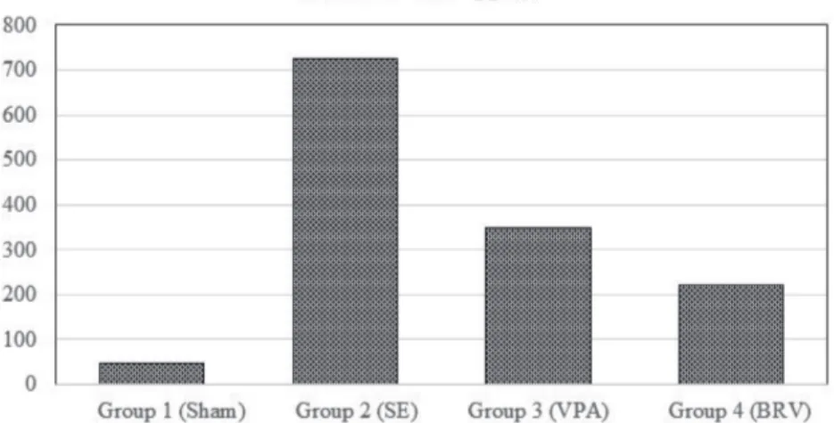
Annexin V levels were statistically higher in the SE group than in the other groups (P < 0.001, Figure 1). Following the treatment with VPA and BRV, a substantial decrease was observed compared to the SE group (P < 0.001). Furthermore, there was a statistically significant decrease in annexin V levels in the BRV group compared to the VPA group (P < 0.008, Figure 1).
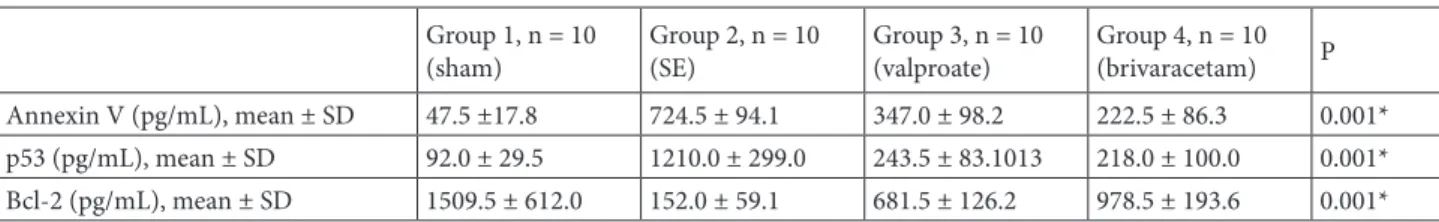
Table 1. Comparison of the mean annexin V, p53, and Bcl-2 levels across the groups (one-way ANOVA tests were used).
Group 1, n = 10
(sham) Group 2, n = 10 (SE) Group 3, n = 10 (valproate) Group 4, n = 10 (brivaracetam) P
Annexin V (pg/mL), mean ± SD 47.5 ±17.8 724.5 ± 94.1 347.0 ± 98.2 222.5 ± 86.3 0.001*
p53 (pg/mL), mean ± SD 92.0 ± 29.5 1210.0 ± 299.0 243.5 ± 83.1013 218.0 ± 100.0 0.001*
Bcl-2 (pg/mL), mean ± SD 1509.5 ± 612.0 152.0 ± 59.1 681.5 ± 126.2 978.5 ± 193.6 0.001*
p53 levels were statistically higher in the SE group than in any other group (P < 0.001, Figure 2). Following the treatment with VPA and BRV, a substantial decrease was observed compared to the SE group (P < 0.001). When p53 levels were compared between the BRV and VPA groups, no statistically significant difference was observed (P < 0.98).
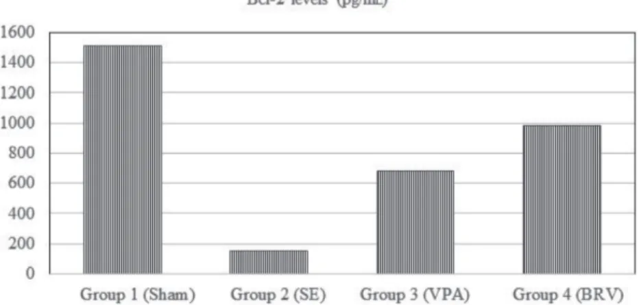
Bcl-2 levels were significantly lower in the SE group than in the sham group and the BRV group (P < 0.001). This measure was significantly lower in the BRV group than in the VPA group as well (P < 0.005). A statistically significant increase was detected after the treatment with VPA (P < 0.005) or BRV (P < 0.001) compared to the SE group. There was no statistically significant difference in Bcl-2 levels between the BRV and VPA groups (P < 0.19, Figure 3).
The mean annexin V, p53, and Bcl-2 levels in the groups are presented below, along with the statistical results (Table 1). In the VPA and BRV groups, the mean Racine seizure staging scores were 1.30 ± 2.11 and 1.90 ± 2.46, respectively. Statistical comparison showed no significant difference (P < 0.5, Table 2).
4. Discussion
The tumor suppressor p53 is among the first identified apoptosis-regulating elements affected by seizure activity (20,21) and continues to be among the active cell death pathways in seizure-induced neuronal death regulation (22,23). Annexin V is a marker that increases in the apoptotic process (16). In our study, the levels of these markers were significantly higher in the SE group than in the other groups. Our results support the apoptotic process after SE in the rats. Importantly, in our study, an
increase in annexin V was demonstrated in the seizure-induced apoptotic process. Apoptosis is a complex process of programmed cell death. The Bcl-2 gene family plays an important role in regulating apoptosis. Bcl-2 plays a role in inhibiting the apoptosis and prolonging cell longevity, whereas the Bax gene promotes cell death (24). Gillardon et al. reported that the proapoptotic protein Bax increased and Bcl-2 decreased after a seizure (9). In our study, Bcl-2 levels in the SE group were significantly lower than those in other groups.
VPA is a broad-spectrum antiepileptic drug used in the treatment of various types of seizures. It is also used for the treatment of bipolar disorder, schizoaffective disorder, social phobia, and neuropathic pain, as well as for the prophylaxis and treatment of migraine-type headaches (25). VPA is one of the antiepileptic drugs shown to reduce the neuronal damage associated with epileptic activity. In an experimental SE model, VPA was shown to significantly reduce neuronal damage in the hippocampal formation and improve neurological and memory function (26). Studies show that treatment with VPA supports neurogenesis and nerve sprouting and that it is neuroprotective in rats (27–29).
In our study, Bcl-2 levels were higher and p53 and annexin levels were lower in the antiepileptic drug groups than in the SE group. Our results overlap with those of previous studies. The decrease in annexin V level in the BRV group was found to be much greater than the decrease in the VPA group. Luo et al. failed to detect a significant change in the apoptosis of hippocampal neurons in epileptic rats after VPA administration compared to the control group, but showed an increase in Bcl-2/Bax ratio and thus the inhibition of apoptosis (24). Dash et al.
Figure 1. Annexin V levels: following the treatment with VPA and BRV, a substantial
decrease was observed compared to the SE group (P < 0.001). The BRV group had a statistically greater decrease compared to the VPA group (P < 0.008).
reported that VPA treatment may have beneficial effects on the blood–brain barrier and cognitive recovery, as well as a neuroprotective effect, and also VPA may be useful for the treatment of traumatic brain injury (30).
When recently administered antiepileptic therapies are examined, BRV has proven to be a safe and
pharmacokinetically efficient drug compared to the preexisting drugs (31). For this reason, we compared VPA, a drug with known efficacy, to a current drug called BRV, examining their effects on apoptosis as well as their antiepileptic effects. BRV is a selective, high-affinity ligand for synaptic vesicular protein 2A (SV2A) and has
Figure 2. p53 levels: following the treatment with VPA and BRV, a substantial decrease
was observed compared to the SE group (P < 0.001). When p53 levels were compared between BRV and VPA groups, no statistically significant difference was observed (P < 0.98).
Figure 3. Bcl-2 levels: Bcl-2 levels were significantly lower in the SE group than in the
sham group (P < 0.001). A statistically significant increase was detected after treatment with VPA (P < 0.005) or BRV compared to the SE group (P < 0.001). No statistically significant difference in Bcl-2 levels was found between BRV and VPA groups (P < 0.19).
Table 2. Comparison of Racine seizure staging scores across the groups
Group 3, n = 10) (valproate) Group 4, n = 10 (brivaracetam) P
Racine seizure scores (mean ± SD) 1.30 ± 2.11 1.90 ± 2.46 0.5
been approved as an adjuvant therapy for focal seizures in adults with epilepsy (32). BRV has been shown to be antiepileptogenic in several experimental models (33). Although the antiepileptic mechanism of BRV has been shown to be similar to that of levetiracetam, affecting sodium channels, the cause of the clinical difference between the two therapeutics remains unexplained (34).
In our study, the seizure behaviors exhibited in a kainic acid-induced SE model and treatment with antiepileptic drugs were scored using the Racine staging scale (19). The comparison shows that the anticonvulsant effects of the two antiepileptic drugs are not significantly different.
Although only a brief procedure, the anesthetics used can alter kainic acid’s effect. For this reason, it is important to choose the preferred anesthetic carefully. When ketamine/xylazine is used, it is usually not necessary to reanesthetize the animal because the effect lasts 20–30 min once deep anesthesia is reached. Whichever is chosen, the same anesthesia procedure should be used for all animals
in each experiment. In general, ketamine is an N-methyl-d-aspartate receptor blocker and has a neuroprotective effect in rats, reducing neuronal degeneration occurring after SE (35,36). However, Less reported that a single high dose of ketamine administered (150–180 mg/kg i.p.) for neuronal damage caused by kainic acid did not reduce the neuronal damage in his study (37). In our study, ketamine, used as a single dose, was not high in doses and the same procedure was used in all groups.
In our results, annexin V and p53 levels were substantially elevated in rats in which SE was induced with kainic acid. This result is supported by neuronal damage observed after SE. It has also been shown that VPA, a potent antiepileptic drug, and BRV, a new-generation antiepileptic drug, exert protective effects against neuronal damage by increasing Bcl-2 and that the anticonvulsant effects of the two drugs are similar. Our study suggests that BRV is also effective against apoptosis, unlike other antiepileptic drugs, and can prevent the epileptic focus from developing apoptotic injury.
References
1. Rice AC, DeLorenzo RJ. NMDA receptor activation during status epilepticus is required for the development of epilepsy. Brain Res 1998; 782: 240-247.
2. Kim JE, Ryu HJ, Yeo SI, Seo CH, Lee BC, Choi IG, Kim DS, Kang TC. Differential expressions of aquaporin subtypes in astroglia in the hippocampus of chronic epileptic rats. Neuroscience 2009; 163: 781-789.
3. Bengzon J, Kokaia Z, Elmér E, Nanobashvili A, Kokaia M, Lindvall O. Apoptosis and proliferation of dentate gyrus neurons after single and intermittent limbic seizures. P Natl Acad Sci USA 1997; 94: 10432-10437.
4. Liou AK, Clark RS, Henshall DC, Yin XM, Chen J. To die or not to die for neurons in ischemia, traumatic brain injury and epilepsy: a review on the stress-activated signaling pathways and apoptotic pathways. Prog Neurobiol 2003; 69: 103-142. 5. Fujikawa DG. Prolonged seizures and cellular injury:
understanding the connection. Epilepsy Behav 2005; 7: S3-S11. 6. Sloviter RS, Dean E, Sollas AL, Goodman JH. Apoptosis and
necrosis induced in different hippocampal neuron populations by repetitive perforant path stimulation in the rat. J Comp Neurol 1996; 366: 516-533.
7. Baille V, Clarke PG, Brochier G, Dorandeu F, Verna JM, Four E, Lallement G, Carpentier P. Soman-induced convulsions: the neuropathology revisited. Toxicology 2005; 215: 1-24. 8. Reed JC. Bcl-2 family proteins. Oncogene 1998; 17: 3225-3236. 9. Gillardon F, Wickert H, Zimmermann M. Up-regulation of
bax and down-regulation of bcl-2 is associated with kainate-induced apoptosis in mouse brain. Neurosci Lett 1995; 192: 85-88.
10. Bargonetti J, Manfredi JJ. Multiple roles of the tumor suppressor p53. Curr Opin Oncol 2002; 14: 86-91.
11. Morrison RS, Wenzel HJ, Kinoshita Y, Robbins CA, Donehower LA, Schwartzkroin PA. Loss of the p53 tumor suppressor gene protects neurons from kainate-induced cell death. J Neurosci 1996; 16: 1337-1345.
12. Reutelingsperger CP, Hornstra G, Hemker HC. Isolation and partial purification of a novel anticoagulant from arteries of human umbilical cord. Eur J Biochem 1985; 151: 625-629. 13. Gatti R, Belletti S, Orlandini G, Bussolati O, Dall’Asta V,
Gazzola GC. Comparison of annexin V and calcein-AM as early vital markers of apoptosis in adherent cells by confocal laser microscopy. J Histochem Cytochem 1998; 46: 895-900. 14. Lizarbe MA, Barrasa JI, Olmo N, Gavilanes F, Turnay J.
Annexin-phospholipid interactions. Functional implications. Int J Mol Sci 2013; 14: 2652-2683.
15. Wang J, He L, Chen D, Pi Y, Zhou W, Xiong X, Ren Y, Lai Y, Hua Z. Quantitative analysis of annexin V-membrane interaction by flow cytometry. Eur Biophys J 2015; 44: 325-336. 16. Kizmazoglu C, Aydin HE, Sevin IE, Kalemci O, Yuceer N,
Atasoy MA. Neuroprotective effect of resveratrol on acute brain ischemia reperfusion injury by measuring annexin V, p53, Bcl-2 levels in rats. J Korean Neurosurg S 2015; 58: 508-512.
17. Reutelingsperger CP, van Heerde WL. Annexin V, the regulator of phosphatidylserine-catalyzed inflammation and coagulation during apoptosis. Cell Mol Life Sci 1997; 53: 527-532.
18. Ben-Ari Y. Limbic seizure and brain damage produced by kainic acid: mechanisms and relevance to human temporal lobe epilepsy. Neuroscience 1985; 14: 375-403.
19. Racine RJ. Modification of seizure activity by electrical stimulation. II. Motor seizure. Electroen Clin Neuro 1972; 32: 281-294.
20. Sakhi S, Bruce A, Sun N, Tocco G, Baudry M, Schreiber SS. p53 induction is associated with neuronal damage in the central nervous system. P Natl Acad Sci USA 1994; 91: 7525-7529. 21. Sakhi S, Sun N, Wing LL, Mehta P, Schreiber SS. Nuclear
accumulation of p53 protein following kainic acid-induced seizures. Neuroreport 1996; 7: 493-496.
22. Tan Z, Sankar R, Shin D, Sun N, Liu H, Wasterlain CG, Schreiber SS. Differential induction of p53 in immature and adult rat brain following lithium-pilocarpine status epilepticus. Brain Res 2002; 928: 187-193.
23. Tan Z, Sankar R, Tu W, Shin D, Liu H, Wasterlain CG, Schreiber SS. Immunohistochemical study of p53-associated proteins in rat brain following lithium-pilocarpine status epilepticus. Brain Res 2002; 929: 129-138.
24. Luo Z, Fang Y, Zhang L. The effects of antiepileptic drug valproic acid on apoptosis of hippocampal neurons in epileptic rats. Pak J Pharm Sci 2015; 28: 319-324.
25. Wadzinski J, Franks R, Roane D, Bayard M. Valproate-associated hyperammonemic encephalopathy. J Am Board Fam Med 2007; 20: 499-502.
26. Brandt C, Gastens AM, Sun M, Hausknecht M, Loscher W. Treatment with valproate after status epilepticus: effect on neuronal damage, epileptogenesis, and behavioral alterations in rats. Neuropharmacology 2006; 51: 789-804.
27. Hao Y, Creson T, Zhang L, Li P, Du F, Yuan P, Gould TD, Manji HK, Chen G. Mood stabilizer valproate promotes ERK pathway-dependent cortical neuronal growth and neurogenesis. J Neurosci 2004; 24: 6590-6599.
28. Laeng P, Pitts RL, Lemire AL, Drabik CE, Weiner A, Tang H, Thyagarajan R, Mallon BS, Altar CA. The mood stabilizer valproic acid stimulates GABA neurogenesis from rat forebrain stem cells. J Neurochem 2004; 91: 238-251.
29. Yuan PX, Huang LD, Jiang YM, Gutkind JS, Manji HK, Chen G. The mood stabilizer valproic acid activates mitogen-activated protein kinases and promotes neurite growth. J Biol Chem 2001; 276: 31674-31683.
30. Dash PK, Orsi SA, Zhang M, Grill RJ, Pati S, Zhao J, Moore AN. Valproate administered after traumatic brain injury provides neuroprotection and improves cognitive function in rats. PLoS One 2010; 5: e11383.
31. Orinx C, Legros B, Gaspard N. Recent antiseizure medications in the intensive care unit. Minerva Anestesiol 2017; 83: 878-887.
32. Klitgaard H, Matagne A, Nicolas JM, Gillard M, Lamberty Y, De Ryck M, Kaminski RM, Leclercq K, Niespodziany I, Wolff C et al. Brivaracetam: rationale for discovery and preclinical profile of a selective SV2A ligand for epilepsy treatment. Epilepsia 2016; 57: 538-548.
33. Matagne A, Margineanu DG, Kenda B, Michel P, Klitgaard H. Anti-convulsive and anti-epileptic properties of brivaracetam (ucb 34714), a high-affinity ligand for the synaptic vesicle protein, SV2A. Brit J Pharmacol 2008; 154: 1662-1671. 34. Kappes JA, Hayes WJ, Strain JD, Farver DK. Brivaracetam:
an adjunctive treatment for partial-onset seizures. J Clin Pharmacol 2017; 57: 811-817.
35. Loss CM, Cordova SD, de Oliveira DL. Ketamine reduces neuronal degeneration and anxiety levels when administered during early life-induced status epilepticus in rats. Brain Res 2012; 1474: 110-17.
36. Fujikawa DG. Neuroprotective effect of ketamine administered after status epilepticus onset. Epilepsia 1995; 36: 186-195. 37. Less GJ. Effects of anaesthetics, anticonvulsants and glutamate
antagonists on kainic acid-induced local and distal neuronal loss. J Neurol Sci 1992; 108: 221-228.