See discussions, stats, and author profiles for this publication at: https://www.researchgate.net/publication/305257702
CD38 Expression and Variation as a Prognostic Factor Chronic Lymphocytic
Leukemia
Article in Clinical laboratory · January 2013 DOI: 10.7754/Clin.Lab.2015.151125 CITATION 1 READS 97 6 authors, including:
Some of the authors of this publication are also working on these related projects:
Gonadotoxic Effects Of Nilotinib In Chronic Myeloid Leukemia Treatment Dose In Mouse Model View project yes my articleView project
Mesude Falay
Ankara Numune Training and Research Hospital 22PUBLICATIONS 58CITATIONS
SEE PROFILE
Ahmet Kürşad Güneş 2PUBLICATIONS 1CITATION
SEE PROFILE
Meltem Aylı
Gulhane Military Medical Academy 44PUBLICATIONS 226CITATIONS
SEE PROFILE
Gulsum Ozet
Ankara Numune Training and Research Hospital 55PUBLICATIONS 506CITATIONS
ORIGINAL ARTICLE
CD38 Expression and Variation as a Prognostic Factor Chronic
Lymphocytic Leukemia
Mesude Falay
1, Funda Ceran
1, Ahmet K. Gunes
1, Simten Dagdas
1, Meltem Ayli
2, Gulsum Ozet
11 Ankara Numune Education and Resarch Hospital, Hematology, Ankara, Turkey
2 Ufuk University Medical Faculty, Hematology, Ankara, Turkey
SUMMARY
Background: In this study, we aimed to determine a cutoff level for CD38 that would aid us in identifying chronic
lymphocytic leukemia patients in need of early therapy and predicting patients at sufficiently low risk who would likely exhibit a rapid improvement; we also aimed to find out if CD38 expression would show variability during disease course and determine the extent of CD38 expression.
Methods: 124 patients were diagnosed with CLL. CD38 and ZAP-70 expression levels were measured with four
color flowcytometry. Time from diagnosis to initial therapy was calculated for all patients. CD38 expression was studied for a second time during follow-up in 50 patients.
Results: For cutoff levels of 7%, 20%, and 30%, CD38 expressions were 61.3%, 25%, and 24.2%, respectively. At
all three cutoff levels there were significant correlations with all parameters except age between CD38+ vs. CD38- groups (p < 0.001). The comparative rates of starting therapy for cutoff levels of 7%, 20%, and 30% in CD38+ and CD38- groups were 77.5% vs. 6.25%; 100% vs. 30.7%, and 100% vs. 31.5%, respectively (p < 0.001). Multi-ple Cox Proportional Hazards Regression analysis: for a cutoff level of 7%, survival was affected by STAGE, ZAP70, and CD38.
Conclusions: A CD38 cutoff level of 7% determined by standardized laboratory techniques is an important
prog-nostic marker. However, the number and frequency of repeat measurements of CD38 expression, and cutoff level of CD38 expression that significantly predict disease prognosis should be further determined by future cohort studies.
(Clin. Lab. 2016;62:1287-1293. DOI: 10.7754/Clin.Lab.2015.151125)
Correspondence: Mesude Falay
Ankara Numune Education and Resarch Hospital Hematology Ankara, Turkey
Phone: +90 3125084646
Fax: +90 3124268561
Email: [email protected]
_____________________________________________
Manuscript accepted December 1, 2015
KEY WORDS
chronic lymphocytic leukemia, CLL, CD38, prognosis, flow cytometry
INTRODUCTION
Chronic lymphocytic leukemia (CLL) is the most com-mon type of leukemia in adults. The clinical presenta-tion of the disease is highly heterogenous, with some forms not requiring any treatment for years while others require rapid therapy. Starting therapy early in the course cannot achieve cure nor does it prolong survival [1]. A timely started therapy, on the other hand, delays the natural course of the disease and prolongs survival. Many studies have been conducted so far to explain the
M. Falay et al.
biology of CLL. These studies have clarified the patho-genesis of the disease and identified novel prognostic parameters that apparently affect prognosis. The first prognostic factor defined in CLL was the stage of the disease. There are two main staging systems currently used for staging CLL (RAI and BINET) [2,3]. Although there is a significant correlation between disease stage and survival, this relationship has some deficiencies. Available staging systems do not aid clinicians in dif-ferentiating patients with a potentially progressive dis-ease from those with a relatively stable one and deter-mining treatment responses of patients at the same stage. Thus, markers that predict prognosis indepen-dently of disease stage have been developed for CLL. In 1999 Hamblin et al. and Damle et al. reported for the first time the relationship between the variable zone of the immunoglobulin heavy chain gene (IgVH) somatic hypermutation and prognosis in CLL [4,5]. Both studies linked the absence of IgVH mutation to a worse progno-sis and its presence to a favorable prognoprogno-sis. However, IgVH mutation analysis is a technically demanding, ex-pensive, and time-consuming process. As a result, its routine use as a diagnostic laboratory test is not feasible in clinical practice. Damle et al. demonstrated a correla-tion between CD38 expression, which is technically easier to demonstrate, and IgVH mutation [5]. While CD38 expression is lower in patients with IgVH muta-tion, it was found to be higher (> 30%) in those free of IgVH mutation [6]. Some of the subsequent studies have also shown inconsistent results between CD38 expression and IgVH mutations, making clinicians con-sider CD38 and IgVH independent prognostic factors [7,8]. CD38 is a transmembrane glycoprotein expressed in hematopoietic cells. While it is highly expressed by activated B and T lymphocytes, natural killer cells, and dendritic cells, its expression is relatively lower in pe-ripheral B cells. It plays a role in cell adhesion, signal transduction, and calcium regulation. Increased CD38 expression in lymph nodes, along with increased vascu-lar density, correlates with increased lymphocyte prolif-eration and disease progression [9]. CD38 expression is easily measured at diagnosis by flow cytometry. IgVH mutation and cytogenetic studies are expensive tests that cannot be performed at every laboratory; however, CD38 expression study by flow cytometry is relatively easier and a less expensive test. However, CD38 expres-sion has some limitations for use as a prognostic factor, including its heterogenous nature, temporal variability, and unclear cutoff levels [11-13].
Many cutoff levels ranging from 5% to 30% have been proposed for CD38 expression. Traditionally, a CD38 expression of greater than 30% has been linked to pro-gressive disease [5,8,10,14]. Some studies have consid-ered a cutoff level of 20% significant [15-17]. Kröber et al. reported that a cutoff level of 7% effectively distin-guished separate prognostic groups [13,18,19]. Hock et al., in a study on 130 patients with newly diagnosed CLL, failed to detect any significant clinical difference between CD38 positivity of > 5% and > 95% [20].
Re-cently, any level of CD38 positivity has been consid-ered as a prognostic disadvantage [21,22].
Some studies have suggested that CD38 expression is a dynamic process that shows variability over time [10-27], that this change is usually not great (10% at most), and that it is not possible to argue that CD38 expression is associated with more or less favorable clinical out-comes [23,24]. However, if a change should occur, it is generally accepted that a decrease in CD38 expression is a favorable sign. Indeed, cells with increased CD38 expression signal for a more aggressive state in clone evolution. Hence, it is argued that the serial analysis of CD38 can be a real-time indicator of leukemic cell pro-liferation and may be a useful tool for the evaluation of clonal behavioral change [25]. However, it has been ob-served that cases which were CD38 negative at the time of diagnosis never expressed CD38 later in the disease course [22].
In this study, we aimed to determine a cutoff level for CD38 that would aid us in identifying patients in need of early therapy and predicting patients at sufficiently low risk who would likely exhibit a rapid improvement; we also aimed to find out if CD38 expression would show variability during disease course and determine the extent of CD38 expression.
MATERIALS AND METHODS Patients
This study retrospectively reviewed the medical records of 124 patients (39 females, 85 males) aged 31 - 87 years who were diagnosed with CLL at Ankara Numune Training and Research Hospital, Hematology Clinic be-tween October 2009 and July 2011 according to the Na-tional Cancer Institute-sponsored Working Group guidelines for chronic lymphocytic leukemia (NCI-WG) criteria [28]. An ethics committee approval was obtain-ed from Ankara Numune Training and Research Hospi-tal, Scientific Committee. A peripheral blood smear was prepared and morphologically examined for each pa-tient at the time of diagnosis. Immunophenotypic studies were done with flow cytometric study. White blood cell count, lymphocyte percentage, hemoglobin concentration, and platelet count were studied, and clin-ical stage was determined according to RAI staging sys-tem. Serum β2 microglobulin (β2MG) level was also measured. CD38 and ZAP70 expressions were studied from fresh peripheral blood samples using flow cytome-try. Time from diagnosis to initial therapy was calculat-ed for all patients. CD38 expression was studicalculat-ed for a second time during follow-up in 50 patients.
Flow cytometry
The monoclonal antibodies (MoAb) used for labeling in flow cytometry were obtained from Beckman Coulter (BC, USA) company. CD45 Fluorescein Isothiocyanate (FITC), CD79b Phycoerythrin (PE), CD11c Phycoery-thrin-Cyanin 5 (PC5), CD19 Phycoerythrin-Texas Red
(ECD), CD5 FITC, CD10 PE, CD14 PC5, CD20 ECD, CD22 PE, CD23 PE, CD38 PC5, CD103 FITC, CD43 PE, Kappa FITC, Lambda PE, ZAP70 FITC, and CD2 PC5 MoAbs were used for this purpose. The Fluores-cence Minus One Control (FMO) is a type of control used to properly interpret flow cytometry data.
For the four color flow cytometric study, fresh peripher-al blood samples were put into K3 EDTA containing tubes and studied without delay. Peripheral blood mononuclear cells were separated from freshly drawn anticoagulated blood by Ficoll-Paque density gradient centrifugation. Cells were preincubated for 5 minutes with mouse serum before the staining in order to block the nonspecific binding. Surface staining was performed by direct immunfluorescence in a standard four color flow cytometry approach. 100 µL (1 x 10⁶ cells)
periph-eral blood mononuclear cells were incubated in 200 µL PBS at room temperature for 30 minutes in the dark with 10 µL monoclonal antibody. After two wash steps with phosphate buffered saline (PBS), cell acquisition and analysis were performed in a FC 500 (BC, USA) flow cytometry device using the CXP program. All samples were of CD5+CD19+, CD20+, CD23+, Ig light chain κ or λ immunophenotype. The study for CD38 ex-pression was carried out by gating from CD5+CD19+ cell population.
Intracellular ZAP70 expression was determined using a commercial fixation/permebealization kit. Briefly, 1 x 10⁶ MNCs were first surface stained with CD2/CD16/
CD19 and then fixed, permeabilized, and stained with ZAP70 FITC moAb. ZAP70 was evaluated in CD19+ B cells by excluding its expression in T and NK cells. A cutoff level of 20% was determined for ZAP70. Sepa-rate analyses were carried out for CD38 cutoffs of 7%, 20%, and 30%.
Statistical analysis
Data analysis was performed by using Statistical Pack-age for Social Sciences (SPSS) version 11.5 software (SPSS Inc., Chicago, IL, USA). Treatment-free survival time was defined as the time from diagnosis to treat-ment or last contact for those who did not have any treatment. Treatment-free survival (TFS) times were computed by the method of Kaplan-Meier and were compared using the log-rank test. Cox proportional haz-ards regression analyses were performed to identify the best cutoff point of CD38 levels after adjustment for all potential risk factors. Any variable whose univariable test had a p-value less than 0.25 was accepted as a can-didate for the multivariable model along with all vari-ables of known clinical importance. Hazards ratios and 95% confidence intervals for all independent variables were also calculated. Categorical data were analyzed by Pearson’s Chi-square or Fisher’s exact test, where appli-cable. A p-value less than 0.05 was considered statisti-cally significant.
RESULTS
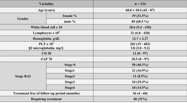
This study included a total of 124 subjects (39 females, 85 males) with a mean age of 68 ± 10.4 years. The clini-cal and laboratory characteristics of the study subjects were summarized in Table 1. Mean white blood cell count was 38.6 x 10³, mean lymphocyte count 31 x 103,
mean hemoglobin level 12.7 ± 2.27 g/dl, mean throm-bocyte count 202 x 103, and β2MG level 3.4 (1.2 -
16.1) mg/L. Fifty (40.3%) patients had stage 0 disease, 21 (16.9%) had stage 1 disease, 11 (8.9%) had stage 2 disease, 24 (19.4%) had stage 3 disease, and 18 (14.5%) had stage 4 disease. The total duration of follow-up was 4 - 68 months (mean 36 months). During treatment free follow-up, 4 patients were excluded from follow-up, 3 patients died from other causes, and 55 (47.4%) patients were started on therapy due to progressive disease. Six-ty-one (52.6%) patients are still followed without any treatment at the time of the writing of this manuscript. The proportion of CLL cells expressing CD38 above the isotype control level ranged from 0.2% to 97.4%, with a median of 12%. ZAP70 expression was 0% to 97%, with a median of 18.5%.
For cutoff levels of 7%, 20%, and 30%, CD38 expres-sions were 61.3%, 25%, and 24.2%, respectively. At all three cutoff levels there were significant correlations with all parameters except age between CD38+ vs. CD38- groups (p < 0.001).
The mean follow-up duration was significantly longer for the CD38 cutoff level of 7% compared to other
cut-off levels (mean 43 months (9 - 68 months)) (p < 0.001). As for therapy need, 47.4% of the subjects
were started on therapy during follow-up. The compara-tive rates of starting therapy for cutoff levels of 7%, 20%, and 30% in CD38+ and CD38- groups were 77.5% vs. 6.25%; 100% vs. 30.7%, and 100% vs. 31.5%, respectively (p < 0.001).
The likelihood of 3- and 5-year treatment free follow-up was 93.75% for CD38- group and 17% for CD38+ group when a cutoff level of 7% was selected for CD38 positivity. When the cutoff level selected was 20%, the likelihood of 3- and 5-year treatment free follow-up was 66.6% for CD38- group and 10.7% for the CD38+ group for one-year. As for the cutoff level of 30%, the likelihood of 3- and 5-year treatment free follow-up was 65.9% for the CD38- subjects and 11.1% for the CD38+ subjects for one-year.
A univariate Kaplan-Meier survival analysis was per-formed to evaluate the individual effects of certain fac-tors such as age, gender, CD38, and ZAP70 on treat-ment free follow-up duration. There were significant correlations between ZAP70, stage, hemoglobin, lym-phocyte count, thrombocyte count, β2 microglobulin, and survival for all cutoff levels of CD38 expression (p < 0.001); on the other hand, age and gender were not correlated to surival (Table 2).
Combined effects of laboratory parameters, STAGE, and other prognostic factors on the differentitation of the group followed without treatment and the group
M. Falay et al. Table 1. Demographic features.
Variables n = 124
Age (years) 68.0 ± 10.4 (42 - 87)
Gender female % 39 (31.5%)
male % 85 (68.5 %)
White blood cell x 10 38.6 (9.4 - 430)
Lymphocyte x 103 31 (6.8 - 420) Hemoglobin. g/dL 12.7 ± 2.27 PLT x 103 β2 microglobulin. mg/L 202 (19 - 403) 3.8 (2.0 - 9.2) CD 38 12 (0 - 97) ZAP 70 18.5 (0 - 97) Stage RAI Stage 0 50 (40.3%) Stage1 21 (16.9%) Stage2 11 (8.9%) Stage3 24 (19.4%) Stage4 18 (14.5%)
Treatment free of follow-up period (months) 36 (4 - 68)
Requiring treatment 40 (32%)
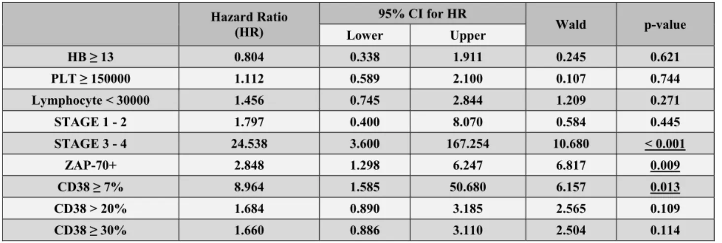
started on treatment were analyzed using the multiple Cox Proportional Hazards Regression analysis. RAI 0 cases were also in the analysis; as the HR value of RAI0 is 1.000, it was not shown in Table 3.
For a cutoff level of 7%, survival was affected by STAGE, ZAP70, and CD38. For cutoff levels of 20% and 30%, survival was affected by STAGE and ZAP70 only. While the rate of starting therapy was significantly different (increased 24.538-fold (95% CI: 3.600 - 167.254) (p < 0.001)) in stage 3 - 4 compared to stage 0 for the cutoff level of 7%, no such significant difference was detected for the cutoff levels of 20% and 30% (it increased 5.257-fold (95% CI: 1.502 - 18.402) for the cutoff level of 20% (p = 0.009) and 5.239-fold (95% CI: 1.496 - 18.347) for the cutoff level of 30%) (p = 0.010). For a cutoff level of 7%, the rate of starting therapy showed a significant increase in CD38+ group compar-ed to CD38- group (HR: 8.964, 95%CI: 1.585 - 50.680, p = 0.013). For the cutoff levels of 20% and 30%, on the other hand, the two groups did not differ significant-ly (p = 0.109 and p = 0.114, respectivesignificant-ly). According to the available results, the most significant difference was achieved for a cutoff level of 7%.
We studied CD38 expression for a second time at a mean of 28 months (12 - 50 months) during the follow-up in 50 patients. Of these, 17 had progressive disease, 11 were at post-tretment period, and 22 were at treat-ment free follow-up. In 48 (90%) patients CD38 expres-sion remained stable during disease course. We only de-tected increased CD38 expression in 1 (2%) patient with progressive disease and 1 (2%) patient receiving
treat-ment subsequent to the treattreat-ment. The first patient had a disease stage of RAI 1 and a CD38 expression of 16% at the time of diagnosis; CD38 expression increased to 41% and disease stage advanced to RAI 3 at 18 months after the diagnosis. In the patient who was receiving therapy, CD38 expression before and 22 months after the therapy were 26% and 40%, respectively. None of the 22 subjects who were at treatment free follow-up had a CD38 expression that turned positive from nega-tive or neganega-tive from posinega-tive or showed fluctuation of more than 5%.
DISCUSSION
Despite being defined as a poor prognostic sign by many studies, the importance of CD38 expression is controversial, particularly in respect to the definition of a positive result. Different cutoff levels have been pro-posed by various studies. While a cutoff level of 20 - 30% is generally accepted, cutoff levels of 5% or 7% have also been proposed [13,18]. Any level of CD38 expression has recently been regarded as a poor prog-nostic sign [21,22].
In the present study we evaluated the extent of CD38 expression by four color flow cytometry, its correlation with clinical and laboratory features, variability of CD38 expression during disease course, its optimum cutoff levels, and treatment free survival (TFS) in CLL. All analyses were performed in samples from fresh pe-ripheral blood samples at a single center. In 124
sub-Table 2. Clinic and laboratory features: a univariate Kaplan-Meier survival analysis.
n/N CSR TFST (95 % CI) Log-Rank p-value
Age < 65 14/32 56.2 33.3 (24.3 - 42.3) 0.47 0.4922 ≥ 65 41/84 51.2 35.3 (28.2 - 42.4) Gender female 20/36 44.4 25.5 (17.4 - 33.5) 1.26 0.2623 male 35/80 56.2 39.2 (32.1 - 46.3) Hemoglobin < 13 37/48 22.9 10.2 (4.7 - 15.7) 49.85 < 0.0001 ≥ 13 18/68 73.5 52.1 (45.8 - 58.4) Platelet < 150000 17/18 5.6 4.6 (0.0 - 10.5) 32.82 < 0.0001 ≥ 150000 38/98 61.2 42.7 (36.5 - 49.0) Lymphocyte < 30000 14/56 75.0 51.1 (43.5 - 58.7) 20.24 < 0.0001 ≥ 30000 41/60 31.7 24.2 (16.6 - 31.8) RAI Stage 0 2/47 95.7 65.3 (61.6 - 69.0) 124.46 < 0.0001 1 - 2 16/30 46.7 30.2 (22.6 - 37.9) 3 - 4 37/39 5.1 1.4 (0.3 - 2.5) CD-38 < 7% 2/45 65.5 (62.2 - 68.8) 59.80 < 0.0001 ≥ 7% 53/71 16.5 (10.8 - 22.2) CD-38 ≤ 20% 27/88 69.3 47.9 (41.6 - 54.2) 64.5 < 0.0001 > 20% 28/28 0.0 3.3 (1.1 - 5.5) CD-38 < 30% 28/89 68.5 47.4 (41.1 - 53.7) 60.93 < 0.0001 ≥ 30% 27/27 0.0 3.4 (1.1 - 5.7) ZAP-70 < 20% 11/64 82.8 57.1 (51.2 - 63.0) 65.45 < 0.0001 ≥ 20% 44/52 15.4 9.5 (5.1 - 13.9)
Table 3. The results of multiple Cox’s proportional hazard regression analyses. Hazard Ratio (HR) 95% CI for HR Wald p-value Lower Upper HB ≥ 13 0.804 0.338 1.911 0.245 0.621 PLT ≥ 150000 1.112 0.589 2.100 0.107 0.744 Lymphocyte < 30000 1.456 0.745 2.844 1.209 0.271 STAGE 1 - 2 1.797 0.400 8.070 0.584 0.445 STAGE 3 - 4 24.538 3.600 167.254 10.680 < 0.001 ZAP-70+ 2.848 1.298 6.247 6.817 0.009 CD38 ≥ 7% 8.964 1.585 50.680 6.157 0.013 CD38 > 20% 1.684 0.890 3.185 2.565 0.109 CD38 ≥ 30% 1.660 0.886 3.110 2.504 0.114
jects with CLL, the rate of CD38 expression was found to be 61.3% for a cutoff level of 7%, which was signifi-cantly higher compared to the rates with cutoff levels of 20% and 30%. Previous studies have generally reported CD38 positivity rates of 27 - 55% for cutoff levels of
20% to 30% [4,5,8,13,19]. We found CD38 positivity rates of 25% and 24.2% for cutoff levels of 20% and 30%, respectively. Letestu et al. found a positivity rate of 32% for a cutoff level of 7% in BINET 0 cases [29]. Kröber et al. followed 325 CLL cases for a mean of 69
M. Falay et al.
months and reported a 52% positivity rate for a cutoff level of 7% and 36% for a cutoff level of 30% [8]. The number of RAI 3 - 4 cases was higher (28%) in our study compared to other studies; we therefore found a higher CD38 positivity rate for a cutoff level of 7%. For all three cutoff levels, CD38 positive subjects, as compared to CD38 negative ones, had a significantly higher percentage of men, a higher proportion of RAI intermediate [1,2] and high-risk (RAI 3 - 4) cases, a lower Hb concentration and platelet count, a higher lymphocyte count, a higher rate of ZAP70 positivity, and a higher β2MG level. Our work thus confirms other studies suggesting that CD38 positivity parallels poor prognostic markers [4,5,8,13,19,30].
One of the main reasons for determining prognostic fac-tors is to identify patients in need of treatment. The most significant difference in the rate of starting treat-ment in CD38+ and CD38- groups occurred for a cutoff level of 7%. A CD38 expression greater than 7% ap-pears as a good cutoff level for identifying patients in need of early therapy. These results overlap those re-ported by Chevallier et al., Durig et al., Dal Poeta et al., Gentile et al., and Thornton et al. [13,14,16,19,30]. Time from diagnosis to first treatment was significantly longer for all cutoff levels in CD38 negative subjects compared to the CD38 positive ones. The likelihood of 5-year TFS for CD38 < 7% and CD38 ≥ 7% patients were 93.75% and 17%, respectively. Gentile et al. found a 3-year TFS rate of 59% for CD38 ≥ 7% group, which was greater than the figure we found [13], because the majority of our subjects had advanced disease stage (RAI 3,4). When regarding the effects of the prognostic value of CD38 expression on overall survival, it is still possible to argue that a cutoff level of 7% is a good cut-off level to identify patients having a poor prognosis. Gentile et al. observed that a cutoff level of 7% was not so successful in identifying patients with a poor progno-sis; in contrast, CD38 negative subjects had a greater likelihood of survival at the cutoff levels of 20% and 30%. The authors attributed this difference to a short follow-up period (32 months) [13]. However, although the follow-up period in our study was nearly as short as in the Gentile et al. study (36 months), we found that 7% was a more significant cutoff level. This difference between the two studies may have resulted from our study containing a lower number of early-stage patients than advanced-stage patients.
Chang et al., Chevalier et al., Hamblin et al., and Ryan D et al., and Nipp et al. found that CD38 expression showed variability with disease progression or after chemotherapy [6,25,26,27,30]. We therefore made a second determination of CD38 expression in 50 patients we followed for a mean of 28 months after the diagno-sis, but we did not detect any variation. We observed in-creased CD38 expression in only two patients who had progressive disease after therapy. Our results were com-patible with those of Damle et al., D’arena et al., Ghia et al., Thorton et al., and Gentile et al. who reported that CD38 expression remained stable during disease course
in 94 - 100% of CLL patients [5,13,19,31]. We did not observe CD38 expression turning from negative to posi-tive or from posiposi-tive to negaposi-tive. Ryan D Nipp et al. re-ported that CD38 expression varied during disease course in a quarter of patients but patients who were negative at the time of diagnosis never expressed CD38, and patients with increased CD38 expression were sistant to treatment. The authors therefore suggested re-peating CD38 measurements during follow-up. Nipp et al. observed a shorter time to treatment and overall sur-vival in patients with significant fluctuations in CD38 expression compared to those without over time [27]. Thus, CD38 measurement intervals may offer further information. The limitation of the present study was that it assessed CD38 expression in only 50 patients and just once during follow-up. If we had repeated CD38 ex-pression measurement more than once during follow-up, we would perhaps have detected variability of CD38 expression in a greater number of patients.
The prognostic markers of CLL are usually measured at the time of diagnosis and they do not reflect the chang-ing nature of CLL. Repeated measurements of CD38 expression during disease course can both verify the ini-tial measurements and provide additional prognostic in-formation. This may be especially important for patients with very low initial measurements. However, the fre-quency of measurements should be addressed by future studies. We are of the opinion that this information may be very useful for the diagnosis and treatment of CLL patients.
CONCLUSION
The results of the current study suggest that, along with ZAP70 and other parameters, a CD38 cutoff level of 7% determined by standardized laboratory techniques is an important prognostic marker. However, the number and frequency of repeat measurements of CD38 expression and cutoff level of CD38 expression that significantly predicts disease prognosis should be further determined by future cohort studies. The confirmation of the vari-ability of CD38 expression may lead putting less impor-tance on the initial CD38 expression and cutoff level during disease course and considering CD38 a dynamic marker in the biology of CLL.
Declaration of Interest:
Clin. Lab. 7/2016 1293
References:
1. Foon KA, Rai KR, Gale R. Chronic Lymphocytic Leukemia: In-sights into Biology and Therapy. Ann Intern Med. 1990;113(7): 529-39.
2. Rai KR, Sawitsky A, Cronkite EP, ChananaAD, Levy RN, Pas-ternack BS. Clinical staging of chronic lymphocytic leukemia. Blood. 1975;46(2):219-34.
3. Binet JL, Auquier A, Dighiero G, et al. A new prognostic classifi-cation of chronic lymphocytic leukemia derived from a multivari-ate survival analysis. Cancer 1981;48(1):198-206.
4. Hamblin TJ, Davis Z, Gardiner A, Oscier DG, Stevenson FK. Un-mutated Ig V(H) genes are associated with a more aggressive form of chronic lymphocytic leukemia. Blood. 1999;94(6):1848-54.
5. Damle RN, Wasil T, Fais F, et al. Ig V gene mutation status and CD38 expression as a novel prognostic indicators in chronic lym-phocytic leukemia. Blood. 1999;94 (6):1840-7.
6. Hamblin TJ, Orchard A, Gardiner A, Oscier DG, Davis Z, Ste-venson F. Immunglobulin V genes and CD38 expression in CLL. Blood. 2000;9(7):2455-7.
7. Matrai Z. CD38 as a prognostic marker in CLL. Hematology. 2005;10(1):39-46.
8. Kröber A, Selier T, Benner A, et al. V(H) mutation status, CD38 expression level, genomic aberrations, and survival in chronic lymphocytic leukemia. Blood. 2002;100(4):1410-6.
9. Deaglio S, Aydın S, Grand MM, et al. CD38/CD31 interactions activte genetic pathways leading to proliferation and migration in chronic lymphocytic leukemia cells. Mol Med. 2010;16(3-4):87-91.
10. Hamblin TJ, Orchard JA, Ibbotson RE, et al. CD38 expression and immunoglobulin variable region mutations are independent prognostic variables in chronic lymphocytic leukemia, but CD38 expression may vary during the course of the disease. Blood. 2002;99(3):1023-9.
11. Crespo M, Bosch F, Villamor N, et al. ZAP-70 expression as a surrogate for immunoglobulin-variable-region mutations n chron-ic lymphocytchron-ic leukemia. N Engl J Med. 2013; 348(18):1764-75. 12. Ghia P, Guida G, Stella S, et al. The pattern of CD38 expression
defines a distinct subset of chronic lymphocytic leukemia (CLL) patients at risk of disease progression. Blood. 2003;101(4):1262-9.
13. Gentile M, Mauro FR, Calabrese E, et al. The prognostic value of CD38 expression in chronic lymphocytic leukemia patients stud-ied prospectively at diagnosis: a single institute experience. Br J Hematol. 2005;130:549-57.
14. Del Poeta G, Maurillo L, Venditti A, et al. Clinical significance of CD38 expression in chronic lymphocytic leukemia. Blood. 2001;98(9):2633-9.
15. Ibrahim S, Keating M, Do KA, et al. CD38 expression as an im-portant prognostic factor in B-cell chronic lymphocytic leukemia. Blood. 2001;98(1):181-6.
16. Durig J, Naschar M, Schmucker U, et al. CD38 expression is an important prognostic marker in chronic lymphocytic leukaemia. Leukemia. 2002;16(1):30-5.
17. Morabito F, Mangiola M, Oliva B, et al. Peripheral blood CD38 expression predicts survival in B-cell chronic lymphocytic leuke-mia. Leukemia Research. 2001;25(11):927-32.
18. Wiestner A, Rosenwald A, Barry TS, et al. ZAP-70 expression identifies a chronic lymphocytic leukemia subtype with unmu-tated immunoglobulin genes, inferior clinical outcome, and dis-tinct gene expression profile. Blood. 2003;101(12):4944-51. 19. Thornton PD, Fernandez C, Giustolisi GM, et al. CD38
expres-sion as a prognostic indicator in chronic lymphocytic leukaemia. Hematol J. 2004;5(2),145-51.
20. Hock BD, McKenzie JL, McArthur L, Tansley S, Taylor KG, Fernyhough LJ. CD38 as a prognostic marker in chronic lympho-cytic leukemia at a single New Zealend centre: patient survival in comparison to age and sex matched population data. Intern Med J. 2010;40(12):842-9.
21. Rosenquist R, Cortese D, Bhoi S, Mansouri L, Gunnarsson R. Prognostic markers and their clinical applicability in chronic lym-phocytic leukemia: where do we stand? Leukemia&Lymphoma. 2013;54(11):2351-64.
22. Ghia P, Guida G, Stella S, et al. The pattern of CD38 expression defines a distinct subset of chronic lymphocytic leukemia (CLL) patients at risk of disease progression. Blood. 2003;101(4):1262-9.
23. Ghia P, Guida G, Scielzo C, Geuna M. Caligaris-Cappio F. CD38 modifications in chronic lymphocytic leukemia; are they rele-vant? Leukemia. 2004;18(10):1733-5.
24. Malavasi F, Deaglio S, Damle R, Cutrona G, Ferrarini M, Chiora-zzi N. CD38 and chronic lymphocytic leukemia: a decade later. Blood. 2011;118(13):3470-8.
25. Chang CC, Cleveland RP. Conversion of CD38 and/or myeloid – associated marker expression status during the course of B-CLL: association with a change to an aggressive clinical course. Blood. 2002;100(3):1106.
26. Thompson PA, Tam CS. CD38 expression in CLL: a dynamic marker of prognosis. Leuk Lymphoma. 2014;55(1):1-2.
27. Nipp RD, Volkheimer AD, Davis ED, Chen Y, Weinberg B, Fri-edman DR. CD38 variation as a prognostic factor in chronic lym-phocytic leukemia. Leuk Lymphoma 2014;55(1):191-4. 28. ChesonBD, BennettJM, GreverM, et al. National Cancer
Insti-tute-sponsored Working Group guidelines for chronic lympho-cytic leukemia: revised guidelines for diagnosis and treatment. Blood 1996;87(12):4990-7.
29. Letestu R, Levy V, Eclache V, et al. Prognosis of Binet stage A chronic lymphocytic leukemia patients: the strenght of routine parmeters. Blood. 2010;116(22):4588-90.
30. Chevallier P, Penther D, Avet-Loiseau H, et al. CD38 expression and secondary 17p deletion are important prognostic factors in chronic lymphocytic leukaemia. BrJ Haematol 2002;116(1):142-50.
31. D'Arena G, Nunziata G, Coppola G, et al. CD38 expression does not change in B-cell chronic lymphocytic leukemia. Blood 2002; 100(8):3052-3.