Original Article
Evaluation of in vitro anti-cancer effects of Styphnolobium
japonicum root extract in human colon (HT-9), brain
(U-87), and prostate (PC-3) cancer cell lines
Mehmet Evren Okur
1, Nihal Karakaş
2,3, Ayşe Esra Karadağ
4,5, Nurşah Öztunç
3,
İbrahim Serkut Tosyalı
3, Fatih Demirci
61University of Health Sciences, Faculty of Pharmacy, Department of Pharmacology, Uskudar, Istanbul, Turkey 2Istanbul Medipol University, School of Medicine, Department of Medical Biology, Istanbul, Turkey
3Istanbul Medipol University, Regenerative and Restorative Medicine Research Center (REMER), Istanbul, Turkey 4Istanbul Medipol University, School of Pharmacy, Department of Pharmacognosy, Beykoz, Istanbul, Turkey 5Anadolu University, Graduate School of Health Sciences, Tepebaşı, Eskişehir, Turkey
6Anadolu University, Faculty of Pharmacy, Department of Pharmacognosy, Eskişehir, Turkey
ORCID IDs of the authors: M.E.O. 0000-0001-7706-6452; N.K. 0000-0002-9096-1512; A.E.K. 0000-0002-3412-0807; N.Ö. 0000-0002-2518-047X; İ.S.T. 0000-0001-6080-2185; F.D. 0000-0003-1497-3017
Cite this article as: Okur, M.E., Karakas, N., Karadag, A.E., Oztunc, N., Tosyali, İ. S., & Demirci, F. (2020). Evaluation of in vitro anti-cancer effects of Styphnolobium japonicum root extract in human colon (HT-9), brain (U-87), and prostate (PC-3) cancer cell lines. İstanbul Journal of Pharmacy, 50(2), 103-110.
ABSTRACT
Background and Aims: Styphnolobium japonicum (L.) Schott. (Sophora japonica) is a medicinal plant applied for various diseases, in the traditional medicine field. The evaluation of methanol extract of S. japonicum root derived from the Pharma Grade plant drug, was performed in terms of various in vitro biological activities.
Methods: The LC-MS analysis was used for the chemical characterization of the methanol extract. The anti-cancer activity was evaluated in colon (HT-9), brain (U-87), and prostate (PC-3) cancer cells by Cell Titer Glo viability assay (Promega) and western blot analysis of PARP (Poly ADP-ribose polymerase) cleavage.
Results: The relative amounts of matrine and oxymatrine in the extract were found as 0.49±0.006 mg/mL and 0.27±0.016 mg/ mL, respectively. The S. japonicum extract showed 53.17±0.97 mg of gallic acid (GA)/g corresponding to the total phenolic amounts, resulting in relatively moderate antioxidant activity (1.94±0.23 and 2.79±0.15 mg/mL) on the in vitro2,2’-azino-bis(3-ethylbenzothiazoline-6-sulfonic acid (ABTS•) and 2,2-diphenyl-1-picrylhydrazyl (DPPH•) assays. Treatment with 10 mg/mL
S. japonica root extract for 24h resulted in a significant decrease in cell viability. The cell viability of U-87, HT-29, and PC-3
cancer cell lines was determined as 35±2.21%, 14±2.11%, and 46±5.67%, respectively. The extract showed 5.104, 5.012 and 0.555 mg/mL IC50 values for HT-29, U-87, and PC-3 cell lines, respectively. Particularly, the IC50 value of PC-3 cancer cell line was significantly lower than the healthy human fibroblast cells. In further, the apoptosis in S. japonicum root extract treated PC-3 cells was detected through flow cytometry analysis of Annexin V positive cells and western blot analysis of PARP cleavage. Conclusion: It can be concluded that the methanol extract in determined doses induces the apoptosis of the PC-3 cancer cells, without any significant cytotoxic effect on healthy human fibroblast cells. In addition, the LCMS analysis showed the presence of matrine and oxymatrine, which are known for their anticancer activity. To the best of our knowledge, these are the first preliminary results indicating the possible use of S. japonicum root extract. Thus, the methanol extract can be further studied for its therapeutic potential of primarily prostate and other cancer types.
Keywords: Cytotoxicity, Styphnolobium japonicum (Sophora japonica), antioxidant, cancer cell lines, western blotting, flow cytometry analysis
Address for Correspondence:
Mehmet Evren OKUR, e-mail: [email protected]
This work is licensed under a Creative Commons Attribution 4.0 International License.
Submitted: 03.03.2020 Revision Requested: 26.05.2020 Last Revision Received: 28.05.2020 Accepted: 18.06.2020
INTRODUCTION
Cancer is a serious health burden and is responsible for the second leading cause of death worldwide. A World Health Organization (WHO) report in 2003 stated that cancer rates could further increase by 50% to 15 million during the year 2020 (Verma & Singh, 2020). Traditional medicinal plants and their natural components have been under special scientific research interests during recent years. Application of aromatic and medicinal plants in phytotherapy is typically due to their numerous biological activities such as antiviral, antibacte-rial, anticarcinogenic, and antioxidant properties. (Nasrollahi, Ghoreishi, Ebrahimabadi, & Khoobi, 2019). Naturally derived anticancer chemotherapeutic products being currently used in cancer management such as vincristine, vinblastine, irino-tecan, etoposide, paclitaxel, camptothecin, and epipodophyl-lotoxin occupy a crucial position because of their limited side effects and anti-multidrug resistance (Cragg & Newman, 2005; Nobili et al., 2009).
Styphnolobium japonicum (Sophora japonica) is a plant known as Chinese Scholar Tree which also grows in Asian countries such as Korea, and Japan. It belongs to the Fabaceae family and is used in Traditional Chinese Medicine. Monographs of the herbal drug have been published in the Materia Medica as well as the European Pharmacopoeia. Fruits, roots, and bark preparations are commonly used to treat haemorrhoids, hae-maturia, arteriosclerosis, headache, hypertension and as well as a haemostatic agent in Korean traditional medicine (He et al., 2016; Kim & Yun-Choi, 2008).
More than 150 chemical compounds have been characterised from S. japonicum such as isoflavonoids, flavonoids, alkaloids, triterpenes, and other compounds (He et al., 2016). Especially the roots are rich in quinolizidine alkaloids. Matrine and oxy-matrine are the characteristic constituents of the root extract and were reported for their diverse biological and pharmaco-logical activities (Pelletier, 1991). There are studies on the seda-tive, inotropic, antipyretic, antitumor effects, antinociceptive activity among others (Ding, Liao, Huang, Zhou, & Chen, 2006; Higashiyama et al., 2005; Ma et al., 2008). Clinically, oxymatrine is known to be more active than matrine. In previous studies, it has been recorded that oxymatrine can regulate cardiac arrhythmias. Matrine is used against eczema, psoriasis, and neurodermatitis in combination with other anti-inflammatory combinations (Ting, Ruwei, Guoyong, Meizhen, & Songhua, 2002). In previous studies, matrine showed in vitro activities in cervical cancer research (Zhang, Jiang, Yan, Liu, & Zhang, 2015). In another study, it was found that matrine inhibited the growth of MCF-7 breast cancer cells with MTT assay. It was determined that the MCF-7 cell cycle changed 48 hours after the administration of matrine and is more effective in S-G0-G1 phases in this cell cycle (Shi, Shen, Fang, Xu, & Hu, 2015). The aim of the present study is to detect possible anticancer activity of the methanol extract of S. japonicum root. For this aim, in the present study, the extract was analysed by LC tech-niques to confirm its matrine and oxymatrine content. S. ja-ponicum root methanol extract was evaluated in respect to its in vitro cytotoxic, apoptotic and antioxidant activities. To
anal-yse cell death, upon treatment with S. japonicum root extract, the viability was measured in vitro in various cancer cell lines including colon (HT-9), brain (U-87), and prostate (PC-3) cancer cells in comparison with healthy human fibroblasts. Further-more, apoptosis was analysed by Annexin V/PI staining and western blot detection of PARP cleavage as a final downstream biochemical indicator of apoptosis (Fischer, Jänicke, & Schulze-Osthoff, 2003; Kaufmann, Desnoyers, Ottaviano, Davidson, & Poirier, 1993; Tewari et al., 1995).
MATERIAL AND METHODS Materials
The standard chemicals were provided from Sigma Chemical Co. (USA) and the HPLC-grade solvents were obtained from Merck.
Plant material and extraction
The Pharma Grade Styphnolobium japonicum root was ac-quired from Germany. For the extraction procedure, the roots were ground to a powder (100 gr), and then macerated with methanol (3x 100 mL) for 48 h. The extract was filtered, and the filtrate was concentrated using a rotary evaporator (Heidolph, Germany). The prepared extract (21g) was stored at 4°C until the experiments.
Antioxidant activity
DPPH• scavenging assay
The antioxidant capacity of the extract was detected using DPPH• by its capability to bleach the stable radical (Blois, 1958). The reaction mix contained 100 µM DPPH• in crude extract and methanol. After 30 min, absorbances were measured at 517 nm by using a UV–Vis spectrophotometer (UV-1800, Shi-madzu, Japan) at 25±2°C and the radical scavenging activity (RSA) was determined as follows:
DPPH• RSA % = [(Absorbance control – Absorbance test sample) / Absorbance control)] x 100
ABTS• scavenging assay
The other method for determining antioxidant capacity of the extract was the ABTS• method (Re et al., 1999). ABTS• solution (7 mM) was mixed with potassium persulfate (2.45 mM). The mixture was kept for 12-16 h in the dark at 25±2°C. To regulate its absorbance at 734 nm, this mixture was diluted. To calcu-late the absorbance of the extract, 990 µL ethanol was used instead of ABTS• in the control. Trolox was used as the positive control standard (Okur et al., 2018). The outcomes were signi-fied as IC50 as follows:
ABTS• RSA % = [(Absorbance control – Absorbance test sample)/ Absorbance control)] x 100
Total phenolic content of the extract
Folin-Ciocalteu method was used for determination of total phenolics content. Folin-Ciocalteau’s reagent (0.25 mL) and Na2CO3 (0.2 mL) were mixed with the extract (5 mL) and al-lowed to incubate at 45ºC for 15 min. The absorbance was de-termined at 765 nm at 25±2°C. The total phenolic content was measured from a linear calibration curve (R2 = 0.9892) (Spanos
LC-MS analysis
The LC analyses were studied on a Shimadzu (LC 2040c, Japan). LC was run on an Agilent C18 column (4.6 x 250 mm x 5 µm, Zorbax, Agilent, Japan) and the column temperature was kept at 40°C. The mobile phase was methanol/water/diethylamine (50:50:0.07, v/v/v) at pH 10.5. The flow rate was 0.8 mL min-1.
Injection volume was 50 µL and total run time was 22 min for each test sample.
An MSD mass spectrometer system (Shimadzu 2020, America) was equipped with electrospray ionisation (ESI) source for the mass analysis and detection. MS data were acquired in the pos-itive mode with selective ion monitoring (SIM). The drying gas (nitrogen) flow rate was 12.0 L min-1 and gas temperature was
maintained at 300°C (Wu, Chen, & Cheng, 2005). Matrine and oxymatrine were analysed by matching their retention times and mass spectra against those of the standards analysed un-der the same conditions.
Preparation of stock solutions
For the preparation of calibration curves of matrine and oxy-matrine, 2 mg of each compound was dissolved in water and filtered. By diluting the stock solutions, three different (0.7, 0.4, 0.1 mg/mL) concentrations of matrine and oxymatrine were prepared. Each concentration was applied triplicate to the system. The regression equation of the calibration curve was obtained as 0.9927.
Cell culture
HT-29; colon cancer, U-87-GBM; brain cancer (ATCC, #HTB-14), (ATCC, #HTB-38), PC-3; prostate cancer (ATCC, #CRL-1435) and human primary dermal fibroblast cells (HDFa) (ATCC, #CRL-PCS-201-012) were purchased from ATCC (U.S.). Then the cells were grown and expanded in DMEM (Gibco) medium with 10% fetal bovine serum (Gibco), 1% antibiotics (penicillin/ streptomycin) at 37°C in a 5% CO2 incubator. The cells were then removed from the flask with Trypsin/EDTA 0.25% (Gibco) and seeded at a density of 5x10³ cells/well into 96 black well
plates (Corning) for cell viability assays.
Cell viability assay
Extracts were dissolved in methanol to prepare stock solu-tions, and serial dilutions were made using 1% methanol as a final concentration to normalise measurements. After seeding into 96 well plates, cells were incubated at 37°C in 5% CO2 for 48 h. Then the culture medium was discarded, and cells were treated with 1, 5 and 10 mg/mL of S. japonicum extract as triplicates. After 24 h of treatment, Cell Titer Glo reagent (Promega) was added into each well and the percentage of vi-able cells was determined by reading the luminescence signal by SpectraMax i3x Multi-Mode Detection Platform (Tomani et al., 2018).
Western blotting
For western blot sample collection, cells were seeded at 2x10⁵
cells/well into 6 well plates. Then the next day, cells were in-cubated at 37°C in 5% CO2 for 24 h. Then the culture medium was discarded and cells were treated with 0 mg/mL (control) or 1 mg/mL of S. japonicum root methanol extract. (Control wells were treated with an equal amount of extract solvent;
DMSO). After 24 h of treatment, protein lysates were obtained from each well using Ripa lysis buffer (Thermo Fischer Scien-tific; #89900).
Equal amounts of protein samples were run on SDS-PAGE and Bio-Rad semi-dry western blotting protocol was applied. As for primary antibodies, cPARP (CST; #9542), and anti-β-actin (CST #4970) were used. As for secondary antibodies, anti-rabbit (CST; #7074), and anti-mouse (GenDEPOT; #W3903) were used.
Flow cytometry analysis
After seeding into 100 mm × 20 mm culture dishes, cells were incubated at 37°C in 5% CO2 for 24 h. Then the culture me-dium was discarded, and cells were treated with 0 mg/mL (control) and 1 or 5 mg/mL, of S. japonicum extract. (Control wells were treated with an equal amount of extract solvent; DMSO). After 24 h treatment with S. japonicum extract, An-nexin V-FITC/Propidium Iodide (PI) early apoptosis double staining protocol was applied according to manufacturer’s instructions (CST #6592 Annexin V-FITC Early Apoptosis Kit). Then, the percentages of apoptotic cells were determined by flow cytometry analysis.
Statistics
Statistical comparisons were performed by unpaired Student’s t-test assuming equal variance. Differences were considered as statistically significant at 0.001<p*< 0.005; p**< 0.0005; and p***< 0.0001. Data are the mean ± standard error (SE).
RESULTS AND DISCUSSION
According to our results, the S. japonicum methanol extract showed relatively less antioxidant activity against DPPH (IC50=2.79±0.15 mg/mL) and ABTS (IC50=1.94±0.23 mg/mL) radicals compared to the standards ascorbic acid and trolox, respectively. The total phenolic content (TPC) of the S. japoni-cum MeOH extract was measured by using the Folin-Ciocal-teu technique and calculated as a gallic acid (GA) equivalent amount. The S. japonicum extract showed 53.17±0.97 mg of GA/g corresponding to the total phenolic amounts, resulting in relatively moderate antioxidant activity on the in vitro ABTS• and DPPH• assays.
Our results also indicate that the S. japonicum root extracts have remarkable antioxidant activity. In previous antioxidant activity studies on S. japonicum extracts, it was observed that S. japonicum extracts showed different and varying results. It can be concluded that this may be due to the differences in the locations and extraction methods of the plant material (Mi-haylova & Schalow, 2013; Tang, Li, Hu, & Lou, 2002). The results suggest that TPC is present in a relatively good amount in the extract. Based on the data obtained from performed experi-ments, a high correlation was found between the total phe-nolic content and antioxidant activity for methanol extract of S. japonicum.
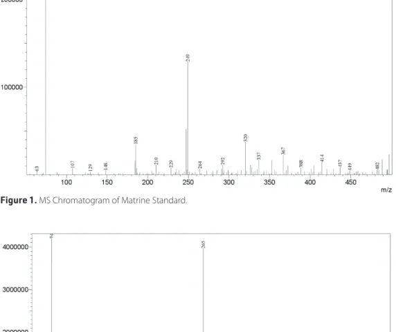
As shown in Figures 1-3, matrine and oxymatrine standards and crude methanol extract were analysed and quantified by LCMS. According to the results obtained from LCMS analysis,
Figure 1. MS Chromatogram of Matrine Standard.
Figure 2. MS Chromatogram of Oxymatrine Standard.
matrine (0.49±0.006 mg/mL) and oxymatrine (0.27±0.016 mg/ mL) were determined in S. japonica root methanol extract. Therefore, it can be asserted that matrine and oxymatrine alka-loids in the extract are responsible for the anticancer activity of the extract. It can be thought that the extract shows a signifi-cant cytotoxic effect against the tested cell lines due to these alkaloids (Li et al., 2011; Ma et al., 2008; Shi et al., 2015; Yu et al., 2009; Zhang et al., 2015 .
In previous studies, S. japonicum leaf and bud extracts have been investigated for their anti-cancer activity against breast and colon cancer cell lines (Abdelhady, Kamal, Othman, Mubarak, & Hadda, 2015; Lee et al., 2015). The obtained re-sults were quite significant in terms of anti-cancer activity. Besides, matrine and its derivatives are molecules for which their anticancer activity is well documented (Li et al., 2011; Yu et al., 2009); matrine is the main alkaloid of S. japonicum root extracts. In past studies, matrine and oxymatrine have been assessed for their remarkable anticancer activity. Matrine and its derivatives have been reported as antineoplastic agents since they can inhibit proliferation and induce apoptosis of cancer cells. Besides, matrine could synergistically improve the efficacy of chemotherapy when it is used in combination with other anticancer drugs (Rashid, Xu, Muhammad, Wang, & Jiang, 2019).
Cancer is defined as uncontrolled or abnormal growth and proliferation of cells as a result of errors in DNA division dur-ing cell division (Hassanpour & Dehghani, 2017). In this pres-ent study, the effect of S. japonicum root MeOH extract on the viability of various cancer cell lines was investigated. The cytotoxic effects of the root of S. japonicum methanol extract was tested on HT-29, U-87 and PC-3 cell lines by measuring metabolically active cells using a luciferase-based assay, Cell Titer Glo (Promega). According to the obtained results, the cell viability was decreased at varying concentrations (1 mg/mL, 5 mg/mL and 10 mg/mL) in a concentration-dependent man-ner for all tested cancer cell lines. The results are in accordance with previous studies (Chang et al., 2013; Coussens et al., 2018; Huang et al., 2018; Rodenhizer, Dean, Xu, Cojocari, & McGuigan, 2018). Previous studies have shown the superior effects of ma-trine on the growth of HT29 cell lines, and the expression of the related proteins. MTT assay indicated that matrine consid-erably inhibited the HT29 cells proliferation in vitro in a dose- and time-dependent manner. MTT assay was used to study the inhibitory effects of matrine on the proliferation of HT29 cells; the treatment of cells was performed using different concen-trations (2–32 mg/mL) for 24, 36 and 48 h. Consequently, when the matrine dose increased, the proliferation of the HT29 cells was significantly suppressed in vitro in a dose- and time-de-pendent way. In conclusion, matrine has strong antitumor ac-tivity against HT29 cells and can act as an alternative agent to treat colon cancer (Chang et al., 2013). Huang et al. studied the efficacy of matrine on prostate cancer lines (DU145 and PC3 cell lines). The results showed that matrine and GADD45B over-expression synergistically inhibited the proliferation, migration, and invasion of prostate cancer cells. Additionally, the apopto-sis of prostate cancer cells was also synergistically enhanced by matrine and GADD45B overexpression (Huang et al., 2018).
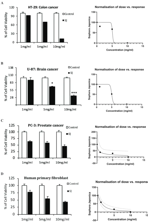
Treatment with 10 mg/mL S. japonicum extract resulted in a 35±2.21% viability of cells on U-87 cells, while PC-3 cells dis-played 46±5.67% cell viability, respectively. Additionally, the extract showed the lowest viability against the HT-29 colon cancer cell line as 14%. Cell viability of S. japonicum root ex-tract-treated HT-29, U-87 and PC-3 cell lines at 24 hours are given in Figure 4 A-C. Accordingly, the tested methanol extract showed 5.104 and 5.012 mg/mL IC50 values for HT-29, and U-87 cell lines, respectively (Figure 4A and Figure 4B) while the IC50 value of PC-3 cell line was determined as 0.555 mg/mL (Figure 4C). Furthermore, among all the cancer lines tested, at the low-est dose of 1 mg/mL of extract treatment, only the PC-3 cell line showed significant cell death with a decreased viability to 64% (Figure 4C).
To analyse the anti-cancer effects of S. japonicum root extract, as a healthy control, human primary fibroblast cells were in-cluded in cell viability experiments. As a result, fibroblasts showed 1.320 mg/mL IC50 value which is approximately two-fold higher than the IC50 of PC-3 cancer line and therefore, fi-broblasts did not show a significant decrease in cell viability when treated with 1mg/mL (Figure 4D).
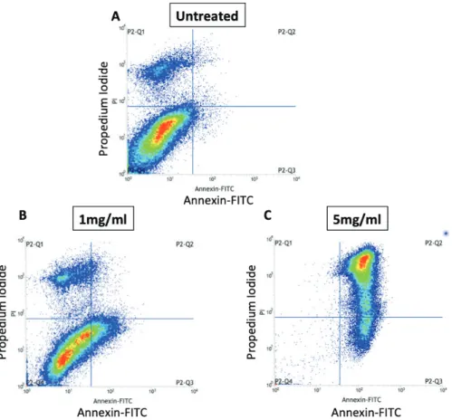
This data indicated the PC-3 cancer cell line could be treated with S. japonicum root methanol extract while the same dose is not cytotoxic to the healthy cells. Then, further analysis of the cell death was followed with biochemical verification of cell death and staining of apoptosis in PC-3 prostate cancer cells upon treatment with S. japonicum root extract for 24h. For detection of apoptosis, we stained the extract-treated PC-3 cells (0, 1 and 5 mg/mL) with Annexin V and Propidium Iodide (PI) then analysed by flow cytometry. According to our results, 1 mg/mL and 5 mg/mL extract treatments showed in-creased apoptosis in PC-3 with 16.3% and 99.6% respectively while untreated cells showed 1.89% apoptotic cells (Figure 5). This data indicates that S. japonica root extract treatment causes enhanced apoptotic cell death following increased doses.
Poly (ADP-ribose) polymerases (PARPs) are enzymes that can catalyse the transfer of ADP-ribose to target proteins. They take an important role in various cellular processes, including transcription, replication, recombination, and DNA repair. Cas-pase mediated apoptosis occurs through the cleavage of sev-eral key proteins required for cellular functioning and survival (Fischer et al., 2003) and PARP-1 cleavage by caspases is con-sidered to be a hallmark of apoptosis (Kaufmann et al., 1993; Tewari et al., 1995). One of the main biochemical indicators of cell death is the cleavage of PARP (Boulares et al., 1999). For this reason, we analysed cPARP levels of extract-treated PC-3 cells (1mg/mL), as well as DMSO treated control cells (DMSO was added instead of an equal volume of the extract). The re-sults indicated significantly increased cPARP (89kda) levels in the extract-treated PC-3 cells (p=0.0002) (Figure 6). This reveals that cell death is triggered upon treatment and possibly by the matrine and oxymatrine compounds residing in S. japonicum extract.
In conclusion, the results revealed that S. japonicum root meth-anol extract could trigger apoptosis in cancer cells and it is
A
0 5 10 15 0 50 100 150Normalisation of dose vs. response
Concentration (mg/ml) Soph or a J apon ic a
B
0 5 10 15 0 50 100 150Normalisation of dose vs. response
Concentration (mg/ml) Soph or a J apon ic a
C
5 10 15 -50 0 50 100 150 200Normalisation of dose vs. response
Concentration (mg/ml) Soph or a J apon ic a
D
5 10 15 -50 0 50 100 150Normalisation of dose vs. response
Concentration (mg/ml) Soph or a J apon ic a
Figure 4. In vitro anti-cancer effects of S. japonicum root extract in various cancer cell lines and relative IC50 graphs. Effects of S. japonicum root extracts in cell viability were tested on A) HT-29 (colon cancer), B) U-87 (brain cancer; glioblastoma) C) PC-3 (prostate cancer), cell lines and D) Human primary fibroblasts (healthy control) and their IC50 calculations were plotted. Data are expressed as ± SE and considering the differences, statistical significance was determined as 0.001< p*< 0.005; p**< 0.0005; and p***< 0.0001.
highly cytotoxic to the PC-3 prostate cancer cell line while cells of healthy tissue (human fibroblasts) were not affected at the same concentrations.
To sum up, the obtained methanol extracts from S. japoni-cum roots can be recognised as a potential anticancer can-didate, especially against prostate cancer while sparing healthy tissue. The obtained results are quite remarkable and the in vitro and in vivo anti-cancer effectiveness of S. ja-ponicum extracts can be further studied, in detail. Therefore, this study can be considered as the first alternative report focusing on a pharma-grade S. japonicum root extract in the cancer field. The preliminary data could be used to demon-strate the potential of the S. japonicum root and can lead the way to future studies.
Peer-review: Externally peer-reviewed.
Author Contributions: Conception/Design of Study- M.E.O., N.K.; Data Acquisition- M.E.O.; N.K.; Data Analysis/Interpretation- M.E.O, A.E.K., N.Ö., İ.S.T.; Drafting Manuscript- F.D., M.E.O.; Critical Revision of Manuscript- F.D.; Final Approval and Accountability- M.E.O., N.K., A.E.K., N.Ö., İ.S.T., F.D.; Technical or Material Support-M.E.O., N.K., F.D.; Supervi-sion- F.D.
Conflict of Interest: The authors have no conflict of interest to de-clare.
Financial Disclosure: Authors declared no financial support.
Figure 5. Flow cytometry analysis of cell death in S. japonicum root extract-treated PC-3 cancer cell line. Plots indicate Annexin
V and Propidium Iodide (PI) stainings of A) Untreated, B) 1mg/mL and C) 5mg/mL extract-treated PC-3 cells.
Figure 6. Western blot analysis of cell death in PC-3 prostate
cancer cells treated with 1mg/mL S. japonicum root extract for 24h. Cleaved PARP (cPARP) is a biochemical marker of cell death and plot indicates significantly increased cPARP levels in S. japonicum root extract-treated PC-3 cells as compared to DMSO treated control group (p**= 0.0002). Experiments were performed as triplicates and band densities were calculated using ImageJ analysis tool.
REFERENCES
• Abdelhady, M. I. S., Kamal, A. M., Othman, S. M., Mubarak, M. S., & Hadda, T. B. (2015). Total polyphenolic content, antioxidant, cy-totoxic, antidiabetic activities, and polyphenolic compounds of Sophora japonica grown in Egypt. Medicinal Chemistry Research,
24(2), 482–495.
• Blois, M. S. (1958). Antioxidant determinations by the use of a stable free radical. Nature, 181(4617), 1199–1200.
• Boulares, A. H., Yakovlev, A. G., Ivanova, V., Stoica, B. A., Wang, G., Iyer, S., & Smulson, M. (1999). Role of Poly(ADP-ribose) Polymerase (PARP) Cleavage in Apoptosis. Journal of Biological Chemistry,
274(33), 22932–22940.
• Chang, C., Liu, S-P., Fang, C-H., He, R-S., Wang, Z., Zhu, Y-Q., & JI, S-W. (2013). Effects of matrine on the proliferation of HT29 human colon cancer cells and its antitumor mechanism. Oncology Letters,
6(3), 699–704.
• Coussens, N. P., Sittampalam, G. S., Guha, R., Brimacombe, K., Gross-man, A., Chung, T. D. Y., Weidner, J. R. … Austin, C. P. (2018). Assay Guid-ance Manual: Quantitative Biology and Pharmacology in Preclinical Drug Discovery. Clinical and Translational Science, 11(5), 461–470. • Cragg, G. M., & Newman, D. J. (2005). Plants as a source of
anti-cancer agents. Journal of Ethnopharmacology, 100(1–2), 72–79. • Ding, P-L., Liao, Z-X., Huang, H., Zhou, P., & Chen, D-F. (2006).
(+)-12alpha-Hydroxysophocarpine, a new quinolizidine alkaloid and related anti-HBV alkaloids from Sophora flavescens.
Bioor-ganic & Medicinal Chemistry Letters, 16(5), 1231–5.
• Fischer, U., Janicke, R. U., & Schulze-Osthoff, K. (2003). Many cuts to ruin: A comprehensive update of caspase substrates. Cell Death
and Differentiation, 10(1), 76–100.
• Hassanpour, S. H., & Dehghani, M. (2017). Review of cancer from perspective of molecular. Journal of Cancer Research and Practice,
4(4), 127–129.
• He, X., Bai, Y., Zhao, Z., Wang, X., Fang, J., Huang, L., Zeng, M., … Zheng, X. (2016). Local and traditional uses, phytochemistry, and pharmacology of Sophora japonica L.: A review. Journal of
Ethno-pharmacology, 187, 160–182.
• Higashiyama, K., Takeuchi, Y., Yamauchi, T., Imai, S., Kamei, J., Ya-jima, Y., Narita, M., & Suzuki, T. (2005). Implication of the Descend-ing Dynorphinergic Neuron ProjectDescend-ing to the Spinal Cord in the (+)-Matrine- and (+)-Allomatrine-Induced Antinociceptive Ef-fects. Biological and Pharmaceutical Bulletin, 28(5), 845–848. • Huang, H., Wang, Q., Du, T., Lin, C., Lai, Y., Zhu, D., Wu, W., ... Li, Q.
(2018). Matrine inhibits the progression of prostate cancer by promoting expression of GADD45B. Prostate, 78(5), 327–335. • Kaufmann, S. H., Desnoyers, S., Ottaviano, Y., Davidson, N., &
Po-irier, G. (1993). Specific Proteolytic Cleavage of poly(ADP-ribose) Polymerase: An Early Marker of Chemotherapy-Induced Apopto-sis -PubMed. Cancer Research, 53(17), 3976–3985.
• Kim, J. M., & Yun-Choi, H. S. (2008). Anti-platelet effects of flavo-noids and flavonoid-glycosides from Sophora japonica. Archives
of Pharmacal Research, 31(7), 886–890.
• Lee, J. W., Park, G. H., Eo, H. J., Song, H. M., Kim, M. K., Kwon, M. J., Koo, J. S., Lee, J. R., Lee, M. H., & Jeong, J. B. (2015). Anti-Cancer Activity of the Flower Bud of Sophora japonica L. through Up-regulating Activating Transcription Factor 3 in Human Colorectal Cancer Cells. Korean Journal of Plant Resources, 28(3), 297–304. • Li, Y., Li, Y., Chen, X., Liu, T., Chen, Y., He, W., Zhang, Q., & Liu, S. (2011).
Autophagy is involved in anticancer effects of matrine on SGC-7901 human gastric cancer cells. Oncology Reports, 26(1), 115–124. • Ma, L., Wen, S., Zhan, Y., He, Y., Liu, X., & Jiang, J. (2008). Anticancer
Effects of the Chinese Medicine Matrine on Murine Hepatocel-lular Carcinoma Cells. Planta Medica, 74(3), 245–251.
• Mihaylova, D., & Schalow, S. (2013). Antioxidant and stabilization activity of a quercetin-containing flavonoid extract obtained from Bulgarian Sophora japonica L. Brazilian Archives of Biology
and Technology, 56(3), 431–438.
• Nasrollahi, S., Ghoreishi, S. M., Ebrahimabadi, A. H., & Khoobi, A. (2019). Gas chromatography-mass spectrometry analysis and antimicrobial, antioxidant and anti-cancer activities of essential oils and extracts of
Stachys schtschegleevii plant as biological macromolecules. Interna-tional Journal of Biological Macromolecules, 128, 718–723.
• Nobili, S., Lippi, D., Witort, E., Donnini, M., Bausi, L., Mini, E., & Ca-paccioli, S. (2009). Natural compounds for cancer treatment and prevention. Pharmacological Research, 59(6), 365–378.
• Okur, M. E., Ayla, Ş., Cicek Polat, D., Gunal, M. Y., Yoltaş, A., & Biceroğlu, O. (2018). Novel insight into wound healing properties of methanol extract of Capparis ovata Desf. var. palaestina Zohary fruits. Journal of Pharmacy and Pharmacology, 70(10):1401–1413. • Pelletier, S.W. (1991). Alkaloids: Chemical and Biological
Perspec-tives. New York, NY: Springer New York.
• Rashid, H ur., Xu, Y., Muhammad, Y., Wang, L., & Jiang, J. (2019). Research advances on anticancer activities of matrine and its derivatives: An updated overview. European Journal of Medicinal
Chemistry, 161, 205–238.
• Re, R., Pellegrini, N., Proteggente, A., Pannala, A., Yang, M., & Rice-Evans, C. (1999). Antioxidant activity applying an improved ABTS radical cation decolorization assay. Free Radical Biology and
Medi-cine, 26(9–10), 1231–1237.
• Rodenhizer, D., Dean, T., Xu, B., Cojocari, D., & McGuigan, A.P. (2018). A three-dimensional engineered heterogeneous tumor model for assessing cellular environment and response. Nature
Protocols, 13(9), 1917–1957.
• Shi, Y., Shen, G., Fang, H., Xu, C., & Hu, S. (2015). Method for quanti-tative determination of matrine in Sophora alopecuroides L. and its inhibitory effect on breast cancer MCF-7 cell proliferation.
Bio-medical Research, 26(3).
• Spanos, G. A., & Wrolstad, R. E. (1990). Influence of processing and storage on the phenolic composition of Thompson Seedless grape juice. Journal of Agricultural and Food Chemistry, 38(7), 1565–1571. • Tang, Y-P., Li, Y-F., Hu, J., & Lou, F-C. (2002). Isolation and
identi-fication of antioxidants from Sophora japonica. Journal of Asian
Natural Products Research, 4(2), 123–128.
• Tewari, M., Quan, L. T., O’Rourke, K., Desnoyers, S., Zeng, Z., Beidler, D. R., Poirier, G. G., Salvesen, G. S., & Dixit, V. M. (1995). Yama/ CPP32β, a mammalian homolog of CED-3, is a CrmA-inhibitable protease that cleaves the death substrate poly(ADP-ribose) poly-merase. Cell, 81(5), 801–809.
• Ting, D., Ruwei, W., Guoyong, Z., Meizhen, C., & Songhua, L. (2002). The manufacture and clinical application of compound matrine.
Acta Chinese Medicine and Pharmacology, 30(2), 47–48.
• Tomani, J. C. D., Gainkam, L. O. T., Nshutiyayesu, S., Mukazayire, M. J., Ribeiro, S. O., Stevigny, C., Frederich, M., Muganga, R., & Souop-gui, J. (2018). An ethnobotanical survey and inhibitory effects on NLRP3 inflammasomes/ Caspase-1 of herbal recipes’ extracts tra-ditionally used in Rwanda for asthma treatment. Journal of
Ethno-pharmacology, 227, 29–40.
• Verma, A. K., & Singh, S. (2020). Phytochemical analysis and in vitro cytostatic potential of ethnopharmacological important medici-nal plants. Toxicology Reports, 7, 443–452.
• Wu, Y., Chen, J., & Cheng, Y. (2005). A sensitive and specific HPLC-MS method for the determination of sophoridine, sophocarpine and matrine in rabbit plasma. Analytical and Bioanalytical
Chem-istry, 382(7), 1595–1600.
• Yu, P., Liu, Q., Liu, K., Yagasaki, K., Wu, E., & Zhang, G. (2009). Matrine suppresses breast cancer cell proliferation and invasion via VEGF-Akt- NF-κB signaling. Cytotechnology, 59(3), 219–229.
• Zhang, G-L., Jiang, L., Yan, Q., Liu, R-H., & Zhang, L. (2015). Anti-tumor effect of matrine combined with cisplatin on rat models of cervical cancer. Asian Pacific Journal of Tropical Medicine, 8(12), 1055–1059.