Clin. Lab. 6/2018 1075 Clin. Lab. 2018;64:1075-1078
©Copyright
CASE REPORT
A Rare Non-Hemolytic Case of Idiopathic Cold Agglutinin Disease
Edip Erkus
1, Mehmet Z. Kocak
1, Gulali Aktas
1, Mehmet Ozen
2, Burcin M. Atak
1,
Tuba T. Duman
1, Buket K. Tekce
3, Haluk Savli
1 1 Abant Izzet Baysal University Hospital, Department of Internal Medicine, Bolu, Turkey2 Ufuk University Hospital, Department of Hematology, Ankara, Turkey
3 Abant Izzet Baysal University Hospital, Department of Medical Biochemistry, Bolu, Turkey
SUMMARY
Background: Cold agglutinin disease is a very rare condition associated with agglutination of erythrocytes in cold
environment usually due to IgM type antibodies. Other than hemolytic anemias, it may interfere with routine he-mogram tests due to miscalculation of red blood cell count (RBC) and other hehe-mogram parameters calculated with involvement of RBC. Awareness of the condition is important to overcome laboratory errors.
Methods: We studied a peripheral blood smear and repeated the hemogram test at 37°C to establish the diagnosis
of cold agglutinin disease.
Results: Initial hemogram test results of the fifty-eight year-old man was as follows: RBC: 1.34 M/µL, hemoglobin
(Hb): 12.4 g/dL, hematocrit (Htc): 11.8%, mean corpuscular hemoglobin (MCH): 92.4 pg, and mean corpuscular hemoglobin concentration (MCHC): 105 gr/dL. Despite the standard indirect Coombs test being negative, repeat-ed tests at room temperature was 4+. We suspectrepeat-ed cold agglutinin disease and repeatrepeat-ed the hemogram test using the Bain-Marie method at 37°C and the test results showed RBC: 3.4 M/µL, hemoglobin: 12.6 g/dL, hematocrit: 30.2%, MCH: 31.7 pg, and MCHC: 41.8 g/dL.
Conclusions: Inappropriate hemogram results may be a sign of underlying cold agglutinin disease. Hemolytic
ane-mia not always accompanies the disease; however, cold exposure may trigger erythrocyte agglutination in vitro and may cause erratic laboratory results.
(Clin. Lab. 2018;64:1075-1078. DOI: 10.7754/Clin.Lab.2018.180114)
Correspondence:
Gulali Aktas, Assoc. Prof.
Abant Izzet Baysal University Hospital Department of Internal Medicine 14280, Bolu Turkey Phone: +90 3742534656 Fax: +90 3742534615 Email: [email protected] _____________________________________________
Case Report accepted January 29, 2018
KEY WORDS
cold agglutinin disease, red blood cell, hemogram, indi-rect Coombs
INTRODUCTION
Cold agglutinin disease (CAD) is a member of autoim-mune hemolytic anemias. It is a very rare disorder af-fecting only one per million, annually [1]. CAD is driven by IgM antibodies in more than 90% of the cases; however, other causes could be IgG and IgA anti-bodies [2]. Polyclonal cold agglutinins are usually caus-ed by post-infectious or rheumatologic conditions; how-ever, monoclonal cold agglutinins are a consequence of an underlying lymphoproliferative disorder. Causes of post-infectious CAD include mycoplasma, Epstein-Barr virus, legionella, varicella, and influenza [2-6].
Post-in-E. Erkus et al.
Clin. Lab. 6/2018
1076 fectious CAD is often a self-resolving disorder, howev-er, CAD driven by hematologic diseases may be persis-tent and relapsing [2].
CAD driven by monoclonal IgM is a long-term, chronic disorder characterized with cold agglutinins against I/i carbohydrate antigens of the erythrocyte membrane or rarely to Pr antigens [2]. CAD is usually diagnosed in older adults (7th decade) and more prevalent in women [7].
The hemolysis during the course of CAD often causes mild or moderate chronic anemia [7]. Although spleno-megaly is not usual in CAD [8], acrocyanosis, livedo re-ticularis and Raynaud phenomenon may accompany to the disease [4].
We aimed to present a rare idiopathic non-hemolytic cold agglutinin disease case in a 58-year-old man.
CASE REPORT
A 58-year-old man admitted to our clinic after detection of mild anemia during a routine 6-month control evalu-ation of aortic valve replacement. He complained of shortness of breath and purple discoloration on finger-tips in cold weather. He had not described hematuria, hematochezia or melena. He had an aortic valve re-placement 6 months ago and a history of coronary ar-tery disease.
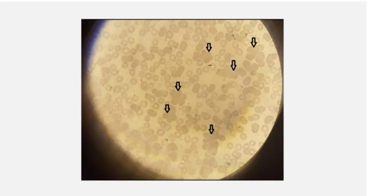
On physical examination, his general appearance was well, body temperature was 36°C, blood pressure was 118/72 mmHg, heart rate was 84 beats per minute, and respiratory rate was 16 per minute. Except for an opera-tion scar on the sternum, the entire physical examina-tion of the patient was normal. His medicaexamina-tions include warfarin 5 mg daily, atorvastatin 10 mg daily, and biso-prolol 5 mg a day. The results of a hemogram test were as follows: red blood cell count (RBC): 1.34 M/µL (ref-erence range: 3.9 - 5.5 M/µL), hemoglobin (Hb): 12.4 g/dL, hematocrit (Htc): 11.8% white blood cell count: 6.8 K/µL, platelet count: 150 K/µL, mean cor-puscular hemoglobin (MCH) 92.4 pg (reference range: 27 - 31.2 pg), and mean corpuscular hemoglobin con-centration (MCHC): 105 gr/dL (reference range: 31.8 - 40). These inappropriate results were confirmed with a repeated hemogram test in order to exclude a laboratory error. Medical data of the patient retrospectively evalu-ated from database and similar laboratory results in RBC, Htc, MCH, and MCHC were also detected 4 years earlier, when he first showed up in our institution. However, surgeons examining the patient did not realize the importance of these laboratory results and operated him. Blood samples in the hemogram tube demonstrat-ed clots as a sign of agglutination in room temperature (Figure 1). A peripheral blood smear revealed multiple, grape shaped groups of aggregated erythrocytes in each field (Figure 2).
We figured out that red blood cell counting was mis-taken due to aggregation of erythrocytes. Htc, MCH, and MCHC were calculated by involvement of RBC,
therefore, these calculations were also erroneous. Typi-cal erythrocyte aggregation in peripheral smear and a history of dyspnea and cyanosis of fingertips led to a preliminary diagnosis of cold agglutinin disease. Another hemogram test was run, but this time using the Bain-Marie method at 37°C and the test results showed RBC: 3.4 M/µL, hemoglobin: 12.6 g/dL, hematocrit: 30.2%, MCH: 31.7 pg, and MCHC: 41.8 g/dL. A reticu-locyte count was 1.2%. Although standard indirect Coombs test was negative, indirect Coombs at room temperature was ++++, direct Coombs (with IgG) test was positive. Serologic tests for Epstein-Barr virus (EBV) were as follows: EBV viral capcid antigen (VCA) IgM: (-), EBV early antigen (EA): (-), EBV VCA IgG Avidity: (+++).
In serum biochemistry, along with total bilirubin (0.26 mg/dL) and direct bilirubin (0.14 mg/dL), serum lactate dehydrogenase (LDH) (215 µ/L) was normal. The rest of the biochemical tests were also in normal range. Other blood tests were as follows: erythrocyte sedimentation rate: 16 mm/h, serum iron: 61 µg/dL (ref-erence range: 31 - 144 µg/dL), ferritin 25 ng/mL (refer-ence range: 22 - 275 ng/mL), serum iron binding capac-ity: 223 µg/dL (reference range: 69 - 240 µg/dL), vita-min B12: 323 pg/mL (reference range: 187 - 883 pg/ mL), folic acid: 4.3 ng/mL (reference range: 3.1 - 20.5 ng/mL), total serum protein: 7.1 g/dL (reference range: 6.4 - 8.3 g/dL), serum albumin: 3.8 g/dL (refer-ence range: 3.5 - 5 g/dL). A coagulation test revealed an INR value of 2.28 (reference range: 0.8 - 1.2).
To exclude rheumatologic diseases, we ordered a couple of tests and anti-nuclear antigen (ANA) and ds-DNA were both negative. In addition, SSA, anti-SSB, anti-SM, anti-Scl 70 were all negative, too. A se-rum protein electrophoresis revealed a peak in the beta-gamma band with polyclonal gammopathy. Serum and urinary immune fixation electrophoresis were normal. Serum levels of IgG, IgM, IgA were 1,619 mg/dL (ref-erence range: 540 – 1,822), 152 mg/dL (22 - 240), 206 mg/dL (63 - 484), respectively. Abdominal and thoracic computerized tomography results, to exclude lympho-prolipherative disorders, were all normal without lymphadenopathy or splenomegaly. Patient was dis-charged without any event and advised for routine fol-low up.
DISCUSSION
Cold agglutination disease is a group of autoimmune hemolytic anemias, mostly due to an infectious, rheu-matologic and herheu-matologic lymphoproliferative disor-der. We presented here an idiopathic cold agglutination case with unclear aetiology. As with anemia and retic-ulocytosis, patients with CAD present with elevated in-direct bilirubin and LDH levels. However, reticulocyte count, serum bilirubin, and LDH were not elevated in the present case, probably due to the absence of cold hemolysins [9].
A Case of Cold Agglutinin Disease
Clin. Lab. 6/2018 1077
Figure 1: Clots on the inner side of hemogram tube suggesting agglutination.
Figure 2: Grape shaped agglutination of erythrocyte in peripheral blood smear (x1000).
Routine hemogram assays directly measure Hb and RBC count; however, RBC tends to be lower than the actual amount due to agglutinated erythrocytes in the present case. Unlike RBC and Hb which are directly
measured, Htc, MCH and MCHC are both obtained by a calculation. Htc is calculated by the formula: RBC x mean corpuscular volume/10. MCH is calculated by the formula: Hb/RBC x 10. Finally, MCHC is calculated by
E. Erkus et al.
Clin. Lab. 6/2018
1078 the formula: Hb/Htc x 100. An incorrectly measured RBC value might cause erratic calculations of Htc, MCH, and MCHC. Abnormal MCHC results and the discordance between Hb and Hct should be a warning for physicians.
Patients with CAD usually present with symptoms that are provoked by cold exposure. Berentsen et al. reported a 90% rate of cold associated symptoms in CAD [1]. However, a recent review denoted the cold symptoms as low as 35% in subjects with CAD [2]. In the present case, purple discoloration on fingers, possibly acrocya-nosis, after exposure to cold could be one of such cold induced symptoms. However, we did not notice that discoloration in repeated physical examinations during hospitalization of the patient, probably due to season ef-fect (summer).
Anemia and acrocyanosis were two of the most com-mon symptoms presenting in CAD subjects in a study in literature [2]. Authors defined CAD as a chronic anemia with severity mild to moderate [7], but there are other studies that reported CAD was associated with severe anemia [1,10]. Although the patient in the present case described acrocyanosis, his Hb was at only mild anemic level.
Indirect Coombs test shows the circulating free antibod-ies to erythrocytes. Normal 37°C in vivo prevents the binding of antibodies to red blood cells. However, after venous sampling into an anticoagulant containing tube for the hemogram test, they cause agglutination of the erythrocytes at room temperature. Therefore, the initial standard indirect Coombs test was negative, and the re-peated test at room temperature was 4+ positive. These results are associated with cold agglutinin disease. Ebstein Barr virus infection causes polyclonal produc-tion of immunoglobulins; such as cold agglutinins. De-spite it generally occurring in the acute phase of infec-tion, cold agglutinins may persist in the chronic phase after the infection resolves. The EBV VCA IgG Avidity was 3+ in the serum of the present case, suggesting past EBV infection at least more than three months ago. However, his history was negative for a recent condi-tion of sore throat, fever, enlarged spleen or lymph nodes. In addition, his CBC results were compatible with CAD 4 years ago and may exclude any EBV effect on his CAD.
Red cell agglutination makes it difficult to determine ABO blood type in CAD; therefore, when necessary, O packed red cells could be transfused if ABO typing is not definitive [11]. The patient we presented did not re-quire blood transfusion; however, his blood type was detected as B Rh +, without any challenge.
In literature, it is reported that 78% of the CAD cases were caused by a lymphoproliferative disorder, by an autoimmune condition, or by an infectious disease [11]. That means remaining cases are either idiopathic or dif-ficult to establish a diagnosis. The present case should be an idiopathic CAD, due to failing to establish the un-derlying cause of the disease, despite great effort. We did not advise any special treatment to the patient
be-cause of the absence of cold hemolysins, and life threatening symptoms, and summer season [9].
CONCLUSION
CAD, even when it was not associated with hemolysis, may cause inappropriate hemogram test results. Physi-cians should be aware of the disease in such patients with a typical history of cold induced symptoms and with abnormal RBC, MCH, and MCHC levels.
Funding:
This study has not received any funds or grants.
Declaration of Interest:
None to declare.
References:
1. Berentsen S, Ulvestad E, Langholm R, et al. Primary chronic cold agglutinin disease: a population based clinical study of 86 pa-tients. Haematologica. 2006;91(4):460-6 (PMID: 16585012). 2. Swiecicki PL, Hegerova LT, Gertz MA. Cold agglutinin disease.
Blood. 2013;122(7):1114-21 (PMID: 23757733).
3. Khan FY. Mycoplasma pneumoniae associated with severe auto-immune hemolytic anemia: case report and literature review. Braz J Infect Dis. 2009;13(1):77-9 (PMID: 19578637).
4. Karunarathne S, Weerasinghe S, Govindapala D, Fernando H, Jayaratne B. Cold autoimmune haemolytic anaemia secondary to Epstein Barr virus infection presenting with peripheral gangrene; case report. Thromb J. 2012;10(1):4 (PMID: 22513360). 5. Tanaka Y, Masuya M, Katayama N, et al. Development of
mix-ed-type autoimmune hemolytic anemia and Evans’ syndrome fol-lowing chicken pox infection in a case of low-titer cold agglutinin disease. Int J Hematol. 2006;84(3):220-3 (PMID: 17050195). 6. Schoindre Y, Bollée G, Dumont M-D, Lesavre P, Servais A. Cold
agglutinin syndrome associated with a 2009 influenza A H1N1 infection. Am J Med. 2011;124(2):e1-e2 (PMID: 20843499). 7. Petz LD. Cold antibody autoimmune hemolytic anemias. Blood
Rev. 2008;22(1):1-15 (PMID: 17904258).
8. Nydegger U, Kazatchkine M, Miescher P, editors. Immunopatho-logic and clinical features of hemolytic anemia due to cold agglu-tinins. Semin Hematol1991 Jan; 28(1):66-77 (PMID: 1708169). 9. Sokol R, Booker D, Stamps R, Walewska R. Cold
haemaggluti-nin disease: clinical significance of serum haemolysins. Clin Lab Haematol. 2000;22(6):337-44 (PMID: 11318799).
10. Gertz MA. Management of cold haemolytic syndrome. Br J Hae-matol. 2007;138(4):422-9 (PMID: 17561970).
11. Gertz MA. Cold hemolytic syndrome. Hematology Am Soc He-matol Educ Program. 2006;2006(1):19-23 (PMID: 17124034).