陨灶贼 允 韵责澡贼澡葬造皂燥造熏 灾燥造援 9熏 晕燥援 3熏 Mar.18, 圆园16 www. ijo. cn 栽藻造押8629原愿圆圆源缘员苑圆 8629-82210956 耘皂葬蚤造押ijopress岳员远猿援糟燥皂
Effect of dexamethasone intravitreal implant in a corneal
graft rejection
窑Letter to the Editor窑
1Department of Cornea, Bascom Palmer Eye Institute,
University of Miami Miller School of Medicine, Miami, Florida 33136, USA
2Ankara Training and Research Hospital, Ankara 06340,
Turkey
3Baskent University Hospital, Ankara 06490, Turkey
Correspondence to: Nilufer Yesilirmak. Bascom Palmer Eye Institute, University of Miami Miller School of Medicine, 900 NW 17 St., Miami, Florida 33136, USA. [email protected]
Received: 2015-02-10 Accepted: 2015-06-16 DOI:10.18240/ijo.2016.03.28
Yesilirmak N, Ozdemir ES, Altinors DD. Effect of dexamethasone intravitreal implant in a corneal graft rejection.
2016;9(3):475-477 Dear Sir,
I
am Dr. Nilufer Yesilirmak, from the Department of Cornea, Bascom Palmer Eye Institute, Miami, Florida, USA. We wrote to report a reduction of graft rejection with dexamethasone intravitreal implant (Ozurdex誖 ) in a case with aphakia.Ozurdex誖 (Allergan Inc., Irvine, CA, USA) is an intravitreal implant containing a biodegradable material combined with the active drug dexamethasone. It is a small rod-shaped, 0.46 mm in diameter and 6 mm in length. Inside the eye, it provides sustained delivery of 0.7 mg of preservative-free dexamethasone to the vitreous cavity and retina for up to 180d[1]. This implant is proved to be effective in the treatment
of macular edema following branch or central retinal vein occlusions, posterior non-infectious uveitis, diabetic macular edema, pseudophakic macular edema and neovascular age-related macular degeneration[2].
Herein we report a different usage of Ozurdex® implant for
51-year-old female diagnosed with corneal graft rejection in her right eye. The patient had been followed at our clinic with chronic visual loss in her right eye for 4y and undergone a penetrating keratoplasty 4y ago due to a fungal keratitis scar. Simultaneously a cataract extraction had been performed without implanting an intraocular lens. The outside surgeon probably decided to implant it on another occasion after making sure that the cornea survived. Because
of the rejection, the patient had undergone her second keratoplasty 2y ago. After the second keratoplasty her best-corrected visual acuity (BCVA) was 0.5, however during the follow-up period, her vision decreased again due to an epiretinal membrane and she underwent a pars plana vitrectomy.
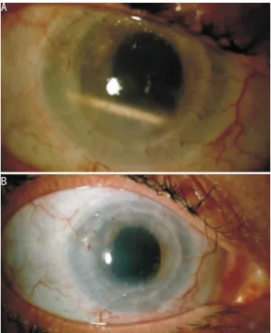
On her last examination, the BCVA was 0.1 and intraocular pressure (IOP) was 15 mm Hg, in the right eye. The anterior segment showed 1+ cells, 1+ flare and fine Khodadoust line which was considered as a rejection (Figure 1A). The posterior segment was normal. Topical steroids were given 4 times a day and subtenon corticosteroid injections were applied twice a month for 3mo. She responded well to each steroid injection, however, because of frequent injections, her eyelids were bruised and became edematous as well as her conjunctiva (Figure 1B). In order to reduce the number of injections, an Ozurdex®implantation was performed through
the superior temporal quadrant of right eye, approximately 3-to 5-mm posterior 3-to the limbus (through the pars plana). After first week of implantation, the Ozurdex ® implant
migrated into the anterior chamber (AC) and wandered between the AC and vitreous cavity with head movements of the patient without any corneal touch (Figure 2A). Therefore, patient was advised not to lean forward too much and she learnt to keep the implant inside the eye by adjusting her body movements. At 4wk post-implantation, the BCVA was 0.2, IOP was 15 mm Hg and rejection regressed significantly (Figure 2B). At 8wk post-implantation, the BCVA was still 0.2, IOP was 18 mm Hg, cornea was clearer and the eye (conjunctiva and eyelids as well) was quiescent with the implant in the vitreous cavity. On her last visit, at 11wk post-implantation, the BCVA was 0.1, IOP was 12 mm Hg, cornea was not clear and the implant was not seen in the vitreous cavity or AC. At almost 3mo time, the implant was absorbed and the rejection seemed to be started again. Penetrating keratoplasty is usually a successful surgical procedure but some factors may cause graft rejection. According to literature immunologic rejection is one of the most common causes for graft failure [3]. Guilbert [4]
observed that lens status influenced the risk of rejection as well. Besides, severe allograft rejection episodes results in endothelial cell density decrease that leads to a lower reversibility rate [5]. Our case had both aphakia and rejection
episodes that might have increased the endothelial failure. 475
Corticosteroids are usually strongest tools to reverse graft rejection usually. Even though our case received intensive topical and subtenon corticosteroid therapy, rejection progressed. Due to the fact that the combination of subconjunctival and topical corticosteroids was reported [6]as
more effective in reversing rejection than treatment with a single pulse of methylprednisolone (500 mg) in combination with the topical corticosteroids, we decided to use Ozurdex誖 due to its long lasting effect. Besides the patient could not tolerate immunomodulator therapies. One week after the implantation, the implant dissolved and passed to anterior chamber easily due to the aphakic and vitrectomized eye. At the following examination, the rejection was significantly
regressed. In contrast this, according to literature there are some studies showing the Ozurdex誖 implant migration to the anterior chamber in aphakic eyes, which resulted with corneal decompensation due to the direct contact of the implant to the endothelium[7-9]. Even, Mar侏n-Lamb侏es [10]
considered that the implant of these devices in aphakic patients should be contraindicated.
Chang-Lin [11] showed the concentration of
dexamethasone in the vitreous and retina raised to high levels within 60d of implantation, extending the therapeutic period to 6mo and Myung [12]showed the duration of its effect
was approximately 3-4mo. It has been mentioned that, in vitrectomized eyes the half-life of drugs after intravitreal injection reduces and the drug clearance increases from the vitreous [13]. Resembling to afore mentioned study, the
Ozurdex®reached its maximum effect within 2mo and instead
of 6mo, it completely dissolved within 3mo, in our case. In long-term usage of corticosteroids, adverse effects such as glaucoma and cataract may appear. It is reported that Ozurdex誖 can cause lower incidence of IOP elevation requiring surgery and cataract extraction than the other intravitreal corticosteroids [14]. In this study, we did not
observe any long-term adverse effect associated with the implantation. The patient did not complain of eyelid swelling as seen in the photo before subtenon steroid injections. Therefore, this swollen eyelid appearance was attributed to our repeated steroid injections. Due to Ozurdex誖 could provide higher and stable corticosteroid levels without obvious adverse effects we might consider a second implantation since the rejection started to be occurred again after the implant dissolution.
In conclusion, dexamethasone intravitreal implantation is a minimally invasive treatment modality that might be an effective treatment option in anterior segment inflammations, especially in aphakic and vitrectomized eyes. Although there are no cases of dexamethasone intravitreal implantation for corneal graft rejection in aphakic and vitrectomized eyes in the literature, this case report provides an insight for future long-term studies. Future comparative clinical studies with a large sample size and long-term follow-up will able to provide better results and guidelines.
ACKNOWLEDGEMENTS
Conflicts of Interest: Yesilirmak N, None; Ozdemir ES, None;Altinors DD, None.
REFERENCES
1 Haller JA, Bandello F, Belfort R Jr, Blumenkranz MS, Gillies M, Heier J, Loewenstein A, Yoon YH, Jacques ML, Jiao J, Li XY, Whitcup SM; OZURDEX GENEVA Study Group. Randomized, sham-controlled trial of dexamethasone intravitreal implant in patients with macular edema due to
retinal vein occlusion. 2010;117(6):1134-1146.
2 Cabrera M, Yeh S, Albini TA. Sustained-release corticosteroid options. 2014;2014:164692.
Figure 1 Anterior segment and external eye photography A: The appearance of patient's cornea upon arrival which showed 1+ cells, 1+ flare, pearl like infiltrates (Arrow: Khodadoust line), corneal edema; B: Lid swelling and tissue damage related to multiple steroid injections.
Figure 2 Anterior segment photography A: The appearance of the Ozurdex®implant in the anterior chamber; B: Regressed pearl
like lesions, flare, cells and diminished corneal edema can be seen. Dexamethasone implant for corneal graft rejection
陨灶贼 允 韵责澡贼澡葬造皂燥造熏 灾燥造援 9熏 晕燥援 3熏 Mar.18, 圆园16 www. ijo. cn 栽藻造押8629原愿圆圆源缘员苑圆 8629-82210956 耘皂葬蚤造押ijopress岳员远猿援糟燥皂
3 Price MO, Thompson RW Jr, Price FW Jr. Risk factors for various causes
of failure in initial corneal grafts. 2003;121 (8):
1087-1092.
4 Guilbert E, Bullet J, Sandali O, Basli E, Laroche L, Borderie VM. Long-term rejection incidence and reversibility after penetrating and
lamellar keratoplasty 2013;155(3):560-569.e2.
5 Lee HS, Kim MS. Influential factors on the survival of endothelial cells
after penetrating keratoplasty. 2009;19(6):930-935.
6 Costa DC, de Castro RS, Kara-Jose N. Case-control study of
subconjunctival triamcinolone acetonide injection intravenous
methylprednisolone pulse in the treatment of endothelial corneal allograft
rejection. 2009;23(3):708-714.
7 Pardo-L佼pez D, Franc佴s-Mu觡oz E, Gallego-Pinazo R, Diaz-Liopis M. Anterior chamber migration of dexametasone intravitreal implants
(Ozurdex誖 ). 2012;250(11):1703-1704.
8 Bansal R, Bansal P, Kulkarni P, Gupta V, Sharma A, Gupta A.
Wandering Ozurdex(誖 ) implant. 2012;2(1):
1-5.
9 Khurana RN, Appa SN, McCannel CA, Elman MJ, Wittenberg SE, Parks DJ, Ahmad S, Yeh S. Dexamethasone implant anterior chamber migration:
risk factors, complications, and management strategies. 2014;121(1):67-71.
10 Mar侏n-Lamb侏es C, Gallego-Pinazo R, Garc侏a-Delpech, Diaz-Liopis M.
Ozurdex (誖 ) and aphakia: a combination to avoid.
2012;87(6):191-192.
11 Chang-Lin JE, Attar M, Acheampong AA, Robinson MR, Whitcup SM, Kupperman BD, Welty D. Pharmacokinetics and pharmacodynamics of the sustained-release dexamethasone intravitreal implant.
2011;52(1):80-86.
12 Myung JS, Aaker GD, Kiss S. Treatment of noninfectious posterior
uveitis with dexamethasone intravitreal implant. 2010;4:
1423-1426.
13 Chang-Lin JE, Burke JA, Peng Q, Lin T, Orilla WC, Ghosn CR. Pharmacokinetics of a sustained-release dexamethasone intravitreal implant in vitrectomized and nonvitrectomized eyes.
2011;52(7):4605-4609.
14 Lowder C, Belfort R Jr, Lightman S, Foster CS, Robinson MR, Schiffman RM, Li XY, Cui H, Whitcup SM; Ozurdex HURON Study Group. Dexamethasone intravitreal implant for noninfectious intermediate or
posterior uveitis. 2011;129(5):545-553.