CLINICAL STUDY
Determination of CEBPA mutations by next generation
sequencing in pediatric acute leukemia
Akin DF
1, Oner DA
2, Kurekci E
2, Akar N
3Nigde Ömer Halisdemir University, Faculty of Medicine, Medical Biology, Nigde, Turkey. [email protected]
ABSTRACT
OBJECTIVES: The CCAAT/enhancer-binding protein-alpha (CEBPA) is lineage-specifi c transcription factor in the hematopoietic system. In this study, we aimed on the clinical features and the prognostic signifi cance as-sociated with CEBPA mutations in 30 pediatric patients with acute leukemia.
METHODS: In addition, the association between found variants and mutations of Ten-Eleven-Translocation 2 (TET2), Kirsten rat sarcoma viral oncogene homolog (KRAS), and Casitas B-cell lymphoma (CBL), FLT3 (Fms-Related Tyrosine Kinase), JAK2 (Januse Kinase-2) and Nucleophosmin 1 (NPM1) were analyzed, which are important prognostic risk factors for pediatric acute leukemia patients. The entire CEBPA coding region was screened using the NGS method.
RESULTS: CEBPA mutations were detected in 16 (53.3 %) of 30 patients. In total, ten distinct of nucleotide changes were identifi ed in 30 patients, including 6 novel and 4 known mutations by sequencing the entire CEBPA gene. We found 6 frame shift mutations, 1 missense mutation, 3 synonymous variants. The most common mu-tation was the c.487del G resulting p.Glu163Ser in 5 cases. Three patients carried CEBPA double mumu-tations. CONCLUSION: The detected variants in this article seemed to be the fi rst screening results of genes studied by NGS in pediatric acute leukemia patients. Our results also showed some degree of association between FLT3-ITD, TET2, KRAS, CBL and CEBPA mutations (Tab. 4, Fig. 1, Ref. 24). Text in PDF www.elis.sk.
KEY WORDS: CEBPA, pediatric acute leukemia, next generation sequencing, molecular marker, mutation.
1Nigde Ömer Halisdemir University, Faculty of Medicine, Medical Biol-ogy, Nigde, Turkey, 2LÖSANTE Children’s and Adult Hospital, Ankara, Turkey, and 3TOBB-ETU Hospital, Ankara, Turkey
Address for correspondence: D.F. Akin, Nigde Ömer Halisdemir Uni-versity, Faculty of Medicine, Medical Biology, Nigde, Turkey. Phone: +90.5363026816
Introduction
Clinical and genetic prognostic markers are important in the classifi cation of leukemia patients. Aberrant chromosomal trans-locations and gene mutations frequently occur in the transcrip-tional factor that lead to uncontrolled proliferation of lymphoid and myeloid progenitors (1–4). The CEBPA gene is a member of the leucine zipper family of the transcription factor family that is essential for the differentiation of myeloid cells (5). The CEBPA gene located on chromosome 19 q13.1 encodes the basic leucin zipper (bZIP) family of the transcription factors (6). It is expressed at high levels during myeloid cell differentiation and binds to the promoters of multiple specifi c genes at different levels of myeloid linage maturation (2). In AML patients, it was reported that two types of mutations in CEBPA gene exist; N terminal and C terminal (2, 5, 7–11). The N terminal mutations are located between the ma-jor translational start site and the second ATG further downstream lead to premature stop of translation of the wild type p 42 CEBPA protein, while preserving translation of a short p30 isoform that
suppresses the function of the full length protein. Mutations in the C terminal are usually in frame and deletions that affect homo-hetero-dimerization and DNA binding (6–8). CEBPA mutations are seen in approximately 4.5–6 % of pediatric Acute Myeloid Leuke-mia (AML) patients. In addition, CEBPA mutations are found in 5–14 % of adult patients with AML (5, 11–13). There is no study, which investigates CEBPA mutations in Turkish pediatric acute leukemia patients. Therefore, in our study, we investigated entire
CEBPA coding region in 30 patients with Turkish pediatric acute
leukemia by using NGS technique and we assessed the frequency of CEBPA mutations and its clinico-hematologic correlation, as well as the cooperating mutations, including FLT3, TET2, CBL,
KRAS, JAK2 and NPM1 mutations. Patients and study design Subjects
The study population consisted of 30 patients aged between 1 and 15 years,who were admitted to Losante Children’s and Adult Hospi-tal with the diagnosis of pediatric acute leukemia. Seventeen patients were diagnosed as AML, 4 as mixed leukemia, 9 as ALL. The study was carried out in accordance with the code of Ethics of the World Medical Association (Declaration of Helsinki) for experiments in-volving humans. The Ankara University, School of Medicine Ethics Committee approved the study protocol (project No.03-107-13/2013) and an informed consent was provided by the patients’ parents.
Cytogenetic techniques
Bone marrow samples were collected with Heparin-containing tubes, and chromosome analysis was performed using G-banding. After the slide preparation, G-banding using Giemsa-staining was carried out according to the standard procedures. On each slide, 20 metaphases were analyzed by a light microscope (Nikon, JAPAN). Karyotypes were described according to International Standing Committee on Human Cytogenetic Nomenclature (ISCN) (14).
Fluorescence in situ hybridization (FISH) was performed on interphase nuclei and metaphase chromosomes of bone marrow cells using dual-color/dual-fusion probes for translocationsof inv (16;16), t(9;22), t(15;17), chromosome 19, and dual-color/deletion probe for del 7q, labeled in green and red spectra according to the manufacturer’s protocol provided by Cytocell, UK. Counterstain-ing was performed with 40,6-dia midino-2-phenylindole (DAPI). At least 100 nuclei were analyzed under the Fluorescence micro-scope, and image capture was performed using Nikon Eclipse 80i equipped with a CCD-camera (CoolCube1), appropriate fi lters and Isis software (MetaSystems).
DNA isolation and Next Generation Sequencing
Blood samples were collected with EDTA-containing tubes and DNA was extracted from peripheral blood and bone marrow leukocytes with Mag-NA Pure automatic DNA isolation instru-ment (Roche Diagnostics, Manheim, Germany).
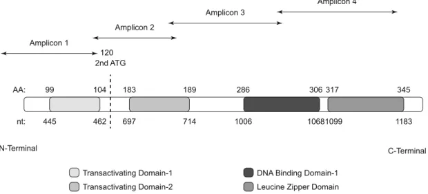
We used NGS to study the entire CEBPA gene region that was presented by 4 overlapping amplicons. Primer sequences lists are shown in Table 1. NGS sample preparation and process stages were generated, previously described by Grossmann et al (8).
NGS was carried out using 454 GS Junior System Instru-ment Roche Applied Science. Data were analyzed using the GS Amplicon Variant Analyzer software version 2.3. (Roche Applied Science). We used it for determination of variances; fi lters were adjusted to show variances in more than 1 % of bidirectional reads per amplicon in at least one patient. Figure 1 summarizes the loca-tion of the amplicons on the CEBPA protein.
Detection of FLT3, TET2, CBL, KRAS, JAK2 and NPM1 muta-tions
FLT3 mutation was analyzed by Real Time PCR on Light Cycler 480 II instrument (Roche Diagnostics, Gmbh, Mannheim, Germany). The results were analyzed with the High -Resolution Melting (HRM) method using genotype profi les. Different plots were created by selecting negative controls as the base-line. There-fore, fl uorescence of all the other samples was diagramed relatively to this sample. Fluorescence signals were analyzed, and signifi -cant differences were used as indicators of the mutations (15–18).
Hot-spot exons of TET2, KRAS and CBL genes were screened using the NGS method. All coding exons of TET2 (ex-ons 3 and 11) were presented by 27 amplic(ex-ons. Besides, two prim-er pairs wprim-ere amplifi ed, known as mutational hotspot regions, to describe the RING fi nger domain and linker sequence for CBL (exons 8 and 9) and KRAS (exons 2 and 3). The analyses were performed as previously described by Kohlmann et al (15). The twelfth of the exons of the JAK2 and NPM-1 genes was ampli-fi ed by polymerase chain reaction (PCR). PCR products were se-quenced using the Beckman DNA Sequencer System. (Beckman Coulter, USA).
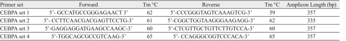
Primer set Forward Tm °C Reverse Tm °C Amplicon Length (bp)
CEBPA set 1 5’- GCCATGCCGGGAGAACT 3’ 62 5’-CCCGGGTAGTCAAAGTCG-3’ 59 357
CEBPA set 2 5’- CCTTCAACGACGAGTTCCTG-3’ 61 5’-CGGCTGGTAAGGGAAGAGG-3’ 62 335
CEBPA set 3 5’-GAGGAGGATGAAGCCAAGC-3’ 60 5’-CTCGTTGCTGTTCTTGTCCA-3’ 60 357
CEBPA set 4 5’-TGGCAGCGCCGTCAAG-3’ 65 5’- CCAGGGCGGTCCCACA-3’ 65 357
Tm = Melting temperature, bp = Base pair
Tab. 1. Primers used for amplifi cation of CEBPA gene.
Characteristic No CEBPA mutation mutationCEBPA valuep Gender Male (n = 21) 11 10 0,33 Female ( n = 9) 3 6 Diagnosis AML (n = 17) 7 10 0,94 ALL (n = 9) 4 5
Biphenotypic Acute Leukemia (n=4) 2 2 Risk Groups High Risk (n = 19) 11 8 0,20 Median Risk (n = 2) 1 1 Standard Risk (n = 9) 2 7 Status Remission (n = 17) 8 9 0,07 Relapse (n = 6) 0 5 Ex (n = 7) 3 4 Genetic Syndrome Down Syndrome (n = 4) – 4 0,52 Fragile X Syndrome (n = 1) – 1 Cytogenetic/ Genetics Normal Karyotype (n = 19) 10 9 1 t(15;17) (n = 1) 1 t(9;22) (n = 1) – 1 t(4;11) (n = 1) 1 t(8;21) (n = 1) 1 t(16;16) (n = 1) – 1 FLT3 mutations (n = 8) 5 3 JAK2 mutations (n = 2) – 2 TET2 mutations (n = 5) – 5 CBL mutations (n = 1) – 1 KRAS mutations (n = 4) – 4 NPM1 mutations (n = 1) 1 –
AML = Acute Myeloid Leukemia, ALL = Acute Lymphoblastic Leukemia, Ex = Exitus Tab. 2. Patient characteristics according to CEBPA mutation status.
Statistics
Statistical analyses were performed with SPSS 15.0 (SPSS IBM, USA). Chi-square test, independent sample t-test, or Mann–Whitney U-t-test, as appropriate for the type of data being analyzed, were used to assess the statistical signifi cance of the difference between the two groups. p val-ues less than 0.05 were considered statistically signifi cant.
Results
Incidence of CEBPA mutations
Among 30 pediatric acute leukemia patients, CEPBA mutations were found in 16 cases (53.3 %). Patient charac-teristics of the 30 pediatric acute leukemia cases are shown in Table 2. Ten distinct nucleotide changes were identifi ed in 30 patients, including 6 novel and 4 known mutations by sequencing the entire gene as shown in Table 3. The frame shift mutation was the most common mutation subtype that was found in 6 types. The remaining mutation subtypes were 3 synonymous variants and 1 missense mutation.
Three patients carried CEBPA double mutations. The other 13 patients carried a single CEBPA mutation. As showed in Table 3, 6 patients had mutations occurring be-tween TAD-1 domain and TAD2 domain. 4 patients had mu-tations in TAD1 domain, 2 patients had the combination of mutations between TAD2 domain and DNA binding domain mutation and a deletion between TAD-1 Domain and TAD-2 Domain. 4 patients had frame shift mutations in TAD1 do-main and N-terminal part of protein.
The novel 487 deletion of G variant was observed in 5 patients (16.6 %) and 3 (10 %) patients present-ed a novel 336 C>T substitution. The 198_201 du-plication of CTAC was detected 3 (10 %) patients. Three (10 %) of 30 patients had the 690 G>T syno-nymous variant. Patient 28 had a 573C>T synosyno-nymous vari-ant. Patient 29 had a 955_961 insertion of TTGACC in DNA binding domain of CEBPA protein. Patient 10 had combined variants of 300 delG and 487 delG.
Cooperating mutations
Patients with CEPBA mutations were also analyzed for mutations of FLT3, NPM1 and JAK2 Exon 12. In the 30 pe-diatric acute leukemia patients examined, FTL3 mutations were detected in two of the sixteen patients with CEBPA mutations and 5 of the 14 patients without CEBPA muta-tions had FLT3 mutamuta-tions. Two patients had both JAK2 and
CEBPA mutations. One patient had NPM1 mutation without CEBPA mutations.
Clinic and hematologic correlation with CEBPA mutations
Karyotype analysis was normal in 19 (63 %) of the 30 patients. Trisomy 14, Trisomy 19, Trisomy 22, monosomy 7, monosomy 14, inv (16;16), t(9;22), t(15;17), t(4;11) were found using chromosome banding and FISH analyses. We screened all the patients during treatment or relapse phases.
Mutation No Type of mutations No of patients Nucleotide change Amino-acid change Localization Mutation Type Rs number Clinical Signi fi cance 1 Nucleotide change 3 g.5800G>T c.690 G>T p.Thr230Thr Between TAD2-DNA binding domain Synonymous variant Rs 34529039 Benign 2 Duplication 3 g.5308_531 1dupCT AC c.198_201dupCT AC p.Ile68LeufsT er41 TAD-1 domain
Frameshift mutation in regulatory region
Rs 137852731 Pathogenic for AML 3 Insertion 1 g.5327_5328insC c.217_218insC p.Phe73SerfsT er35 N-terminal domain Frameshift mutation Rs 137852733 Pathogenic for AML 4 Nucleotide change 1 g.5683C>T c.573C>T p.His191His Between TAD2-DNA binding domain Synonymous variant Rs 192240793 Benign 5 Nucleotide change 3 g.5444 C>T c.336 C>T p.Pro1 12Pro Between TAD1-T AD2 domain Synonymous variant Novel NA 6 Deletion 1 g.5492_5493delC c.382 del C p.Pro128ProsT er31 Between TAD1-T AD2 domain Frameshift mutation Novel NA 7 Deletion 5 g. 5597 c.487 del G p.Glu163Ser Between TAD1-T AD2 domain Frameshift mutation Novel NA 8 Deletion 1 g.5410-541 1delC c.300 del C p.Gly100GlysT er59 TAD-1 domain Frameshift mutation Novel NA 9 Insertion 1 g.1 107-1 113ins TTGACC c.955-961 ins TTGACC p.Ser319T rpsT er13 C-terminal domain Frameshift mutation Novel NA 10 Nucleotide change 1 g.5599 C>A c.489 C>A p.Glu163Asp Between TAD1-T AD2 domain Missense variant Novel NA NA = Not A vailable, Rs = Reference SNP T
ab. 3. Characterization of CEBP
A
Patient No Age/ Gender Diagnosis Risk Group Cytogenetics-Molecular abnormalities CEBP A Mutations FL T3 TET2 CBL KRAS JAK2 ITD TKD 1 5,5/ M Biphenotypic (Mixed Phenotypic) Acute Leukemia HR Ex-46 ,XY c.690 G>T p.Thr230Thr –– – – -c.1641+179_1641+ 183delTCTT A-intronic 2 2/M Biphenotypic (Mixed Phenotypic) Acute Leukemia HR Ex-46 ,XY c.690 G>T p.Thr230Thr c.487 del G p.Glu163Ser –– – -3 12/M AML-M2 SR 46 ,XY c.690 G>T c.487 del G –– – -4 11 /M AML-M1 HR Ex-46 ,XY c.198_201dupCT AC p.Ile68LeufsT er41 +– – -5 4/ M Pre B-ALL SR 46,XY c.336 C>T p.Pro1 12Pro –– – -6 7/ M AML-M4 HR 47, XY ,+ 22[12] Inv (16;16), Fragile X syndrome c.198_201dupCT AC p.Ile68LeufsT er41 –– p.Pro363Leu p.Gly614Gly p.V al218Met c.*128-7564G>A p.L ys1678Glu --7 6/ F AML-NA SR 46,XY ,t(15;17) c.487 delG p.Glu163Ser –– -8 13/M AML-M4 HR 46, XY , Ms,T s 14, c.336 C>T p.Pro1 12Pro – – p.Ile1762V al -9 4/ M AML-M4 HR 46, XY c.382 del C – – – -10 5,5/F AML-M2 SR 46, XX
c.300 del C c.487 delG p.Glu163Ser
– – p.Gly614Gly -p.Ile20Thr 11 8/ F AML-M5 SR 46, XX c.198_201dupCT AC p.Ile68LeufsT er41 –– p.Gly614Gly p.Ser217Ser -p.Ile20Thr 20 6,5/F AML-M5 HR 46, XX c.487 del G p.Glu163Ser c.336 C>T –– – -c.1641+179_1641+ 183delTCTT A-intronic 22 3/ F AML-NA HR Ex, 47, XX (+21) c 21 der(14) (14q1,2 → q 3,2ii 1q21 → q43),der(19)(19qdel) → p13.3: : 1 1q13)1 1qdel [9]. t(9,22) c.217_218insC ++
p.Pro363Leu p.Pro761Leu p.Ser217Ser
c.714-72237T>C p.Ile20Thr 28 3/ M Pre B-ALL MR 47,XY(+21) c.573C>T –– – -29 2,5/M Pre B-ALL SR 47,XY(+21) c.955-961 ins TT -GACC –+ – -30 3/ F Pre B-ALL SR 47,XX(+21) c.489 C>A – – –
-M = -Male, F = Female, HR = High Risk, -MR = -Medium Risk, SR = Standard Risk,
AML
=
Acute Myeloid Leukemia,
ALL
=
Acute L
ymphobla
stic Leukemia, Pre B-ALL
= Precursor B- Acute L ymphoblastic Leukemia, NA = Not A
vailable, Ex = Exitus, Del = Deletion, Ins = Insertion, Dup = Duplication
T
ab. 4. CEBP
A
mutations in sixteen childr
All relapse patients (Patient No 1–4, 9, 20) had CEBPA muta-tions. The one patient with t(15;17)/PML-RARα, 1 patient with t(9;22) BCR-ABL, and 1 with inv(16)/ CBFß-MYH11 had CEBPA mutations.
Four patients including CEBPA mutations did not have cyto-genetic and molecular aberrations. The presence of CEPBA mu-tations with no correlation with prognostic classifi cation were di-vided into “high”, “moderate” and “standard” risk groups of acute leukemia. There was no difference in FAB classifi cation between
CEBPA wild type and CEBPA mutant type. Four patients (Patients
No 22, 28–30), who had Down syndrome had CEPBA mutations. Two mutations (198_201dupCTAC and 217_218insC) reported in the previous study were described as pathogenic for AML. Ten (33.3 %) patients died during the treatment or relapse. The clini-cal and laboratory features of the study group were summarized in Table 4. No statistical signifi cant differences were detected in the two groups.
Discussion
Acute leukemia is a heterogeneous disorder of hematopoietic stem cells, characterized by multiple genetic events, which have an impact on proliferation and differentiation. Some of the genetic and epigenetic alterations play a major role in leukemogenesis; gene mutations, deletions, translocations, and DNA methylation. Compared with adult leukemia, there were fewer studies of gene mutations in pediatric leukemia patients. We previously reported the frequencies of TET2, CBL, and KRAS mutations by using NGS in pediatric AML patients and the frequencies of FLT3-ITD and FLT3-TKD mutations in acute leukemia patients (18).
Although CEBPA mutations have been studied for many years in AML, there were no data about its prevalence and prognostic signifi cance in Turkish patients with AML or ALL. In this study, we investigated CEBPA aberrations in pediatric acute leukemia patients to determine their frequency and prognostic impact. In
previous studies, several common patterns of CEBPA mutations have been reported in AML. In the N-terminus, small out-of-frame insertions or deletions occurred resulting in a premature stop codon, which inhibits transcription of the p42 product (4, 5, 6, 9–13, 20).
CEBPA mutations were detected in 30 pediatric acute
leuke-mia cases (53.3 %), with 9 cases combination of mutations and 7 cases single mutations. As the frequency of CEBPA mutations in this study was quite high, the number of patients was not enough to assess the clinical association and mutations. We found no sta-tistical differences in the clinical parameters. The combined mu-tations (CEBPA, FLT3, NPM1) have been detected in adult AML, their frequencies varied considerably, ranging between 25 and 35 % (21). Mutations in CEBPA have been described in approxi-mately 5–14 % of adult patients with AML (4, 5, 22–24). The frequency of CEBPA mutations have been reported to be lower in pediatric AML when compared to the adult AML (5, 19, 20). The study by Hollink et alshowed CEBPA mutations in 20 out of the 252 (7.9 %), including 14 double mutant and 6 single mutant cases in the pediatric AML patients (5). Fröhling et al showed that frequency of CEBPA was 15 % in young adults with AML. They reported that mutations of CEBPA predicted a favorable progno-sis and improved risk stratifi cation in AML patients with normal cytogenesis (10).
We identifi ed 10 mutations in CEBPA by NGS. Six novel mu-tations were detected in the present study. The remaining 4 types of CEBPA mutations have been reported in the previous studies. We used an amplicon- based sequencing method to fi nd possi-ble new genetic markers for leukemia diagnosis. The c.690G>T, c.198_201dupCTAC, C.217_218insC and c.573C>T variants had been reported in previous studies, whereas we detected 6 novel variants in CEBPA gene in this present study. We detected three types of variants in CEBPA; frame-shift, missense and synonymous (Tab. 3). Totally ten variants were identifi ed involving N-terminal, TADs and C-terminal domains. The most frequent type of variants
in CEBPA in pediatric acute leukemia patients was 487 deletion of G, which led to a glutamin-serin deletion between the TAD1 and TAD2 domain in CEBPA protein.
In our study, we detected that frame-shift mutations (68.75 %) resulted in a premature terminal of the full length 42–kd protein of CEBPA, which is shown in Table 3. The 198_201 duplication of CTAC and 217_218 insertion of C was previously reported as pathogenic mutations for AML. The four of sixteen patients had the pathogenic mutations and one patient had benign muta-tions, which were previously reported. Two types of mutations including c.198_201dupCTAC and c.300delC occurred in TAD1 domain. These two types of mutations could be predicted to loss of the transactivation activity of CEBPA protein. The one type mutation (c.955_961 delTTGAC) in the C terminal domain was caused by truncated the b-ZIP domain of CEBPA protein. In our study as well as in previous studies, the patients with frame shift mutations in encoding regions were associated with favorable clinical outcome (11, 23).
The only patient (patient no 22) in our study group had com-bination mutations of CEBPA, FLT3, TET2, CBL and KRAS genes (Tab. 4). This patient was a three year old girl diagnosed with AML and classifi ed as a high risk group and 18 months later she was diagnosed with a relapse and died during the treatment. The combination of mutations cases were very few. The patients with combination of mutations died in the reported studies either dur-ing treatment or after relapse (11, 13). We screened 6 patients at relapse: All the relapse-screened patients carried CEBPA tions (Tab. 4). Fröhling et al. detected both types of FLT3 muta-tions and found no correlation with prognostic infl uence among AML patients with CEBPA mutations (10). In this study, we found 3 patients, who carried both CEBPA and FLT3 mutations and two of them died during treatment.
The 5-base deletion in the intronic region (1641+179_1641+ 183delTCTTA-intronic) of JAK2 gene was fi rst reported in a Down syndrome patient associated with B-cell precursor ALL was detected with CEBPA synonymous variant in pediatric bipheno-typic acute leukemia and AML patients in this present study. In our study, we found no signifi cant correlation between CEPBA mutations and other gene mutations and clinical parameters. In addition, four patients with CEBPA mutations did not have cyto-genetic and molecular aberrations.
This study is the fi rst report of the frequency of CEBPA mu-tations and its correlation with other genes mumu-tations in Turkey. We think that c.487del G and c.198_201dupCTAC could be im-portant prognostic markers for pediatric acute leukemia patients at relapse. There may be biologic differences between adults and children, which may require another study on a larger group of patients to validate the prognostic signifi cance of CEBPA muta-tions in pediatric acute leukemia.
Conclusion
CEBPA may be potential genetic markers for pediatric acute
leukemia diagnosis. However, these results need to be confi rmed by further studies on a larger number of patients.
References
1. Pabst T, Mueller BU. Transcriptional dysregulation during myeloid transformation in AML. Oncogene 2007; 15: 6829–6837.
2. Kassem N, Fahmy EA, Desoky M, Medhat N, Zawam H. CCAAT/ enhancer binding protein α gene expression in Egyptian patients with acute myeloid leukemia. J Egypt Nat Cancer Inst 2013; 25: 115–120. 3. Roe JS, Vakoc CR. C/EBPα: critical at the origin of leukemic trans-formation. J Exp Med 2014; 13; 1–4.
4. Preudhomme C, Sagot C, Boissel N, Cayuela JM, Tigaud I, de Bot-ton S, Thomas X,Raffoux E, Lamandin C, Castaigne S, Fenaux P, Dombret H; ALFA Group. Favorable prognostic signifi cance of CEBPA mutations in patients with de novo acute myeloidleukemia: a study from the Acute Leukemia French Association (ALFA). Blood 2002; 15: 17–23. 5. Hollink IH, van den Heuvel-Eibrink MM, Arentsen-Peters ST, Zim-mermann M,Peeters JK, Valk PJ, Balgobind BV, Sonneveld E, Kaspers GJ, de Bont ES, Trka J,Baruchel A, Creutzig U, Pieters R, Reinhardt D, Zwaan CM. Characterization of CEBPA mutations and promoter hy-permethylation in pediatric acute myeloid leukemia. Haematologica 2011; 96: 384–392.
6. Fasan A, Haferlach C, Alpermann T, Jeromin S, Grossmann V, Eder C, Weissmann S, Dicker F, Kohlmann A, Schindela S, Kern W, Ha-ferlach T, Schnittger S. The role of different genetic subtypes of CEBPA mutated AML. Leukemia. 2014; 28: 794–803.
7. Liss A, Ooi CH, Zjablovskaja P, Benoukraf T, Radomska HS, Ju C, Wu M, BalastikM, Delwel R, Brdicka T, Tan P, Tenen DG, Alb-erich-Jorda M. The gene signature in CCAAT-enhancer-binding protein α dysfunctional acute myeloid leukemia predicts responsiveness to histone deacetylase inhibitors. Haematologica 2014; 99: 697–705.
8. Grossmann V, Schnittger S, Schindela S, Klein HU, Eder C, Dugas M, Kern W, Haferlach T, Haferlach C, Kohlmann A. Strategy for robust detection ofi nsertions, deletions, and point mutations in CEBPA, a GC-rich content gene, using 454 next-generation deep-sequencing technology. J Mol Diagn 2011; 13: 129–136.
9. Fuchs O, Kostecka A, Provazníková D, Krásná B, Kotlín R, Stanková M, Kobylka P, Dostálová G, Zeman M, Chochola M. CCAAT/enhancer-binding protein alpha (CEBPA) polymorphisms and mutations in healthy individuals and in patients with peripheral artery disease, ischaemic heart disease and hyperlipidaemia. Folia Biol (Praha) 2010; 56: 51–57. 10. Fröhling S, Schlenk RF, Stolze I, Bihlmayr J, Benner A, Kreitmeier S, Tobis K,Döhner H, Döhner K. CEBPA mutations in younger adults with acute myeloid leukemiaand normal cytogenetics: prognostic relevance and analysis of cooperating mutations. J Clin Oncol 2004; 15: 624–633. 11. Pabst T, Eyholzer M, Haefl iger S, Schardt J, Mueller BU. Somatic CEBPA mutations are a frequent second event in families with germline CEBPA mutations and familial acute myeloid leukemia. J Clin Oncol 2008; 1: 5088–5093.
12. Taskesen E, Bullinger L, Corbacioglu A, Sanders MA, Erpelinck CA, Wouters BJ, van der Poel-van de Luytgaarde SC, Damm F, Krauter J, Ganser A, Schlenk RF, Löwenberg B, Delwel R, Döhner H, Valk PJ, Döhner K. Prognostic impact, concurrentgenetic mutations, and gene expression features of AML with CEBPA mutations in a cohort of 1182 cytogenetically normal AML patients: further evidence for CEBPA double mutant AML as a distinctive disease entity. Blood 2011; 24: 2469–2475. 13. Leroy H, Roumier C, Huyghe P, Biggio V, Fenaux P, Preudhom-me C. CEBPA point mutations in hematological malignancies. Leukemia 2005;19: 329–334.
14. Basel S. ISCN An International System for Human Cytogenetic No-menclature. Karger and Cytogenetic and Genome Research 2013. 15. Kohlmann A, Grossmann V, Klein HU, Schindela S, Weiss T, Kazak B, Dicker F,Schnittger S, Dugas M, Kern W, Haferlach C, Haferlach T. Next-generation sequencing technology reveals a characteristic pattern of molecular mutations in 72.8% of chronic myelomonocytic leukemia by detecting frequent alterations in TET2, CBL, RAS, and RUNX1. J Clin Oncol 2010; 28: 3858–3865.
16. Tan AY, Westerman DA, Carney DA, Seymour JF, Juneja S, Do-brovic A. Detection of NPM1 exon 12 mutations and FLT3- internal tan-dem duplications by high resolution melting analysis in normal karyotype acute myeloid leukemia. J Hematol Oncol 2008; 1: 1–10.
17. Murugesan G, Aboudola S, Szpurka H, Verbic MA, Maciejewski JP, Tubbs RR, Hsi ED. Identifi cation of the JAK2 V617F mutation in chronic myeloproliferative disorders using FRET probes and melting curve analysis. Am J Clin Pathol 2006; 125: 625–633.
18. Akin DF, Öner D, Mumcuoğlu M,Bahçe M, Kurekci E, Ezer Ü, Akar N. Detection of TET2, KRAS and CBL variants by Next Genera-tion Sequencing and analysis of their correlaGenera-tion with JAK2 and FLT3 in childhood AML The Egyptian Journal of Medical Human Genetics 2016; 17: 209–215.
19. Ho PA, Alonzo TA, Gerbing RB, Pollard J, Stirewalt DL, Hur-witz C, Heerema NA, Hirsch B, Raimondi SC, Lange B, Franklin JL, Radich JP, Meshinchi S. Prevalence and prognostic implications of CEBPA mutations in pediatric acute myeloid leukemia (AML): a report from the Children’s Oncology Group. Blood 2009; 25: 6558–6566.
20. Liang DC, Shih LY, Huang CF, Hung IJ, Yang CP, Liu HC, Jaing TH, Wang LY,Chang WH. CEBPalpha mutations in childhood acute myeloid leukemia. Leukemia 2005; 19: 410–414.
21. Rubio P, Campos B, Digiorge JA, Gallego MS, Medina A, Rossi JG, Felice MS, Alonso CN. NPM1, FLT3 and CEBPA mutations in pediatric patients with AML from Argentina: incidence and prognostic value. Int J Hematol 2016; 104: 582–590.
22. Barjesteh van Waalwijk van Doorn-Khosrovani S, Erpelinck C, Meijer J, van Oosterhoud S, van Putten WL, Valk PJ, Berna Bever-loo H, Tenen DG, Löwenberg B, Delwel R. Biallelic mutations in the CEBPA gene and low CEBPA expression levels as prognostic markers in intermediate-risk AML. Hematol J 2003; 4: 31–40.
23. Gombart AF, Hofmann WK, Kawano S, Takeuchi S, Krug U, Kwok SH, Larsen RJ, Asou H, Miller CW, Hoelzer D, Koeffl er HP. Mutations in the gene encoding the transcription factor CCAAT/enhancer binding protein alpha in myelodysplastic syndromes and acute myeloid leukemias. Blood 2002; 15: 1332–1340.
24. Snaddon J, Smith ML, Neat M, Cambal-Parrales M, Dixon-McIver A, Arch R, Amess JA, Rohatiner AZ, Lister TA, Fitzgibbon J. Muta-tions of CEBPA in acute myeloid leukemia FAB types M1 and M2. Genes Chromosomes Cancer 2003; 37: 72–78.
Received February 19, 2018. Accepted March 14, 2018.