Multidetector Computed Tomographic Urography For
Evaluation Of Crossed Fused Renal Ectopia
Çapraz Füzyone Renal Ektopinin Deðerlendirilmesinde Multidedektör Bilgisayarlý Tomografik Ürografi
Aysel Türkvatan, Tülay Ölçer, Turhan Cumhur, Pýnar Özdemir Akdur
Türkiye Yüksek Ýhtisas Hastanesi, Radyodiagnostik Anabilim Dalý
Received: 12.11.2007 • Accepted: 17.02.2008
Corresponding author
Uzm. Dr. Aysel Türkvatan
Türkiye Yüksek Ýhtisas Hastanesi, Radyodiagnostik Anabilim Dalý Phone : + 90 (312) 306 10 00
E-mail address : [email protected]
Crossed fused renal ectopia is generally an incidental finding for an otherwise normal kidney, but associated with various urological abnormalities. To date, the diagnosis of this abnormality was made by ultrasonography, excretory urography and computed tomography. Multidetector com-puted tomographic (MDCT ) urography depicts the normal urinary tract anatomy, including both the renal parenchyma, collecting structures and ureters. Congenital fusion anomalies of kidney are easily depicted with MDCT urography than with conventional computed tomography. Herein we report four cases of crossed fused renal ectopia associated with other urological abnormalities that was suspected or diagnosed by ultrasonography and confirmed by MDCT urography and review the available literature. This report underlines the importance of MDCT urography in the diagnosis of congenital anomalies of urinary tract especially renal fusions.
Key Words: crossed fused renal ectopia, renal fusion, ectopic kidney, congenital renal
malformation, MDCT
Çapraz füzyone renal ektopi, genellikle diðer yönlerden normal olan bir böbrekte rastlantýsal bir bulgudur ancak çeþitli ürolojik anomalilerle birliktelik gösterebilir. Bu anomalinin tanýsý, günümü-ze kadar ultrasonografi, eksretuar ürografi ve bilgisayarlý tomografi ile yapýlmýþtýr. Multidetektör bilgisayarlý tomografik (MDBT ) ürografi, normal üriner trakt anatomisini hem renal parankimi hem de toplayýcý sistem ve üreterleri içerecek þekilde gösterir. Böbreðin konjenital füzyon anomalileri, MDBT ürografi ile konvansiyonel bilgisayarlý tomografiye oranla daha kolay olarak gösterilir. Biz burada diðer ürolojik anormallikler ile birliktelik gösteren, ultrasonografide þüphelenilen ya da taný alan ve tanýsý MDBT ürografi ile teyid edilen çapraz füzyone renal ektopili dört olguyu sunduk ve mevcut literatürü gözden geçirdik. Bu rapor, özellikle renal füzyonlar baþta olmak üzere üriner traktýn konjenital anomalilerinin tanýsýnda MDBT ürografinin önemini vurgulamaktadýr.
Anahtar Kelimeler: çapraz füzyone renal ektopi, renal füzyon, ektopik böbrek, konjenital renal
malformasyon, MDBT
Crossed fused renal ectopia is an un-common congenital anomaly in which one kidney crosses over to opposite side and parenchyma of the two kidneys fuse. Most com-monly, the upper pole of the infe-riorly positioned crossed ectopic kidney is fused to the lower pole of the superior, normally positioned kidney. The ureter of the ectopic kidney crosses the midline and enters the bladder on the oppo-site side (1). The condition itself is generally not associated with symptoms, unless it is complicat-ed by obstruction or infection, and diagnosed an incidental finding.
However, when diagnosed, fur-ther investigation is warranted to exclude other correctable pathol-ogy in the urinary tract. We report four cases of crossed fused renal ectopia associated with urological abnormalities, that was suspected or diagnosed by ultrasonography and confirmed by multidetector computed tomographic (MDCT) urography and review the avail-able literature. Our paper under-lines the importance of MDCT urography in the diagnosis of con-genital anomalies of urinary tract especially renal fusions.
Case Reports
Four patients (2 female, 2 male), ranging in age from 23 years to 69 years, were examined for a variety of reasons. All patients were sonographically suspected or diagnosed as having crossed fused renal ectopia. The diagnosis was confirmed in all patients by MDCT urography. MDCT urogra-phy was performed with a 16-row multislice CT (Light-speed Ultra, GE medical System, Wisc., USA) and the following protocol was implented; scans are obtained in three phases: unenhanced, neph-rographic phase and pyelographic phase. Additionally, in the case 3 and 4 delayed pyelographic phase images were obtained. Patient is positioned supine and receive a supplemental infusion of 250 ml normal saline immediately follow-ing the injection of contrast me-dium. Initial unenhanced images are obtained from the diaphragm to the symphysis pubis with 2.5 mm collimation and 1.25 mm re-construction interval. A
nephro-graphic phase acquired with 2.5 mm collimation 100 sec after ad-ministration of 100 cc of iodinated contrast agent (Iodixanol, Visi-paque 320 mgI/ml, GE Healthcare Bio-Sciences, USA) a rate of 3 ml/ sec and a pyelographic phase ac-quired after a 10 min delay with 1.25 mm collimation. For 3-dimen-sional image reconstruction, the raw CT data were processed on a separate workstation (Advanced Workstation 4.2, GE Medical Sys-tem, Wisc., USA) with multiplanar reformatting, maximum intensity projection and volume rendering.
Case 1
A 51-year-old woman with history of multiple urinary tract infections, presented with a left lumbar pain. Urinalysis showed pyuria. The sonogram demonstrated the ab-sence of a right kidney, and the presence of two kidneys in the left renal fossa joined by a parencmal tissue. A mild degree of hy-dronephrosis and a stone 20 mm in diameter in the lower pole of
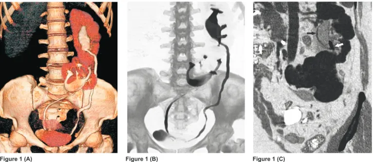
the left superior kidney were de-tected. A MDCT urography con-firmed the ultrasound findings and revealed the fusion of of the lower pole of the normally located left kidney and the upper pole of the right ectopically located kid-ney. Axial images showed contrast enhancement of thickened wall of the renal pelvis and stranding of the fat in the vicinity of the in-flamed renal pelvis in the left su-perior kidney. The attenuation val-ue of the infected fluid was 25 HU. The diagnosis of crossed fused re-nal ectopia with pyonephrosis was made (Figure 1).
Case 2
A 40-year-old male presented with hypertension. Blood pressure was 90/140 mmHg. Labaratory stud-ies revealed mild hypercholester-olemia. The sonogram demon-strated the absence of left kidney, and the presence of two kidneys in the right renal fossa, indistinguish-able from one another. Renal Dop-pler ultrasound showed normal
Figure 1. Coronal volume rendering (A) and maximum intensity projection (B) images show crossed fused renal ectopia on the left side. The
ureter of the right ectopic kidney crosses the midline and enters the bladder on the opposite side. Oblique multiplanar reforma tting image in the nephrographic phase (C) shows a mild degree of hydronephrosis, contrast enhancement of thickened wall of the renal pelvis (white arrow) and a stone (black arrow) in the lower pole of the left superior kidney. In addition, stranding of the fat in the vicinity of the i nflamed renal pelvis in the left superior kidney is also seen.
renal artery in the right superior kidney, but did not demonstrate the renal artery of the left ectopic kidney clearly due to bowel gas su-perposition. Renal MDCT angiog-raphy revealed the renal artery of right superior kidney arose from the aorta at the normal location and it had prehilar branching. Two renal arteries were present at the
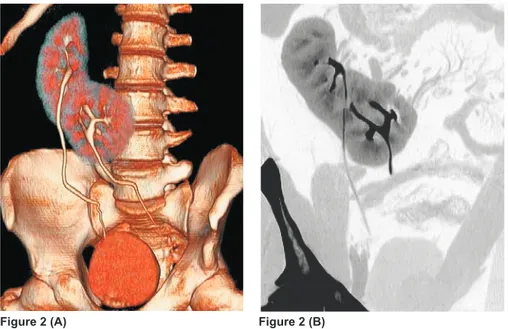
left ectopic inferior kidney. One of them arose from distal abdomi-nal aorta, with prehilar branching, and other arose from left common iliac artery. In all of the renal ar-teries was not detected stenoses. The pyelographic phase images, acquired after a 10 min delay, con-firmed diagnosis of crossed fused renal ectopia (Figure 2).
Case 3
A 69-year-old woman with consid-ered for surgical intervention for pelvic organ prolapsus presented with bilateral lumbar pain. An ultrasound showed two kidneys were located in the right renal fossa which could not be sepa-rated. Severe hydronephrosis was present in both kidneys. MDCT urography revealed crossed fused renal ectopia with severe hydro-nephrosis. No stone was detected both either in collecting systems or ureters. Prolapse of bladder and uterus was detected. The delayed pyelographic phase im-ages clearly demonstrated that both ureters were entrapped by the genital hiatus and obstructed. Crossed fused renal ectopia with severe hydronephrosis secondary to pelvic organ prolapsus was di-agnosed (Figure 3). Ultrasonog-raphy 3 months after operation showed no hydronephrosis in the both kidney.
Figure 3. Oblique multiplanar reformatting image in the nephrographic phase (A) and coronal volume rendering image in the delayed pyelograp-hic phase (B) show crossed fused renal ectopia with severe hydronephrosis. Bladder prolapsus is also seen (arrow). Coronal maximum intensity projection image (C) demonstrate that both ureters entrapped by the genital hiatus and obstructed (arrows).
Figure 3 (A) Figure 3 (B) Figure 3 (C) Figure 2. Coronal volume rendering (A) and maximum intensity projection (B) images show
crossed fused renal ectopia on the right side. The ureter of the left ectopic kidney crosses the midline and enters the bladder on the opposite side.
Case 4
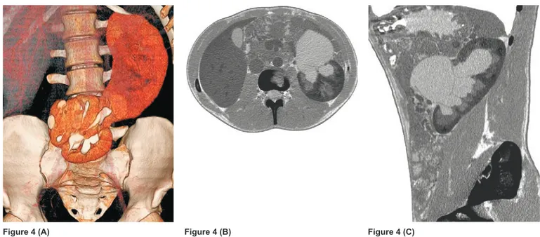
A 23-year-old male presented with a left lumbar pain. The sonogram demonstrated the right kidney was located in the midline and fused to the inferior pole of the left kid-ney with advanced hydronephro-sis. A MDCT urography revealed the right kidney, in a transverse position, it was located in the midline anterior to the fourth and fifth lomber vertebra and fused the lower pole of the left normally located kidney. Advanced hydro-nephrosis of the left kidney was detected. No stone was detected in the left collecting system and ureter. In the pyelographic phase images, were acquired after a 10 min delay, was not seen a pyelo-gram in the left kidney. The de-layed pyelographic phase images after 45 min delay showed a pyel-ogram in the left kidney. L-shaped crossed fused renal ectopia and advanced hydronephrosis of the left kidney secondary to pelvic-ureteric junction obstruction was diagnosed (Figure 4).
Discussion
Renal fusion anomalies are cake kidney (complete renal fusion), crossed fused renal ectopia and horseshoe kidney. Cake kidney is an anomaly characterized by com-plete fusion of both kidneys and accounts for only 2% of these fused kidneys (2). A partial renal fusion is represented by the horseshoe kidney and crossed renal ectopia with fusion. The commonest type of fusion anomaly is the horseshoe kidney which results when the axes of the kidneys are altered by a medial shift of the lower poles (or, rarely the upper poles) which are joined by a preaortic isthmus of renal parenchyma or fibrous tissue in the midline. It accounts for 90% of all renal fusion anoma-lies and occurs with incidence of about 0.25% of the population (1,2).
Crossed fused renal ectopia is the second most common fusion ab-normality of the kidney, with an es-timated incidence of approximate-ly 1:1300-1:7500 (3). In crossed
fused ectopia, the kidneys both lie in one side of the abdomen and one ureter will cross the midline. The left kidney is most frequently ectopic (crossing to the right side of the abdomen) and there is a slightly higher incidence in males (1). Most cases are sporadic but dominant inheritance has been re-ported (3).
Mcdonald and Mc Clellan classified crossed ectopic kidney into four types; crossed renal ectopia with fusion (85%), crossed renal ecto-pia without fusion (10%), solitary crossed renal ectopia and bilater-ally crossed renal ectopia (4). Six variations of crossed fusion have been described. In decreasing or-der of frequency, they are; type 1: inferior crossed fused ectopia, type 2: sigmoid or S-shaped kid-ney, type 3: unilateral lump kidkid-ney, type 4: unilateral disc kidney, type 5: L-shaped kidney, type 6: supe-rior crossed fused ectopia (1). The etiology of crossed fused renal
ectopia has been assumed to be due to a number of possible
fac-Figure 4. Coronal volume rendering image in the pyelographic phase (A) shows crossed fused renal ectopia on the left side. The pyelogram of
the left superior kidney was not seen. Axiel (B) and sagittal multiplanar reformatting image in the pyelographic phase (C) show severe hydro-nephrosis in the left superior kidney.
tors, including; teratogens, aber-rant development of the caudal end of the fetus with abnormal ro-tation, a wandering ureteral bud inducing differentiation of the kidney on the opposite side, and pressure from abnormally located umbilical arteries preventing ce-phaled migration of the renal unit prompting migration to the op-posite side along the path of least resistance. Fusion can occur at any time during the migration or as-cent of the crossed kidney (1). In all of fused kidneys, the arterial
supply and venous drainage are grossly abnormal. This reflects the primitive arrangement variably seen in ectopic kidneys, often be-cause of their very limited rostral migration. Blood supply to the ectopic kidney most frequently arises from the vessels on the ipsi-lateral side, but occasionally from the contralateral side. Both the normally located kidney and its fused mate commonly have abber-ant arterial anatomy, with arterial supply from the upper abdominal aorta in 25% of cases, and the re-mainder from the lower aorta or iliac arteries. While the total num-ber of arteries ranges from one to six, most commonly there are two to four major arteries to the two kidneys (1,5).
Generally, cross fused renal ectopia is typically asymptomatic and is diagnosed as an incidental find-ing when the patient is examined for other medical diseases. When symptoms do occur, the most common symptoms reported are: abdominal or flank pain, a palpa-ble mass, hematuria, dysuria and urinary tract infections. The uro-logical conditions associated with this anomaly are: nephrolithiasis, ureteropelvic junction obstruc-tion, hydronephrosis, reflux, mul-ticystic displasia, ectopic uretero-cele (1). The abnormal position of
the kidney can lead to poor out-flow and a predisposition to hydo-nephrosis, calculi, and infection (1,5). Tumors in crossed ectopic kidneys have been reported spo-radically (6).
Pediatric patients most often present with multiple congenital anoma-lies, especially of the skeletal anomalies of the bony pelvis and vertebra. Other anomalies associ-ated with this condition are car-diovascular, gastrointestinal and other genitourinary anomalies, as previously mentioned (1).
Hypertension has been diagnosed in patients with crossed renal ecto-pia. However a causal link is not usually established (1). In one case report of an infant with renal arte-rial stenosis secondary to crossed renal ectopia, renovascular hyper-tension was present (7). In our case 2 with hypertension was not detected stenoses all of the renal arteries.
Hydronephrosis may be associated with pelvic organ prolapsus as in our case 3. Several theories have been proposed to explain the as-sociation of hydronephrosis with pelvic organ prolapsus. The most recent and widely accepted expla-nation is that the ureters become entrapped by the genital hiatus against the fundus of the uterus (8). In our case 3, MDCT urog-raphy clearly demonstrated that both ureters were entrapped by the genital hiatus and obstructed. To date, the diagnosis of crossed
fused renal ectopia was made by ultrasonography, excretory urog-raphy and computed tomogurog-raphy (CT). Ultrasound is often the initial procedure performed in the work-up of the patient. The sonographic appearance of this anomaly con-sist of a characteristic anterior and/or posterior notch, difference
in orientation of the two collecting systems in the fused kidneys, and absence of a kidney in the contral-ateral renal fossa, or elsewhere in the abdomen or pelvis (9). Ex-cretory urography is often useful, but the absence of fusion may not be apparent unless the two renal masses are widely separated. CT allow accurate diagnosis. The ad-vantage of MDCT urography is its ability to depict the normal urinary tract anatomy, including both the renal parenchyma and collecting structures and ureters. Congenital anomalies of the collecting system and ureters can be visualized bet-ter with MDCT urography than with conventional CT. Congenital anomalies of renal position, num-ber and form are easily depicted with MDCT urography, including renal ectopia, malrotation and fu-sion anomalies (10).
The differential diagnosis includes a solitary kidney with compensatory hyperplasia, a solitary kidney con-taining a mass, a solitary kidney containing a double collecting sys-tem, a horseshoe kidney, and mas-sive displacement of the kidney by an abdominal mass (9).
Crossed fused renal ectopia is usually diagnosed as an incidentally, when patient is examined for other rea-sons, unless it is complicated by obstruction or infection. Yet, when it is diagnosed, further investiga-tion is necessary to rule out other correctable pathologies in the uri-nary tract and concurrent congeni-tal anomalies. MDCT urography in which both axial and 3-dimension-al images are ev3-dimension-aluated are useful for revealing urinary tract anatomy and pathologic conditions and it is promising as an alternative to ex-cretory urography. Especially coro-nally reformatted MDCT urograms can provide good delineation of congenital anomalies of urinary tract especially renal fusions.
REFERENCES
1. Bauer SB. Anomalies of the upper urinary tract. In: Walsh PC, Retik AB, Vaughan ED, Wein AJ, editors. Campbell’s Urology. 8th ed. Phila-delphia: W.B.Saunders Co.; 2002. p. 1898-1906.
2. Kaufmann MH, Findlater GS. An unu-sual case of comlete renal fusion. Gi-ving rise to a ‘cake’ or ‘lump’ kidney. J Anat 2001; 198:501-4.
3. Rinat Choni, Farkas Amicur, Frishberg Y. Familial inheritance of crossed fu-sed renal ectopia. Pediatr Nephrol 2001; 16: 269-70.
4. McDonald JH, McClellan DS. Crossed renal ectopia. Am J Surg. 1957; 93: 995-99.
5. Bailey SH, Mone MC, Nelson EW. Trans-plantation of crossed fused ectopic kidneys into a single recipient. J Am Coll Surg 2002; 194: 147-50. 6. Gerber WL, Culp DA, Brown RC, Chow
KC, Platz CE. Renal mass in crossed-fused ectopia. J Urol 1980; 123: 239-44.
7. Mininberg DT, Roze S, Yoon HJ, Pearl M. Hypertension associated with crossed renal ectopia in an infant. Pediatrics 1971; 48: 455-57.
8. Hadar H, Meiraz D. Total uterine pro-lapse causing hydroureteroneph-rosis. Surg Gynecol Obstet 1980; 150:711-4.
9. Goodman JD, Norton KI, Carr L, Yeh HC. Crossed fused renal ectopia: sonographic diagnosis. Urol Radiol 1986; 8: 13-6.
10. Chow LC, Sommer FG. Multidetec-tor CT urography with abdominal compression and three-dimensional reconstruction. Am J Roentgenol 2001; 177: 849-855.