Determination of presence of equid alpha and gammaherpesvirus
infections in foals with respiratory distress
Seval BİLGE DAĞALP
1, Ali Rıza BABAOĞLU
5, Veysel Soydal ATASEVEN
2, Zeynep KARAPINAR
3,
Mehmet Özkan TİMURKAN
4,
Fırat DOĞAN
2,
Aykut ÖZKUL
1, Feray ALKAN
11Ankara University, Faculty of Veterinary Medicine, Department of Virology, Ankara; 2Mustafa Kemal University, Faculty of
Veterinary Medicine, Department of Virology, Hatay; 3Balıkesir University, Faculty of Veterinary Medicine, Department of
Virology, Van; 4Atatürk University, Faculty of Veterinary Medicine, Department of Virology, Erzurum; 5Sekans Laboratory, Ankara
Turkey.
Summary: The aim of this study was to investigate the presence of equid herpesviruses and to elucidate the possible mutual effects of equid alpha and gammaherpesviruses in an outbreak of respiratory tract disease in foals in a private pension stable (transient residency stable), Turkey. For this purpose, nasal swabs (n=21) and peripheral blood leukocytes (n=28) from 28 foals with respiratory tract diseases, and tissue samples from one dead foal were tested for equid herpesvirus-1, -4, -2 and -5 by multiplex nested PCR targeting the glycoprotein B (gB) gene. Of the 29 sampled animals, 3.4% (1/29), 58.6% (17/29), 58.6% (17/29) and 75.9% (22/29) were found positive for EHV-1, EHV-4, EHV-2 and EHV-5, respectively. Especially, a high ratio of multiple infections (75.9%; 22/29) caused by EHV-4, EHV-2 and EHV-5 were detected in the tested foals. The phylogenetic analysis showed that our equid gammaherpesviruses had a high degree of genetic heterogeneity, in contrast to our EHV-4 strains analyzed.
Keywords: Equid alphaherpesvirus, equid gammaherpesvirus, foal, phylogenetic analysis, respiratory disease.
Solunum sistemi problemli taylarda equid alpha ve gammaherpesvirus enfeksiyonlarının varlığının
belirlenmesi
Özet: Bu çalışmanın amacı, özel bir pansiyon harasında (geçici konaklama yeri) bulunan taylarda bir solunum sistemi hastalığı salgınında equid herpesvirusların varlığını araştırmak ve equid alpha ve gammaherpesvirusların olası etkileşimlerini ortaya koymaktır. Bu amaçla, solunum sistemi hastalığı bulgulu 28 taydan nazal swap (n=21), lökosit örnekleri (n=28) ve ölen bir taydan alınan doku örnekleri, equid herpesvirus-1, -4, -2 ve -5 yönünden glikoprotein B (gB) genini tespit etmeyi hedefleyen multipleks nested PCR ile test edildi. Örneklenen 29 hayvanın %3.4’ü (1/29), %58.6’sı (17/29), %58.6’sı (17/29) ve %75.9’u (22/29) sırasıyla EHV-1, EHV-4, EHV-2 ve EHV-5 yönünden pozitif olarak bulundu. Özellikle söz konusu viruslar tarafından oluşturulan tekli enfeksiyonlardan çok, EHV-1,-2 ve -5 arasında çoklu enfeksiyonların varlığı (%75.9; 22/29) dikkat çekiciydi. Filogenetik analiz sonuçları yerel equid gammaherpesvirusların, EHV-4 suşlarının aksine genetik olarak büyük farklılık gösterdiğini ortaya koydu.
Anahtar sözcükler: Equine alphaherpesvirus, equine gammaherpesvirus, tay, filogenetik analiz, solunum sistemi hastalığı.
Introduction
Equid herpesvirus infections are ubiquitous in all equid populations worldwide. Equid herpesvirus type 1 (EHV-1) and type 4 (EHV-4), members of the Alphaherpesvirinae subfamily. Herpesviridae (7), cause significant economic losses due to respiratory disease, abortion and neonatal foal deaths. EHV-1 is rarely associated with fatal myeloencephalopathy (19, 22). Equid herpesvirus types 2 (EHV-2) and 5 (EHV-5), members of the Gammaherpesvirinae subfamily, are classified as equid gammaherpesviruses (7). EHV-2 has been isolated from cases of respiratory diseases (14). However, the possible role of EHV-5 in equid respiratory diseases has not been proved (8). It has recently been suggested that EHV-5 may be an etiologic agent
associated with serotypes of equid lymphoma (32). Some reports have suggested that EHV-2 plays a predisposing and/or reactivating role for other equid herpesviruses, such as EHV-1 and EHV-4, or bacterial pathogens like
Rhodococcus equi (8, 11, 33).
As in all herpesviral infections, equid herpesviruses establish lifelong latency in their host following initial infection (4), and can be reactivated due to adverse endogen and exogen factors like environmental stress and pharmacological stimulation (such as corticosteroids). In stables, asymptomatic carriers consistently spread the infection to naive animals (24).
EHV-1 and 4 infections are endemic in equid populations worldwide (22), and EHV-1 and 4 has recently been detected in the respiratory tract diseases of
non-vaccinated Turkish equids (2). EHV-1 and EHV-4 DNAs were also detected in abortion cases in mares and their fetuses in the Marmara Region of Turkey, with phylogenetic analysis of Turkish (TR) EHV-1 and EHV-4 strains being performed (31). Equid gammaherpesvirus infections occurring with respiratory tract diseases in horses in Turkey were also characterized based on gB gene partial sequences (3). EHV-2 and EHV-5 infections were determined as positive at a rate of 58.78% and 62.42% in Arabian horses with and without clinical signs respectively in three breeding farms in Turkey (1).
This study was undertaken to investigate the presence of equid herpesviruses and elucidate the possible mutual effects of equid alpha- and gammaherpesviruses in an outbreak of respiratory tract disease in foals in a private pension stable in Turkey. Additionally, the study also reports the molecular characterization based on the partial
gB coding gene sequences of the Turkish EHV-4, EHV-2
and EHV-5 strains.
Materials and Methods
Clinical cases and sample collection: The cases were
reported by the veterinarian of the private pension stable in the Marmara region stabling race horses transiently. All foals were born in this place. The symptomatic foals showed fever (40°C), nasal discharge, inappetence and weakness. The foals, which were 7 to 13 months of age, had been vaccinated against EHV-1 and EHV-4 (Duvaxyn IE Plus, Fort Dodge Animal Health), and equid influenza virus (Equilis Prequenza Te, Intervet). Nasal swabs (n=21) and peripheral blood mononuclear cell leukocytes (n=28) from 28 foals with respiratory disease symptoms, and tissue samples (lung, liver, spleen, intestine and kidney) from one dead foal housed on the same pension stable, were administered to our laboratory.
Multiplex nested polymerase chain reaction: All
samples from live foals and one dead foal were submitted to PCR analysis for detection of genomic DNA of equid herpesvirus types 1, 4, 2 and 5. Total DNA was extracted from samples according to Sambrook and others (26). Multiplex nested PCR was performed as described by Wang and others (33). The PCR reaction mixtures for the first and second round were prepared by adding 3.0 µl of purified DNA template to 27 µl of single-strength Taq-DNA polymerase PCR buffer containing a final concentration of 1 mM MgCl2, 200 µM of each
dinucleotide triphosphate (dNTP), 1 µM of each primer and 1 unit/ml of Taq DNA polymerase (Fermentas, Lithuania). Amplification consisted of an initial denaturation at 94˚C for 5 mins followed by 30 cycles consisting of denaturation at 94˚C for 1 min, annealing at 60˚C for 1 min and extension at 72˚C for 1 min. The reaction was terminated after a final extension at 72˚C for 10 mins. The final specific PCR products were visualized under UV-light by 1.5% agarose gel electrophoresis.
Nucleotide sequencing and phylogenetic analysis:
The PCR products were purified using the High Pure PCR Product Purification Kit (Roche Diagnostic, Germany). Sequencing of the gB amplicons were performed using a GenomeLab DTSC Kit on a CEQ 8000 Genetic Analyzer system in the laboratory (Beckman Coulter, USA). All sequences obtained from this study were submitted to GenBank in order to assign an “accession number”. The sequences from both directions were assembled together for each isolate to obtain consensus sequences, which were used in multiple alignments of sequences from different isolates using BioEdit v.7.0.9.0 (20). A phylogenetic tree was generated using the Neighbour-Joining method with the Maximum Likelihood parameter model within the MEGA v5.2 software (29). Bootstrap values were calculated from 1,000 replicates with 111 random seeds.
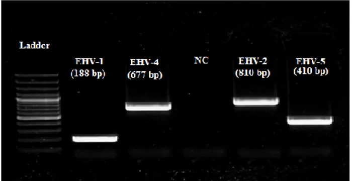
Figure 1. The agarose gel electrophoresis of EHVs 1,4,2,5 multiplex PCR product in clinical samples of foals with respiratory stress: Line1, 100 bp DNA ladder (Fermentas,Lithuania); Line2 for EHV1, Line3 for EHV4, Line4 for negative control, Line5 for EHV2 and Line6 for EHV5 positive samples.
Şekil 1. Solunum sistemi hastalıklı tayların klinik örneklerinde EHV-1,4,2,5 yönünden multipleks PCR ürünlerinin jel elektroforez görüntüsü: Sıra 1, 100 bp DNA merdiven (Fermentas, Lithuania); Sıra 2, EHV-1, Sıra 3, EHV-4; Sıra 4, Negatif kontrol; Sıra 5, EHV-2 ve Sıra 6 EHV-5 pozitif örnekler.
Results
Frequency of EHV infections in foals with respiratory tract diseases: Gel electrophoresis image of
PCR belonging to positive samples in this study is shown in Figure 1. A total of 3.4% (1/29), 58.6% (17/29), 58.6% (17/29) and 75.8% (22/29) of the tested foals were found to be positive for EHVs 1, 4, 2 and 5, respectively (Table 1). While all tissue samples from the dead foal were found to be negative for EHV-1 and EHV-4, the kidney and spleen tissues tested positive for EHV-2 and EHV-5 (Table 2). The positivity rate for nasal swab samples were higher than for the blood samples (Table 1). Regarding the positivity rates for single infections, EHV-1 was found in one leukocyte sample, EHV-4 in one nasal swab sample and 2 leukocyte samples, and EHV-5 in 4 leukocyte samples. EHV-2 was not detected alone in any sampled
animals. Multiple EHV infections were detected in samples obtained from 75.8% (22/29) of sampled animals. Triple (EHV-4,-2,-5; n=9 cases) and double (EHV-2, -5; n=8 cases, and EHV-4, -5; n=5 cases) infections were identified in 22 of the 29 animals included in this study (Table 2).
Table 2. The results of single and multiple infections for EHVs in foals.
Tablo 2. Taylarda EHV yönünden tekli ve çoklu enfeksiyon bulguları.
Combination of single/multiple infections
The number of nasal swab and/or other samples
positive foals EHV-1 1/29 (3,44 %) EHV-4 3/29 (10,34 %) EHV-2 0/29 (0%) EHV-5 0/29 (0%) Subtotal 4/29 (13,79%) EHV-2/EHV-5 8/29 (27,58%) EHV-4/EHV-5 5/29 (17,24%) EHV-2/EHV-5/EHV-4 9/29 (31,03%) Subtotal 22/29 (75,86%)
Total positive foals 26/29 (89,65%) Negative foals (-) 3/29 (10,34%)
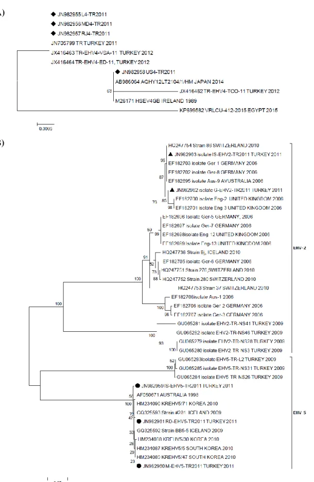
Phylogenetic analysis of EHVs 4, 2 and 5: The
partial sequences obtained from gB-encoding genes of two EHV-2, three EHV-5 and four EHV-4 strains of Turkish horses [TR-EHV-2: G-EHV-2-TR2011 (JN982962), IS-EHV-2-TR2011 (JN982963); TR-EHV-5: IS-EHV-5-TR2011 (JN982959), M-EHV-5-IS-EHV-5-TR2011 (JN982960), RD-EHV-5-TR2011 (JN982961); TR-EHV-4: L4-TR2011 (JN982955), MD4-L4-TR2011 (JN982956), RJ4-TR2011 (JN982957), US4-RJ4-TR2011 (JN982958)] were compared to those published in the GenBank database and previously reported Turkish EHV-2 and EHV-5 field isolates. However, although the leukocyte sample was evaluated as positive for EHV-1, the sequence analysis could not be performed because of poor sample quality.
As the number of EHV-4 gB sequences published in the GenBank is limited, only a few examples were available to compare with the local EHV-4 gB sequences. In this study, all sequences of EHV-4 were found to be almost similar, apart from US4-TR2011. LD4-TR2011, MD4-TR2011 and RJ4-MD4-TR2011 strains clearly differed from the US4-TR2011 strain, being located on a separate branch. Three EHV-4 strains (L4-TR2011, MD RJ4-TR2011 and RJ4-TR2011) were closely related to strains circulating in Turkey in 2011-2012, and one strain (US4-TR2011) was related closely to strains circulating in Japan, Ireland and Turkey (2012 strain) (Figure 2A).
A sequence analysis for the partial gB coding region of all TR EHV-2 strains revealed that these viruses shared 85.7-99.7% and 92.4-99.7% nucleotide similarity with previously reported Turkish EHV-2 field strains. TR EHV-2 strains were found to be identical to EHVs especially detected in Switzerland, Germany and United Kingdom. EHV-5 strain sequences in this study showed less similarity (98.8-99.7%) than previously reported Turkish EHV-5 viruses (99.4-100%). On the other hand, the similarity rate of all EHV-5 viruses used in the analysis varied between 98.5-100%, indicating that the viruses were almost genetically identical in terms of partial gB sequences (Figure 2B).
Discussion and Conclusion
According to some reports, authors suggest that EHV-2, and possibly EHV-5, can play a predisposing role in respiratory diseases caused by viral and bacterial pathogens, such as Rhodococcus equi, Streptococcus equi and Streptococcus zooepidemicus (8, 11, 33). Multiple EHV-2 and/or EHV-4, EHV-5 infections may indicate a possible mutual effect of equid alpha- and gammaherpesviruses in foals with respiratory problems.
Many foals are co-infected with EHV-1 and EHV-4 along with EHV-2 and -5 during the first few months of their life. Foals are known to be predisposed to the infection during the weaning period, shortly before the maternal antibody levels decrease, and show a high rate of subclinical infections (9, 15, 16, 18, 23). Considering the
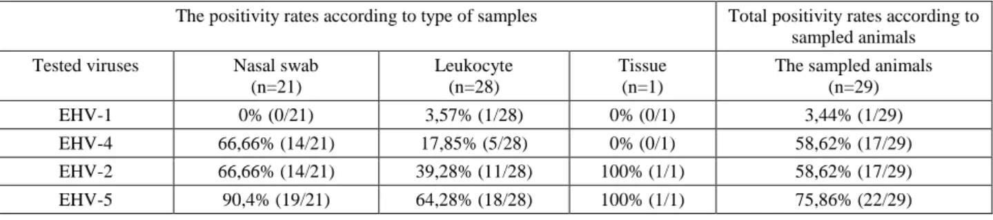
Table 1. The positivity rates for EHVs according to type of samples and animals. Tablo 1. Örnek tipine ve örneklenen hayvanlara göre EHV’lere ait pozitiflik oranları.
The positivity rates according to type of samples Total positivity rates according to sampled animals Tested viruses Nasal swab
(n=21)
Leukocyte (n=28)
Tissue (n=1)
The sampled animals (n=29)
EHV-1 0% (0/21) 3,57% (1/28) 0% (0/1) 3,44% (1/29)
EHV-4 66,66% (14/21) 17,85% (5/28) 0% (0/1) 58,62% (17/29)
EHV-2 66,66% (14/21) 39,28% (11/28) 100% (1/1) 58,62% (17/29)
A)
B)
Figure 2. Phylogenetic analysis of EHV-4 (A) and EHVs-2 and 5 (B) detected in this study.
Solid circles, rectangular and equilaterals in panel A and B indicate Turkish EHV-4, EHV-2 and EHV-5 isolates, respectively. Numbers at nodes indicate bootstrapping values. Bar: represents number of base substitution per position.
Şekil 2. Bu çalışmada tespit edilen EHV- 4‘ün (A), EHV-2 ve EHV-5‘in (B) filogenetik analizi.
A ve B panelinde daire, dikdörtgen ve eşkenar üçgen şeklinde Türk EHV-4, EHV-2 ve EHV-5 suşları sırasıyla görülmektedir. Çizgedeki sayılar bootstrapping değerlerini gösterir. Çubuk: Her konum için nukleotid değişimi sayısını temsil eder.
age distribution of foals tested in the current study, our results agree with existing data (9, 16, 17, 18). EHV-2 is most frequently transmitted horizontally to a newborn foal from its dam via the nasopharyngeal route or later through contact with other foals (4). Persistent infection is established with constant shedding (5), with foals shedding higher viral loads than adult horses (21). This may explain why EHV-2 was frequently detected in the sampled population. Schlocker and others (27) have hypothesized that EHV-2 behaves differently in foals than in adult horses. It could be that EHV-2 is not strictly cell dependent in foals so it can be easily isolated from swab samples obtained from the upper respiratory tract and eyes; on the other hand, the virus is strictly cell-bound in adult horses, being contained in macrophages and lymphocytes (27). In this study, EHV-2 and 5 infections were found to be common in tested foals. The frequent detection of the virus or viral genome of 2 and EHV-5 in the nasal swabs of asymptomatic horses is a serious concern (30). Compatible with this, current study detected a higher positive ratio of EHV-2 and 5 in nasal swab samples than in the blood samples of the tested horses. Although no major diseases have been attributed to EHV-2 and 5, they may be a cofactor in EHV-1 and/or EHV-4 replication by immunosuppression, thereby causing general malaise.
In the current study, although the animals were regularly vaccinated against these agents, EHV-1 and especially EHV-4 were detected in foals. EHV-1 and 4 vaccines are recommended for all foals and adult horses. Initial vaccination begins at 4-6 months of age to be followed by boosters at 4-6 weeks after the first dose, and a third dose at 10-12 months of age. It is assumed that this vaccination protocol allows all foals to be vaccinated properly. Although fully protective vaccines against herpesvirus infections have not yet been developed owing to the latency properties of the viruses, main goal of vaccines widely used for these pathogens is prevention of outbreaks of EHV-associated diseases (22). Although many multivalent EHV-1 and EHV-4 vaccines are used, they cannot prevent equid herpesvirus infection and most only protect against respiratory symptoms due to EHV-1 and 4 (22, 24).
The results of this study also support evidence that EHV-4 continues to circulate in properly vaccinated population. Detection of EHV-4 DNA, especially in nasal discharge samples from sampled animals with symptoms of respiratory system may indicate that vaccination for EHV-1/EHV-4 cannot fully prevent the EHV infections. However, apart from vaccination, environmental and nutritional factors, management practices, seasonal conditions and other infections may influence the prevalence of EHV infections (12). Moreover, animals were only tested for equid herpesviruses (EHV-1, -4, -2,-5) in this study.
In the present study, the partial nucleotide sequences obtained from gB-encoding genes of two EHV-2, three EHV-5 and four EHV-4 strains in Turkish horses were compared to those published in the GenBank database and to previously reported Turkish EHV-2, -4 and 5 field viruses. There is limited data regarding the sequences of the EHV-4 gB-coding gene region (6, 25). In this study, all sequences of EHV-4 were found to be almost similar, apart from US4-TR2011. LD4-TR2011, MD4-TR2011 and RJ4-TR2011 strains clearly differed from the US4-TR2011 strain, being located on a separate branch (Figure 2A). Moreover, a sequence analysis of all TR EHV-2 strains revealed that they shared high nucleotide similarity (at least 85%) with each other. EHV-2 and EHV-5 strain sequences in this study showed 85.7-99.5% nucleotide similarity with those available in the GenBank. All EHV-5 strains (IS-EHVEHV-5-TR2011, M-EHVEHV-5-TR2011 and RD-EHV5-TR2011) detected in this study were also different from each other (93.6-98.9% nucleotide similarity) and clustered in a separate branch, as seen in the case of previously isolated TR EHV-5 field strains (95.4-98.9% nucleotide similarity). Thus, similarly to previous studies (3, 10, 28), the equid gammaherpesviruses studied in the current study had a high degree of genetic diversity, in contrast to Turkish EHV-4 strains.
In conclusion, we consider that as reported by Foote et al. (13) seroepidemiologic and molecular studies provide an aspect about EHV infections, which needs to be carefully considered when implementing a vaccination programme, or in the strategic design of new vaccines or control measures. In addition, this study provides further insights into the potential role of EHV-2, and EHV-5 as predisposing factors for EHV-4 infection. Further studies are needed in order to improve the understanding of the epizootiological factors involved in both gammaherpesvirus and alphaherpesvirus infections in horses of different ages and breeds, and for the investigation of the interaction between gammaherpesviruses and respiratory disease in foals.
Ethical approval: This article does not contain any studies with animals performed by any of the authors.
References
1. Akkutay AZ, Osterrieder NA, Damiani A, et al. (2014):
Prevalence of equid gammaherpesviruses on breeding farms in Turkey and development of a TaqMan-MGB real-time PCR to detect equid herpesvirus 5 (EHV-5). Arch
Virol, 159, 2989-2995.
2. Ataseven VS, Bilge Dağalp S, Güzel M, et al. (2009):
Prevalence of equid herpesvirus-1 and equid herpesvirus-4 infections in equid species in Turkey as determined by ELISA and multiplex nested PCR. Res in Vet Sci, 86,
339-344.
3. Ataseven VS, Bilge Dağalp S, Oğuzoğlu TÇ, et al. (2010):
gammaherpesviruses from horses with respiratory tract disease in Turkey. Transbound Emerg Dis, 57, 271-276.
4. Bell SA, Balasuriya UBR, Gardner IA, et al. (2006):
Temporal detection of equid herpesvirus infections of a cohort of mares and their foals. Vet Microbiol, 116, 249-257.
5. Browning GF, Studdert MJ (1987): Epidemiology of
equid herpesvirus 2 (equid cytomegalovirus). J Clin
Microbiol, 25, 13-16.
6. Cullinane AA, Rixon FJ, Davison AJ (1988):
Characterization of the genome of equid herpesvirus 1 subtype 2. J Gen Virol, 69, 1575-1590.
7. Davison AJ, Eberle R, Ehlers B, et al. (2009): The order
Herpesvirales. Arch Virol, 154, 171-177.
8. Diallo IS, Hewitson GR, Jong A, et al. (2008): Equid
herpesvirus infections in yearlings in South-East Queensland. Arch Virol, 153, 1643-1649.
9. Donald JJ, Wilks CR (1999): A type-spesific ELISA for
equid herpesvirus-1: Prevalence and seroepidemiology in horses in New Zealand. In: Proceedings of the 8th
International Conference Equid Infectious Diseases, Dubai, 537-538.
10. Dunowska M, Holloway SA, Wilks CR, et al. (2002):
Genomic variability of equid herpesvirus-5. Arch Virol,
145, 1359-1371.
11. Dunowska M, Wilks CR, Studdert MJ, et al. (2002):
Equid respiratory viruses in foals in New Zealand. N Z Vet
J, 50, 140-147.
12. Foote CE, Gilkerson JR, Whalley JM, et al. (2003):
Seroprevalence of equid herpesvirus 1 in mares and foals on a large Hunter Valley stud farm in years pre- and postvaccination. Aust Vet J, 81, 283-288.
13. Foote CE, Love DN, Gilkerson JR, et al. (2006): EHV-1
and EHV-4 infection in vaccinated mares and their foals.
Vet Immunol Immunopathol, 111, 41-46.
14. Fortier G, Erck E, Fortier C, et al. (2009): Herpesviruses
in respiratory liquids of horses: Putative implication in airway inflammation and association with cytological features. Vet Microbiol, 139, 34-41.
15. Fu ZF, Robinson AJ, Horner GW, et al. (1986):
Respiratory disease in foals and the epizootiology of equid herpesvirus type 2 infection. N Z Vet J, 34, 152-155.
16. Gilkerson JR, Love DN, Whalley JM (1999a):
Serological evidence of equid herpesvirus-1 (EHV-1) infection in Thoroughbred foals 30-120 days of age. In:
Proceedings of the 8th International Conference Equid Infectious Diseases, Dubai, 546.
17. Gilkerson JR, Whalley JM, Drummer HE, et al. (1999b):
Epidemiology of EHV-1 and EHV-4 in the mare and foal populations on a Hunter Valley stud farm: are mares the source of EHV-1 for unweaned foals. Vet Microbiol, 68, 27-34.
18. Gilkerson JR, Whalley JM, Drummer HE, et al. (1999c):
Epidemiological studies of equid herpesvirus 1 (EHV-1) in Thoroughbred foals: a review of studies conducted in the Hunter Valley of New South Wales between 1995 and 1997.
Vet Microbiol, 68, 15-25.
19. Goehring LS, Landolt GA, Morley PS (2010): Detection
and management of an outbreak of equid herpesvirus type 1 infection and associated neurological disease in a veterinary teaching hospital. J Vet Intern Med, 24,
1176-1183.
20. Hall TA (1999): BioEdit: A user-friendly biological
sequence alignment editor and analysis program for Windows 95/98/NT. Nucleic Acids Symposium Series, 41, 95-98.
21. Hartley CA, Dynon KJ, Mekuria ZH, et al. (2013): Equid
gammaherpesviruses: perfect parasites? Vet Microbiol
167, 186-192.
22. Heldens JGM, Hannant D, Cullinane AA, et al. (2001):
Clinical and virological evaluation of the efficacy of an inactivated EHV-1 and EHV-4 whole virus vaccine (Duvaxyn EHV1,4). Vaccination/challenge experiments in
foals and pregnant mares. Vaccine, 19, 4307-4317.
23. Marenzoni ML, Coppola G, Maranesi M, et al. (2010):
Age-dependent prevalence of equid herpesvirus 5 infection.
Vet Res Commun, 34, 703-708.
24. Patel JR, Heldens J (2004): Equid herpesviruses 1 (EHV1)
and 4 (EHV4)-epidemiology, disease and immunoprophylaxis: A brief review. Vet J, 170, 14-23.
25. Riggio MP, Cullinane AA, Onions DE (1989):
Identification and nucleotide sequence of the glycoprotein gB gene of equid herpesvirus 4. J Virol, 63, 1123-1133.
26. Sambrook J, Fritsch EF, Maniatis T (1989): Isolation of
high molecular-weight DNA from mammalian cells. In:
Molecular Cloning: A Laboratory Manual, Second Ed. Cold Spring Harbor Laboratory Press, New York, USA, 9.14-9.23.
27. Schlocker N, Gerber-Bretscher R, Von Fellenberg R (1995): Equid herpesvirus 2 in pulmonary macrophages of
horses. Am J Vet Res, 56, 749-754.
28. Sharp EL, Farrell HE, Borchers K, et al. (2007):
Sequence analysis of the equid herpesvirus 2 chemokine receptor homologues E1, ORF74 and E6 demonstrates high sequence divergence between field isolates. J Gen Virol, 88,
2450-2462.
29. Tamura K, Peterson D, Peterson N, et al. (2011):
MEGA5: Molecular Evolutionary Genetics Analysis using Maximum Likelihood, Evolutionary Distance, and Maximum Parsimony Methods. Mol Biol Evol, 28, 2731-2739.
30. Torfason EG, Thorsteinsdottir L, Thorsteinsdottir S, et al. (2008): Study of equid herpesviruses 2 and 5 in Iceland
with a type-specific polymerase chain reaction. Res in Vet
Sci, 85, 605-611.
31. Turan N, Yildirim F, Altan E, et al. (2012): Molecular
and pathological investigations of EHV-1 and EHV-4 infections in horses in Turkey. Res in Vet Sci, 93,
1504-1507.
32. Van Der Welf KA, Davis EG, Janardhan K, et al. (2014):
Identification of equid herpesvirus 5 in horses with lymphoma. JEVS, 34, 738-741.
33. Wang L, Raidal SL, Pizzirani A, et al. (2007): Detection
of respiratory herpesviruses in foals and adult horses determined by nested multiplex PCR. Vet Microbiol, 121,
18-28.
Geliş tarihi : 25.07.2016 / Kabul tarihi : 01.12.2016
Address for correspondence:
Prof.Dr. Seval BİLGE DAĞALP
Ankara University, Faculty of Veterinary Medicine, Department of Virology,
06110, Dışkapı, Ankara, Turkey. e-mail: [email protected]