aper
Hepato-Gastroenterology 2015; 62:3-7 doi 10.5754/hge14958 © H.G.E. Update Medical Publishing S.A., Athens
Key Words:
GLP-1, Liver resec-tion, Oxidative damage, Regenera-tion.
Background/Aims: The purpose of our study is researching into impact of glucagon like peptide 1 (GLP 1) analogue on liver regeneration after major hepatectomy. Methodology: 24 wistar albino rats were consecutively divided into 3 groups. Group 1: Control (sham) group day 14 (n=8), Group 2: Liver resection group day 14 (n=8); 70% Liver resection was performed, Group 3: Study group day 14 (n=8); Subsequent to performing 70% liver resection, GLP-1 analogue was administered 2 times a day. (10 μgr/70 kg x 2 times). ��ter 1� day� rats were sa�ri�i�ed. �xi���ter 1� day� rats were sa�ri�i�ed. �xi-dative stress and antioxidant enzymes and
mitochon-drial permeability transition, cytochrome-c, Bax, Bcl-2, caspase-3, caspase-8 and caspase-3 activity were examined. Results: 70% Liver resection induced oxi-70% Liver resection induced oxi- induced oxi-dative stress of liver tissue was ameliorated by GLP 1 induction. Administration of GLP increased Bcl-2 ex-pression. Decreased expression of cytochrome-c was accompanied by a decrease caspase-3, caspase-8, and Bax expression and caspase-3 activity. Conclusions: Glp 1 induction plays a regenerative role in the major hepatectomy. This effect is dependent on modulation of the antiapoptotic and antioxidative pathways by GLP 1 expression.
ABSTRACT
Impact of GLP-1 Analogue on Oxidative
Damage and Hepatic Regeneration in
Experimental 70% Hepatectomy Model
Muharrem Battal
1, Bulent Çitgez
1, Abdulcabbar Kartal
1, Ahu Kemik
2, Pınar Yıldırım
3,
Yasar Ozdenkaya
4, Ahmet Yilmaz
4and Oguzhan Karatepe
41
Hamidiye Etfal Training Hospital, General Surgery Clinic, Istanbul, Turkey
2
Istanbul University� Cerrahpaşa S�hool o� Medi�ine� Department o� Bio�hemistry� Istanbul� Turkey
3Yenimahalle State Hospital, Pathology Clinic, Ankara, Turkey
4
Medipol University, General Surgery Clinic, Istanbul, Turkey
Corresponding author: Oguzhan Karatepe, Medipol University ,Department of Surgery,34718,Kosuyolu,Istanbul, Turkey; E-mail: [email protected]
INTRODUCTION
Major hepatectomy and advanced liver resections may be performed at many centers today. One of most important problems among surgical problems after liv-er surgliv-ery is state of residual livliv-er volume. It is known that if a patient undergoes chemotherapy or existence of chronic liver disease is known, volume up to 40% is sufficient, for those with regular liver tissue up to 25% is sufficient (1). Despite all advanced diagnosis methods, during and after surgery, unexpected liver deficiency may emerge. Many patients may compensate this with early stage liver regeneration. Literature contains a lot of studies with the purpose of increasing liver regenera-tion. However, at the moment there is no effective agent used in our clinical practice.
GLP-1 analogues were discovered in 1923 at pan-creas tissue. Afterwards, it was found that this analogue also exists at gastrointestinal system mucosa. Receptors are particularly located at pancreas islet cells, brain, kid-ney, heart and intestine mucosa. It has various effects and these effects are known in literature as “incritin ef-fect” (2).
The most important effect is increasing concentra-tion of peripheric insulin concentraconcentra-tion. This effect is especially made by reducing insulin uptake of liver after oral glucose intake and increasing insulin quantity in circulation. At the same time, pancreas B cell functions increase. Other than this, incritins inhibit gastrointes-tinal system, pancreas secretion. They reduce intestine and stomach motility. Pancreatitis is reported as the
most serious adverse effect. Today, GLP-1 analogues are used for the purpose of increasing insulin concentration for Type-II diabetes treatment (3-5).
Studies conducted allow us to know that insulin has significant hepatotropic effect on early stage liver re-generation (6). Our purpose in this study is to research impact of incritins, which are so effective at gastrointes-tinal and pancreas tissue, on liver generation and oxida-tive damage.
METHODOLOGY
The study was conducted from December 2012 to March 2013 at Istanbul University, Istanbul School of Medicine Experimental Medicine Research Center Laboratory with ethical review board approval dated 01.11.2012 and no. 2012/156.
Twenty-four male Wistar-albino rats weighing 250- 300g were used in the study. All animals were housed in wiremesh bottomed cages in 12 hours light/12 hours dark cycle at a constant room temperature of 22 ± 2 ºC. They were fed a Standard chow diet and water. The
study was approved by the local ethics review board. Three equally numbered groups were created as fol-lows:
Group 1: Control (sham) group day 14 (n=8). Group 2: Liver resection group day 14 (n=8). 70% Liver resection was performed.
Group 3: Study group day 14 (n=8). Subsequent to performing 70% liver resection, GLP-1 analogue was ad-ministered 2 times a day. (10 μgr/70 kg x 2 times)
4 Hepato-Gastroenterology 62 (2015) Battal M, Citgez B, Kartal A, et al.
Western blot
expressions (control)Group 1
Group 2 (%70 hepatectomy) Group 3 (glp 1 treatment) cytochrome-c in cytosol 11.8±2 147.17±16 95.4 ±11 Bax 101.14±23 157.56±36 56.5±14 Bcl-2 93.5±13 38.7±7 167.56±31 Bax/Bcl-2 1.8±0.3 3.4±1.2 0.4±0.1 Caspase-3 98.4±15 172.44±38 101.34±14 Caspase- 8 87.38±22 156.65±42 98.47±21
TABLE 1. The results of Western Blot analysis.
Values with di��erent letters have signi�i�an�e a��ording to �N�V� test. B�x/��l� The sig-ni�i�an�e between �ontrol and group is p<0.01; group 2 and 3.
Surgical procedure
The surgical procedure was carried out under ster-ile conditions on the 2nd and 14th day of the therapy
re-ceived by the experiment group. All rats were anesthe-tized by intramuscular administration of 80 mg/kg of ketamine hydro�hloride (Ketalar� E�za�ıbası) and 8 mg/ kg of xylazin (Rompun, Bayer). Thus a general anesthe-sia and spontaneous respiration were maintained. The abdominal region was cleaned with povidone iodine. Liver resection technique
After shaving abdomens, laparotomy was performed with midline incision, approximately 3 cm in length. Af-ter freeing pedicles of liver left laAf-teral and median lobs, coronary, left lateral and gastrohepatic ligaments, they were tied with 3/0 silk and 70% hepatectomy was per-formed as described by Higgins and Anderson (7).
For the purpose of daily liquid supplement to all re-sected animals, subcutaneous SF 5 cc was performed. After surgery, all animals were sacrificed at day 2 and 14. Liver tissue samples were collected for bio-chemical and histological examination. 5cc of blood was drawn into an appropriate sample tube suitable for the investi-gation of biochemical parameters. Tissue samples were obtained for biochemical and histopathological inves-tigations and trans�erred into 0.9% NaCl solution or into 10% formaldehyde solution. Blood samples were �entri�ugated and preserved at −80 º C �or bio�hemi�al investigations. Residual liver tissue after surgery was weighed with precision balance. All results were statis-tically compared.
Preparation of postmitochondrial and cytoplasmic extracts
Preparation of postmitochondrial and cytosolic frac-tions was performed aspreviously described (8).
Ho-mogenates were centrifuged at 600 X g for 15min at 4°C, and the supernatants were then centrifuged at 14 000 X g for 15 minute (min) at 4°C. Supernatants were used for HO-1, Bcl-2, Bax, caspase 3, and caspase 8 western blot analysis and oxidative parameters. For cytochrome-c analysis, the remaining supernatants were cytochrome-centrifuged at 100 000 X g for 1 hour at 4°C.
Western blot analysis
The mitochondrial and cytosolic fractions were lysed in modified RIPA buffer (50 mM Tris HCl, pH 7.4, 0.25% Na�deoxy�holate� 10 % SDS�150 mM NaCl� 1 mM EDT�� 1 mM PMSF, 1% Triton X-100, 1% Glycerol, 1 µg/ml of aprotinin, 1 µg/ml leupeptin, 1 µg/ml pepstatin A, 1 µg/ml soybean trypsin inhibitor, 0.5 mM dithiothreitol, and 1 mM NaF). Sodium dode�yl sul�ate (SDS)–poly-acrylamide gel electrophoresis (PAGE) was performed using the Bio-Rad Mini Protean III gel system accord-ing to Laemmli’s method (9). Equal amounts of protein from each sample (50 mg/well) were loaded onto a 10% SDS–P�GE and proteins were then trans�erred to poly-vinylidene difluoride (PVDF) membranes. Following incubation, membranes were washed with PBS contain-ing 0.01% Tween 20 (PBS-T) and then exposed to sec-ondary antibodies. After washing, blots were visualized using the enhanced chemiluminescence (ECL) kit from Amersham (Pharmacia Biotech., NJ, USA) according to the manufacturer’s protocol. The relative densities of the bands were quantified using the Vilber Lourmat-Bio-Profil imaging system. (Vilber Lourmat Biotechnol-ogy, Marne-la-Vallée, France). The following primary antibodies were used: mouse monoclonal anti-Bcl-2 (SC-7382, Santa Cruz Biotechnology, CA), mouse mono-clonal anti-Bax (SC-7480, Santa Cruz Biotechnology, CA), Mouse monoclonal anti-cytochrome-c (GTX13575, Ge-neTex, Inc., SA), rabbit polyclonal anti-caspase 8 (FLICE, Ab-4Lab Vision Corporation, UK), mouse monoclonal anti� α a�tin (SC-32251, Santa Cruz Biotechnology, CA), rabbit polyclonal anti-caspase 3 (CPP32, Ab-4, Lab Vi-sion Corporation, UK). Secondary antibodies were used goat anti-mouse IgG-HRP (SC-2005, Santa Cruz Biotech-nology, CA), goat anti-rabbit IgG-HRP (SC-2030, Santa Cruz, Biotechnology, CA), goat anti-rabbit IgG Peroxidase Conjugated, H+L (AP124P,Chemicon, CA).
Caspase-3 activity assay
Caspase-3 activity was measured with a colorimetric caspase-3 assay kit (Sigma-Aldrich, St. Louis, MO). The activity of the enzyme was expressed as the amount of p-nitroaniline liberated per mg protein per minute.(28) Oxidant and antioxidant status
The levels of malondialdehyde (MDA) and glutathi-one (GSH) and the activities of superoxide dismutase (SOD), glutathione peroxidase (GSH-Px) and glutathi-one transferase (GST) were determined in the post-mi-tochondrial fraction. The levels of MDA were measured with a thiobarbituric acid test (9). GSH levels were mea-sured with 5,5-dithiobis-(2-nitrobenzoate) at 412nm.21
SOD activities were assayed by its ability to augment the effect of riboflavin-sensitized photooxidation of ortho-dianisidine. GSH-Px and GST activities were measured using cumene hydroperoxides and 1-chloro-2,4-dinitro-benzene as substrates, respectively. The carbonyl con-tent, used to determine the extent of oxidative damage to proteins, was measured according to the method by Reznick and Packer based on the spectrophotometric
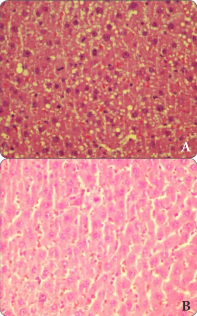
A
B
FIGURE 1. A) Macro and microvesicular fattening at liver, expansion at sinusoids anda great number of mitosis H/E X400 (GLP KC 1). B) Expansion at sinusoids, hyperemia, mitosis H/E X400(SHAM KC 1).
detection of the reaction between 2,4-dinitrophenylhy-drazine and protein carbonyls to form protein hydra-zone (10).
Histopathological Methods
Mitotic index, inflammation and fibrosis level were investigated at liver tissue. During the study, number of mitosis was sought at 10 large growth sites. Ishak scor-ing was used for inflammation and fibrosis at liver (11). Statistical analysis
Data were analyzed using SPSS 16.0 for Windows. Results with normal distribution were expressed as mean ± SD. Comparisons of normal distribution data were done by one�way �N�V� test. Results were �onsid-ered statistically significant when the two tailed p value was less than 0.05.
RESULTS
During first 24 hours before sacrifice, we lost 2 rats from group 2. Early autopsy was performed on these rats. Intense acid was found inside abdomen. These rats were excluded from study and replaced with new ani-mals. No mortality was determined at groups adminis-tered with GLP 1 analogue.
During evaluation of liver tissues which regenerate histopathologically, no significant difference in terms of fibrosis was found.
Upon comparing control group and experiment group with respect to mitosis index, significant differ-ence was determined at mitosis index at group where GLP 1 analogue was used (group 3) in comparison with group where GLP 1 was not used on group 2. (Figure 1)
There was no significant difference in histopatho-logical comparison of inflammation level among groups. Bio-chemical examination results of groups are present-ed in Table 1.
Oxidant and antioxidant status
The levels of MDA and protein carbonyl were found to be significantly higher in group 2 compared with group 1 (3.8±0.5 nmol/mg protein and 4.7±0.7 nmol/ mg protein vs. 2.5±0.1 nmol/mg protein and 2.05±0.4 nmol/mg protein, p<0.01). The activities of GSH, GST, GSH-Px, and SOD were found to be significantly lower in group 2 compared with group 1 (16.1±2.6 nmol/mg protein, 135.2±20 nmol/min/mg protein, , p<0.01). GLP 1 administration significantly increased antioxidant en-zymes and decreased oxidative damage compared with group 2 (Figure 1).
The results of Western Blot analysis
The results of western blot analysis were summa-rized in Table 1. Cytochrome-c expression in the cytosol: Cytochrome-c expression was found to be significantly higher in group 4 compared with group 1, p<0.001. In group 3, cytochrome-c expression was found to be in-creased significantly compared with all other groups including group 3, p<0.001. Bax expression: The expres-sion levels of the proapoptotic protein, Bax, in the liver tissue were found to be significantly higher in group 2 compared with group 1, p<0.001. In group 3, Bax ex-pression was found to be increased significantly com-pared with all other groups including group 2, p<0.001. Bcl-2 expression: The levels of the antiapoptotic pro-tein, Bcl-2 was found to be significantly lower in group 2 compared with group 1, p<0.001. GLP 1
administra-tion significantly increased the Bcl-2 expression in the liver tissue compared with group 2, p<0.05. The ratio of Bax/Bcl-2: The ratio of Bax/Bcl-2 was found to be significantly higher in group 2 compared with group 1, p<0.01. GLP 1 treatment significantly decreased the Bcl-2 expression in the liver tissue compared with all other groups including group 2, p<0.001. Caspase-3 and caspase-8 expression: The expression of caspase-3 and caspase-8 in the liver tissue was found to be significant-ly higher in group 2 compared with group 1, p<0.001. In group 3, caspase-3 and -8 expressions was found to be increased significantly compared with all other groups including group 2, p<0.001.
Caspase-3 activity in the liver
The caspase-3 activity in the liver tissue was found to be significantly higher in group 2 compared with group 1, p<0.001. GLP 1 treatment significantly decreased the caspase-3 activity in the liver tissue compared with
6 Hepato-Gastroenterology 62 (2015) Battal M, Citgez B, Kartal A, et al. group 2, p<0.001. In group 3, caspase-3 activity was
found to be decreased significantly compared with group 2, p<0.001.
DISCUSSION
Hepatic regeneration is an important physiological process at early period, for major liver resections in par-ticular. Regeneration response is typically dependent on proliferation of acinar structure of residual liver tissue. Many factors take a part in this process. It is known that Hepato�yte growth �a�tor� TNF�α� IL�6� Epidermal growth �a�tor (EGF)� TGF� α� Norepinephrine� Insulin� gender hormones, Fibroblast growth factor (FBF), Vas-cular endothelial growth factor (VEGF), retinoic acid, thyroid hormones, growth hormone and many medica-tions have positive effects on liver regeneration (6).
Liver is first transit point of insulin generated at pancreatic tissue. It was reported that liver atrophy develops at cases where direction of portal vein flow is changed with portacaval shunt (12). Development of at-rophy was prevented by administering insulin directly to liver tissue at rats to which experimental portocaval shut was applied (13). Insulin has great impact on liver regeneration but direct mitogen impact was not deter-mined (12).
Even though effects of GLP-1 analogues on liver re-generation are not known, their effects on various sys-tems were researched. These researches concentrate particularly treatment for diabetes. The study by Vered aviv et al emphasizes that GLP-1 receptors could not be determined at liver cells but it is effective on metabo-lism of liver (14, 15, 16).
The study conducted by Daniel J. Cuthbertson et al determined that quantity of liver fattening is reduced at persons treated with GLP-1. In a similar study, Yong Ook Kim et al reported that GLP-1 reduces liver glucose output, oxidative stress and liver insulin resistance and liver fattening (3).
Efficiency of GLP-1 analogues was intensively stud-ied also after pancreas islet cell and pancreas transplant and positive impact on glucose metabolism was clearly revealed (17, 18).
There are question marks regarding reliability of long term use of GLP-1 analogues and impact of cancer.
A study reported that use of GLP-1 may trigger devel-opment of medullary carcinoma of thyroid and multiple endocrine neoplasia Type-2 but it has preventive effects on development of breast and colon cancer. (19, 20)
In the study we found that GLP-1 analogues increase ratio of mitosis of liver cell at early stage after resection in particular (Day 14) and during bio-chemical mea-surements ratio of Bax BCL-2 was found higher at ex-perimental group in comparison with control group and statistically significant.
In the clinic, especially after major liver resection, some patients may present liver deficiency symptoms. This occurs especially at 3-5% patients with previously known chronic liver disease or undergoes intensive chemotherapy within post-operative 48-72 hours. (21) The International Study Group of Liver Surgery recently developed a consensus definition for post-hepatectomy liver failure namely ‘the impaired ability of the liver to maintain its synthetic, excretory, and detoxifying func-tions, which are characterized by an increased interna-tional normalized ratio and concomitant hyperbilirubi-nemia on or after postoperative day 5 (22). This arises �lini�ally with a�id existen�e� INR elevation� hyperbili-rubinemia, encephalopathy and recovers with support therapy (23). In addition to scarcity of residual liver tis-sue after liver resection, may factors like bleeding, sep-sis, ischemia, venous thrombosis may cause this. Other than support therapies� antioxidant N�a�etyl�ysteine may be used to reduce free oxygen radicals that form as a result of ischemia reperfusion damage that occur at liver (24,25).
The study determined that GLP 1 analogue increases liver tissue quantity at early period. The study we con-ducted is a pilot study on this subject; it encouraged us for studies that would be conducted at cellular level later.
In conclusion, GLP-1 analogues increase early period regeneration after liver resection; they make positive contribution to tissue quantity of liver. These effects do not last in the long run. We are of the opinion that in clinical practice early period effects of GLP 1 analogues may be benefited from. For this purpose, cellular and clinical studies with a broad range of case series are needed.
REFERENCES
1. Simon A. W. G. Dello, Ronald M. van Dam, et al.: Liver Volu-metry Plug and Play: Do It Yourself with ImageJ World J Surg. 2007 November; 31(11): 2215–2221.
2. Jens Juul Holst: The Physiology of Glucagon-like Peptide 1, Physiol Rev 87: 1�09–1�39� 2007
3. Daniel J. Cuthbertson, Andrew Irwin: Improved Glycaemia Correlates with Liver Fat Reductionin Obese, Type 2 Diabe-tes, Patients Given Glucagon-Like Peptide-1 (GLP-1) Receptor �gonists� PL�S �NE De�ember 2012 Volume 7 Issue 12 4. Francis S. Willard and Kyle W. Sloop, Physiology and
Emerg-ing Biochemistry of the Glucagon-Like Peptide-1 Receptor, Experimental Diabetes Research Volume 2012, Article ID 470851, 12 pages
5. Alan J. Garber. In�retin Therapy – Present and Future� The Review of DIABETIC STUDIES Vol. 8 ⋅ No. 3 ⋅ 2011
6. Friederike Bohm, Ulrike A. Kohler, Tobias Speicher, Sabine Werner: Regulation of liver regeneration by growth �a�tors and �ytokines� EMB� Mol Med 2� 29�–305
7. Higgins GM and Anderson RM: Experimental pathology of the liver . restoration of the liver of the white rat following partial surgical removal. Arch Pathol 1931; 7: 187-202, 8. Sawle P, Foresti R, Green CJ, Motterlini R. Homocysteine
attenuates endothelial heme oxygenase-1 induction by nitric oxide and hypoxia. FEBS 2001;508:�03– 6.
9. Laemmli UK. Cleavage of structural proteins during the assembly o� the head o� ba�teriophage T�. Nature 1970;227:680-685.
10. Reznick AZ, Packer L. Oxidative damage to proteins: spec-trophotometric method for carbonyl assay method. Methods Enzymol 1994;233:357-63.
11. Ishak K, Baptista A, Bianchi L, et al. Histologic grading and staging of chronic hepatitis. J Hepatol 1995;24:289-293. 12. Bucher NL, Patel U, Cohen S. Hormonal factors concerned
with liver regeneration. Ciba Found Symp1977;55:95–107. 13. Evarts RP, Raab M, Marsden E, Thorgeirsson SS.
Histo-�hemi�al �hanges in livers �rom porta�avalshunted rats. J Natl Can�er Inst 1986;76:731–738
14. Vered Aviv, Irit Meivar-Levy, Itzhak H. Rachmut, et al.: Ex-endin-4 Promotes Liver Cell Proliferation and Enhances the
PDX-1-induced Liver to Pancreas Transdifferentiation Process,
J Biol Chem. 2009 November 27; 28�(�8): 33509–33520 15. Yong Ook Kim and Detlef Schuppan: When GLP-1 hits the
liver: a novel approa�h �or insulin resistan�e and N�SH� �m J Physiol Gastrointest Liver Physiol 302: G759–G761� 2012 16. C. M. Mathes, M. Bueter, K. R. Smith, et al.: Roux-en-Y
gas-tric bypass in rats increases sucrose taste-related motivated behavior independent of pharmacological GLP-1-receptor modulation, Am J Physiol Regul Integr Comp Physiol 302:
R751–R767� 2012.
17. Jill L. Buss, Amer Rajab, Elizabeth D. Essig, et al.: Exenatide Pretreatment Improved Gra�t Fun�tion in Nonhuman Primate Islet Recipients Compared to Treatment after Transplant Only, Journal of Transplantation Volume 2012, Article ID 382518, 10 pages
18. Tatiana Froud, Raquel N. Faradji, Antonello Pileggi, Shari Messinger: The Use of Exenatide in Islet Transplant Re-�ipients with Chroni� �llogra�t Dys�un�tion: Sa�ety� E��i�a�y and Metabolic Effects, Transplantation. 2008 July 15; 86(1): 36–�5.
19. Roman Vangoitsenhoven, Chantal Mathieu and Bart Van der Schueren: GLP1 and cancer: friend or foe? Endocrine-Related Can�er (2012) 19 F77–F88
20. Charles Pyke and Lotte Bjerre Knudsen: The Glucagon-Like Peptide�1 Re�eptor—or Not? Endo�rinology� January 2013� 15�(1):�–8
21. Shahid G Farid, K Rajendra Prasad, Gareth Morris-Stiff: Operative terminology and post-operative management ap-proaches applied to hepatic surgery: Trainee perspectives
World J Gastrointest Surg 2013 May 27; 5(5): 146-155
22. Rahbari NN, Garden OJ, Padbury R, et al.: Post-hepatecto-my haemorrhage: a de�inition and grading by the Internation-al Study Group of Liver Surgery (ISGLS). HPB (Oxford) 2011; 13: 528-535
23. Jarnagin WR, Gonen M, Fong Y, et al.: Improvement in peri-operative outcome after hepatic resection: analysis of 1,803 consecutive cases over the past decade. Ann Surg 2002; 236: 397-406; discussion 406-407
24. Jegatheeswaran S, Siriwardena AK: Experimental and �lini�al eviden�e �or modi�i�ation o� hepati� is�haemia�reper-�usion injury by N�a�etyl�ysteine during major liver surgery.
HPB (Oxford) 2011; 13: 71-78
25. George K. Michalopoulos: Liver Regeneration J Cell Physiol. 2007 November;213(2)
View publication stats View publication stats