Ankara Üniv Vet Fak Derg, 61, 151-152, 2014
Short Communication / Kısa Bilimsel Çalışma
Bilaterally diffuse malignant seminoma in a dog
Nihat YUMUŞAK1, Murat ÇALIŞKAN2, Osman KUTSAL3
1 Harran University,Faculty of Veterinary Medicine, Department of Pathology, Şanlıurfa; Ankara University,Faculty of Veterinary
Medicine, 2Department of Surgery, 3Department of Pathology, Ankara, Turkey.
Summary: In the case, diffuse malignant seminoma was pathologically described in both testicles of 6 years old, male
Terrier. Testicles had firmness consistency and multilobullary appearence. On cut sections, a wide brownish-black colored and haemorragic area were restricted by gray colored necrotic areas. Microscopically, multiple masses in different size which composed of polihedral shaped neoplastic cells that had vesicular nuclei and basophilic cytoplasm.
Key words: Dog, histopathology, malignant seminoma.
Bir köpekte bilateral diffuz malign seminoma
Özet: Bu olguda, terrier ırkı, 6 yaşlı, erkek bir köpeğin her iki testisinde diffuz malign seminom olgusu tanımlandı. Testisler
elastik kıvamlı ve lobuler görünümdeydi. Kesit yüzleri kahverengi-siyah renkte, kanamalı görünümde keskin sınırlı bir alanın çevresinden beyaz yer yer nekrotik görünümlüydü. Mikroskobik incelemelerde diffuz dağılımlı, farklı büyüklüklerde, polihedral şekilli, veziküler çekirdekli ve bazofilik sitoplazmalı hücreler arasında çok sayıda mitotik figürle karşılaşıldı. Bu yapılara geniş kanama ile birlikte nekrotik alanların ve lenfosit infiltrasyonlarının eşlik ettiği görüldü.
Anahtar sözcükler: Histopatoloji, köpek, malign seminom.
Testicle tumors are commonly seen on older dog and other species although the tumors in dogs show low frequency (4.6-6%) but seminomas consist of 33% of all tumors of dog (1, 3, 11). Seminomas, sertoli cell tumors and Leydig cell tumors are most encountered among primary testicle tumors according to World Health Organization (WHO) (1, 4, 6, 7, 9). And histologically evaluated mainly in two types (tubular and diffuse type). It is known that cryptorchidism is an important predisposing factor in the tumors. Seminomas get the risk of cryptorchidism rise to 15 fold, though it is increased to 26 fold in sertoli tumors (1, 3, 8). The testicle tumors were reported unilaterally and bilaterally, however, they are often occured in Right and in Boxer than other breeds (5, 6, 9). In this case, bilaterally diffuse malignant seminoma in a dog has been described.
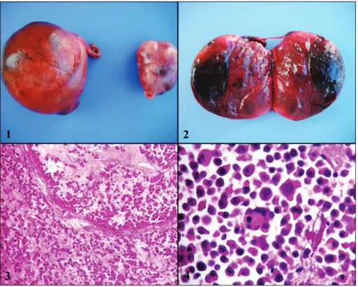
In present case, both testicle, which submitted to Department of Pathology, Faculty of Veterinary Medicine in Ankara University for diagnosis, were evaluated belong to 6 years old and cryptorchidic male Terrier. Tissues were macroscopically evaluated and fixed in 10% formalin. After fixation, the tissues were processed routinely and embedded in paraffin, cut the 5 μ thickness. the sections were deparaffinizated, dehydrated and stained with Hematoxylin-Eosin (HxE). Macroscopically, right testicle was weighed of 317 g, diametered in
11x9x7 cm and left one respectively 64 g and 4x3x4 cm. Both testicles had firmness consistency and lobullary (Figure 1). On their cut sections, a wide brownish-black colored and haemorragic area were restricted by gray colored necrotic areas (Figure 2). Microscopically, multiple masses in different size which composed of polihedral shaped neoplastic cells that had vesicular nuclei and basophilic cytoplasm. And also, nuclei of some neoplastic cells contained mitotic figures (Figure 3, 4). In some areas, widely haemorragic areas and lymphositic infiltration were seen.
In general, tumors are occurred bilaterally or unilaterally in right testicle in elderly dogs (1, 2, 7, 9). In the case, tumor was seen bilaterally in 6 years old Terrier. On the other hand, cryptorchidism is documented as a predispose factor (1, 3, 5, 8, 10). Cryptorchidism on both testicle was informed from anamnesis alike to documents before. Testicle tumors give rise to feminism, gynecomasti and growing in testicles. Moreover, the tumors can cause to prostatic and haematologic disorders, skin diseases and also perianal tumors (4, 6, 8, 11). In the case, general constitution and condition of the dog are well informed in anamnesis. Seminomas are usually documented in right testicle and can be found in solitary or multiple masses. They generally had pulpy consistency (4, 9, 10). Cut sections can be coincided with
Nihat Yumuşak - Murat Çalışkan - Osman Kutsal 152
haemorragie and necrosis. However, both testicles had firmness consistency and right testicle were overgrowth more than other in the case. Furthermore, both cut section contain some necrotic and haemorraghic areas. Seminomas is usually benign and had low malignancy (7, 8, 9). On the other hand, it is mentioned that malignant ones metastasize to mostly sublumbar lymph nodes and rarely internal organs (7). However, the dog is still healthy and it is monitorized periodically in radiography with respects to metastasis. It is not reported encountered up to now.
In conclusion, the case is found original in terms of occurring in middle age, diffuse type and showing malignancy despite of being not metastasize to anywhere.
References
1. Ciaputa R, Nowak M, Kiełbowicz M, Antończyk A, Błasiak K, Madej JA (2012): Seminoma, sertolioma, and
leydigoma in dogs: clinical and morphological correlations. Bull Vet Inst Pulawy, 56, 361-367.
2. Grieco V, Riccardi E, Greppi GF, Teruzzi F, Iermanò V, Finazzi M (2008): Canine testicular tumours: a study
on 232 dogs. J Comp Pathol, 138, 86-89.
3. Grieco V, Rondena M, Romussi S, Stefanello D, Finazzi M (2004): Immunohistochemical characterization of the
leucocytic infiltrate associated with canine seminomas. J
Comp Path, 130, 278-284.
4. Kim O, Kim KS (2005): Seminoma with hyperesterogenemia
in a Yorkshire Terrier. J Vet Med Sci, 67, 121-123.
5. Kutsal O (2003): 1971-2001 yılları arasında incelenen köpek
testis tümörleri. Ankara Üniv Vet Fak Derg, 50, 217-218.
6. Liao AT, Chu PY, Yeh LS, Lin CT, Liu CH (2009): A
12-year retrospective study of canine testicular tumors. J
Vet Med Sci, 71, 919-923.
7. MacLachlan NJ, Kennedy PC (2002): Tumours of the
genital system. 547-573. In: DJ Meuton, (ed), Tumors in
Domestic Animals. 4 th ed. lowa State Press, Iowa. 8. Masserdotti C, Bonfanti U, De Lorenzi D, Tranquillo M,
Zanetti O (2005): Cytologic features of testicular tumours
in dog. J Vet Med A Physiol Pathol Clin Med, 52, 339-346.
9. Ozsoy SY, Kutsal O (2007): Bir köpekte malign seminom. Ankara Üniv Vet Fak Derg, 54, 65-66.
10. Peters MA, de Rooij DG, Teerds KJ, Van De Gaag I, Van Sluijs FJ (2001): Spermatogenesis and testicular
tumours in ageing dogs. J Reprod Fertil Suppl, 57, 419-421.
11. Reimann-Berg N, Murua Escobar H, Nolte I, Bullerdiek J (2008): Testicular tumor in an 25 dog. Cancer Genet Cytogenet, 183, 114-116.
Geliş tarihi: 21.08.2013 / Kabul tarihi: 31.10.2013
Address for correspondence:
Nihat Yumusak
Harran University, Faculty of Veterinary Medicine Department of Pathology,
63000, Sanliurfa-TURKEY e-mail: [email protected]
Figure 1. Macroscopical appearance of both testis. Şekil 1. Her iki testisin makroskobik görünümü.
Figure 2. Cut section of right testis with haemorrhagic and necrotic areas. Şekil 2. Sağ testisin kesit yüzünde nekroz ve kanamalar.
Figure 3. Neoplastic cells both in tubulus seminiferus and its peripeheral invasion, HxE, x100. Şekil 3. Tubulus seminiferus ve çevresine neaplazik hücre invazyonları, HxE, x100.
Figure 4. Neoplastic cells with highly pleomorfic features, HxE, x400. Şekil 4. Pleomorfik özellikler gösteren tümör hücreleri, HxE, x400.