MRI Appearance Of Ectopic Axillary Breast Tissue During Lactational
Period
Ektopik Aksiller Meme Dokusunun Laktasyonel Dönemde MRG ile Değerlendirilmesi
Alper Dilli, İdil Güneș Tatar, Volkan Kızılgöz, Elif Rayegan Koç, Baki Hekimoğlu
Diskapi Yildirim Beyazit Training and Research Hospital, Radiology
Department, ANKARA Supernumerary nipples or breasts are found in 1-5% of the population. A 31-year-old woman presented with an axillary lump which had tenderness during postpartum period. On routine MRI sequences signal intensities similar to normal breast parenchyma but discontinuous with it were demonstrated. It is important to differentiate benign from malignant axillary masses to avoid unnecessary intervention. MRI has been reported to be a valuable tool for the diagnosis of accessory breast tissue especially in the peripubertal–pubertal girls and young patients. Key Words: MRI, ectopic breast tissue, lactational period
Popülasyonun %1-5’inde aksesuar meme dokusuna rastlanmaktadır. Otuzbir yașında bir kadın hasta postpartum dönemde ağrılı aksiller kitle ile bașvurdu. Rutin MRG sekanslarında aksillada normal meme parankim dokusu ile benzer özellik gösteren ancak ayrı olarak izlenen sinyal değișiklikleri belirlendi. Aksiller kitlelerde benign durumların malign lezyonlardan ayrılması gereksiz girișimlerden kaçınılması açısından önem tașımaktadır. MRG özellikle peripubertal-pubertal kız çocuklarında ve genç hastalarda aksesuar meme dokusu tanısında değerli bir modalite olarak bildirilmektedir.
Anahtar Sözcükler: MRG, ektopik meme dokusu, laktasyon dönemi.
In 1-5% of women and men supernumerary nipples (polithelia) and less frequently supernumerary breasts (polymastia, accessory breast) are found. Accessory breast tissue is most commonly found in axilla (1). It may be asymptomatic or present with pain and cosmetic problems. Symptoms often increase in puberty, pregnancy and lactation in response to hormonal stimulation
Case Report
A 31-year-old woman presented with a lump in the left axilla originally noticed 15 years ago. During her pregnancy tenderness occurred in this mass. There was no sign of nipple on the skin. Her mother had died of breast cancer nine years ago. After delivering her baby, she noticed swelling from this lump. With the presumptive diagnosis of lymhadenopathy she underwent
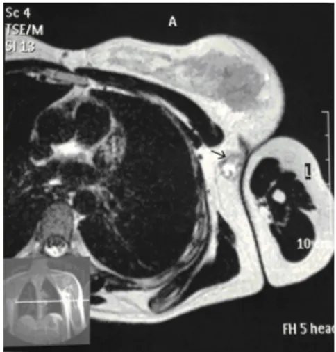
ultrasound examination. An echogenic area with the same appearance of normal breast tissue was demonstrated and accessory breast tissue was diagnosed. Since the patient had a family history of breast cancer, MRI at 1.5 T (Philips, Achieva, The Netherlands), without any intravenous contrast medium injection, was performed. MRI showed a poorly demarcated subcutaneous mass in the left axilla discontinuous with the breast. On T1 and T2-weighted images signal intensities similar to normal breast parenchyma were demonstrated but the mass was separate from the normal breast tissue (Figure 1-3). The dilated ductal structures were obvious on T2-weighted images. MRI appearance of the mass was diagnostic for accessory breast and a pathological condition was not observed.
Ankara Üniversitesi Tıp Fakültesi Mecmuası 2013, 66 (2)
DOI: 10.1501/Tıpfak_000000845
DAHİLİ TIP BİLİMLERİ/ MEDICAL SCIENCES Olgu Sunumu /Case Report
Received : 10.04.2012 Accepted: 10.02.2014 Corresponding Author
Dr. İdil Güneș Tatar
Irfan Bastug Street, Ankara, Turkey Phone : 596 26 16
E-mail Adress: [email protected]
Diskapi Yildirim Beyazit Research Hospital, Department of Radiology
Ankara Üniversitesi Tıp Fakültesi Mecmuası 2013, 66 (2)
MRI Appearance Of Ectopic Axillary Breast Tissue During Lactational Period
76
Discussion
During embryogenesis the galactic band extends from the axillae to the groins. Breast tissue continues to form only in the pectoral region. Failure of regression of the remainder of this galactic band gives rise to ectopic breast tissue. Although they can be located anywhere along the embryonic milk line extending from the axilla to the inguinal line, 60-70% of the accessory breast tissue occur in axilla (2). Since an overlying accessory areola or nipple is usually missing clinical diagnosis of the malignant conditions are often delayed.
Accessory breast tissue should be differentiated from ‘axillary tail of Spence’ which is defined as the extension of the breast tissue to the axilla.
Ectopic breast tissue is subject to the same physiological and pathological changes as in the eutopic breast tissue, including lactational changes, benign and malignant conditions. The most common pathology of the accessory breast is cancer followed by mastopathy and fibroadenoma (3). Cancer of the accessory breast tissue has been reported to be 0.3-0.6% of all breast cancers, occurring in axilla in 70-80% of the cases. The criteria of diagnosis are the discontinuity with the normal breast tissue, existence of normal breast tissue around the carcinoma, absence of metastatic carcinoma and absence of sudoriparous carcinoma (4).
When a female patient is diagnosed with a mass in axilla, first of all metastatic lymphadenopathy from breast cancer should be excluded. Lymph node involvement of lymphoma, and granulomatous
diseases (tuberculosis and sarcoidosis) should be ruled out. For the axillary masses lipoma, sabecceous cyst, hidraadenitis, vascular malformations such as cavernous hemangioma and venous malformation, lymphangioma are also in the differential diagnosis (5). In the differential diagnosis of the masses in the localization of the embryogenic galactic band, especially in the axilla, ectopic breast tissue and its pathologies must be considered. If the patient is in the lactational period galactocele should also be kept in mind (6). Other conditions such as renal anomalies, urogenital defects, vertebral anomalies, pyloric stenosis, congenital cytogenetic syndrome, urologic malignancies have been associated with accessory breasts (7, 8).
Conclusion
It is crucial to differentiate benign from malignant axillary masses to avoid unnecessary intervention. MRI has been reported to be a valuable tool for the diagnosis of accessory breast tissue in the peripubertal–pubertal girls (9). In this age group and in any patient with dense breast parachyme mammography has a low sensitivity in addition to its radiation load. MRI is a useful diagnostic method, which does not require ionizing radiation, to evaluate accessory breast tissue especially in young patients, particularly if they are lactating or have a family history of breast cancer.
Figure 1. Axial T2-weighted image
demonstrates a left axillary mass (arrow) isointense to normal breast parenchyma. Dilated ductal structures are seen inside the mass
Figure 2. Coronal T2-weighted HASTE
image shows an axillary mass (arrow) isointense to breast parenchyma and the dilated ductal structures inside.
Figure 3. Coronal T1-weighted image shows
a mass in the left axilla (arrow) which is isointense to normal breast parenchyma
Journal Of Ankara University Faculty of Medicine 2013, 66 (2)
Alper Dilli, İdil Güneș Tatar, Volkan Kızılgöz, Elif Rayegan Koç, Baki Hekimoğlu 77
REFERENCES
1. Dixon JM, Mansel RE. ABC of breast
diseases. Congenital problems and aberrations of normal breast development and involution. BMJ 1994; 309:797-800.
2. Burdick A, Thomas KA, Welsh E,
Powell J, Elgart GW. Axillary polmastia. J Am Acad Dermatol 2003; 49:1154-1156.
3. Smith GMR, Greening WP.
Carcinoma of aberrant breast tissue. Br J Surg 1972; 59:89-90.
4. Azuma T, Yamamoto K, Kobayashi T,
Nakano H. Accessory breast cancer: A case report of carcinoma originating from aberrant breast tissue in the axillar region. Breast Cancer 1997; 4:49-52.
5. Bertschinger K, Caduff R, Kubik-Huch
RA. Benign intramammary and axillary lesions mimicking malignancy. Eur Radiol 2000; 10:1029-1030.
6. Whang IY, Lee JH, Kim KT.
Galactocele as a changing axillary lump in a pregnant woman. Arch Gynecol Obstet 2007; 276:379-382.
7. Mehes K. Association of
supernumerary nipples with other anomalies. J Pediatr 1979; 94:274-275.
8. Mehes K, Szule E, Torzsok F,
Meggyessy V. Supernumerary nipples and urologic malignancies. Cancer Genet Cytogenet 1987; 24:185-188.
9. Laor T, Collins MH, Emery KH, et al. MRI appearance of accessory breast tissue: a diagnostic consideration for an axillary mass in a peripubertal or pubertal Girl. Am J Roentgenol 2004;183:1779-1781.
Ankara Üniversitesi Tıp Fakültesi Mecmuası 2013, 66 (2)
MRI Appearance Of Ectopic Axillary Breast Tissue During Lactational Period